


Abstract
Adverse drug reactions (ADRs) remain a major public health issue, and genetic factors contribute importantly to interindividual variability in drug response. Pharmacogenetic testing helps reduce ADR risk by optimizing drug selection and dosage, particularly in monogenic disorders. Whole-exome sequencing of 6739 samples from the Russian population was performed on the DNBSEQ-G400 platform (MGI). Variants in 48 genes were examined, focusing on inherited arrhythmias, enzyme deficiencies (Glucose-6-Phosphate Dehydrogenase Deficiency [G6PDD], Porphyrias), Dravet Syndrome (DS) and Malignant Hyperthermia (MH). Variants reported as pathogenic (P), likely pathogenic (LP), or variants of uncertain significance (VUS) in ClinVar were manually re-evaluated using ACMG criteria. A total of 75 unique variants in 18 genes were observed in 119 individuals (1.77%), including 21 carriers and 13 women with a G6PD mutation. Of these, 44 variants were classified as P, 24 as LP, and 7 as VUS. Missense variants accounted for the largest proportion (73.33%). The most affected genes were KCNQ1 (24/119), which exhibited the highest number of unique variants (18), G6PD (20/119), SCN1A (15/119), and RYR1 (14/119). Regarding associated conditions, mutations linked to arrhythmias were found in 51 individuals, MH in 27, G6PDD in 20, DS in 15, and Porphyrias in 6. Integrating common and rare clinically actionable genetic variants with attention to penetrance and clinical validity may improve medication safety, reduce preventable ADRs, and enhance personalized pharmacotherapy.
1. Introduction
An adverse drug reaction (ADR) is defined as a harmful and unintended response to a medication occurring at standard doses with an established causal relationship [1]. Although the risk of ADRs in elderly patients was reported to be four times higher than in younger individuals (16.6% vs. 4.1%), the proportion of preventable cases was 88% and 24%, respectively, in a 2002 meta-analysis [2]. A subsequent analysis of 5707 emergency hospitalizations, conducted twelve years later, indicated that 5% of cases were directly attributable to ADRs, with advanced age and the expected polypharmacy remaining the primary risk factors [3]. However, hospital-based ADR statistics differ: among 5644 cases studied from 2020 to 2023 in a Chinese hospital, severe ADRs (n = 408, 7.2%) were more common in middle-aged patients (46–65 years, 36.77%), predominantly female (two-thirds), frequently associated with intravenous administration (53.92%), and primarily affecting the circulatory system (53.19%) [4]. According to VigiBase data (2010–2019), fatal ADRs accounted for 1.3% (43,685 cases) of all reported reactions, with antineoplastic and immunomodulatory drugs (denosumab, lenalidomide, thalidomide) representing the leading cause [5]. Between Q3 2014 and Q3 2024, approximately 2.7 million ADR reports were registered in the FAERS database, with each report linking an event to a single drug [6]. Given that the FDA approved roughly 494 drugs during this period, the increase in FAERS reports is likely more reflective of changes in pharmacovigilance systems and awareness than of actual incidence [7].
Genetics plays a central role in ADR susceptibility, as UK Biobank data demonstrate that nearly all individuals (99.5%) carry at least one pharmacogenetic variant capable of eliciting an atypical drug response, and 24% of participants had already been exposed to the corresponding medications [8]. While widely implemented pharmacogenetic testing programs (e.g., PharmCAT [9]) focus on common polymorphisms (allele frequency > 3%) to optimize dosing of frequently used medications or guide therapeutic substitutions, the clinical significance of rare variants associated with monogenic syndromes remains insufficiently explored. The presence of such variants implies a life-threatening risk of severe idiosyncratic reactions upon exposure to specific medications during essential treatment courses [10]. ADRs can manifest for the first time in advanced age, as illustrated by a case of malignant hyperthermia (MH) in a 79-year-old woman following succinylcholine administration in the absence of prior genetic testing [11].
The clinical utility of genetic screening for long QT syndrome (LQTS) in asymptomatic carriers has been demonstrated in a large Amish cohort carrying the KCNQ1 p.Thr224Met variant [12]. Among 124 carriers identified through population screening, 5.7% were found to be taking QT-prolonging medications at baseline; after genetic disclosure, 93% received appropriate beta-blocker therapy. Importantly, the oldest syncopal event in a carrier occurred at age 60 years triggered by an antidepressant, highlighting that the elderly, often on multiple medications, are additionally vulnerable. This example supports the value of proactive genotyping for monogenic syndromes, regardless of penetrance.
Motivated by such evidence, and given the increasing accessibility of whole-exome sequencing (WES) in the Russian Federation, it has become feasible to construct a population-level genetic risk profile for severe ADRs, which is particularly relevant for a country with pronounced ethnic diversity. Integration of such data into preoperative screening, targeted genetic testing, and clinical protocols could substantially reduce the risk of fatal outcomes.
2. Results
2.1. Overall Characteristics of Clinically Significant Variants
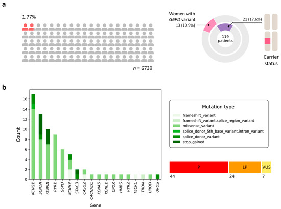
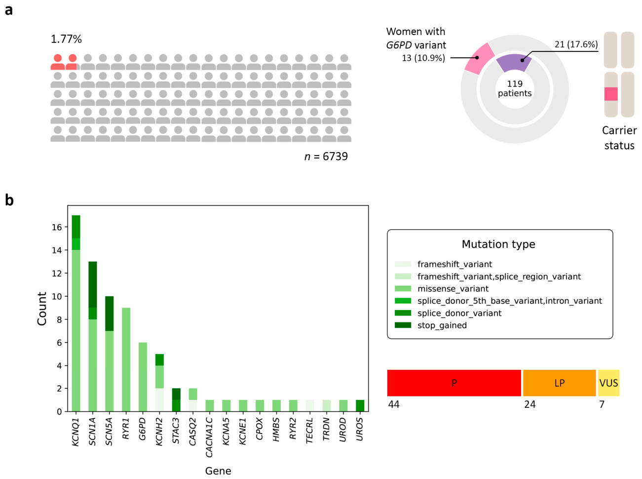
In the studied cohort, 119 individuals were identified with clinically significant rare variants associated with monogenic disorders predisposing to ADRs (Figure 1a). Among them, 21 individuals (17.65% of genotype-positive subjects) carried at least one clinically significant rare variant associated with a monogenic disorder conferring pharmacogenetic risk, corresponding to an overall prevalence of 1.77% (Figure 1a). Additionally, 13 women were carriers of variants in the G6PD gene (X-linked) (10.92%).
Figure 1.
Population distribution and genetic architecture of pharmacogenetically significant monogenic conditions. (a) Proportion of the screened population carrying clinically significant rare variants associated with monogenic syndromes predisposing to disease (n = 119, 1.77% of the total cohort). The distribution of carriers is shown separately: 21 individuals, including 13 women carrying variants in the X-linked G6PD gene. (b) Distribution of unique variants across genes associated with pharmacogenetically significant monogenic disorders: a total of 75 variants, including 44 pathogenic (P), 24 likely pathogenic (LP), and 7 variants of uncertain significance (VUS).
Six distinct G6PD missense variants were identified: Mediterranean, Kerala-Kalyan, Chatham, Nashville, Union, and Kaiping. All variants were classified as pathogenic in ClinVar and by ACMG criteria. According to the 2022 WHO classification, five variants fell into class B (moderate deficiency, associated with acute hemolytic anemia), while Nashville (rs137852316) was assigned to class A (severe deficiency with chronic non-spherocytic hemolytic anemia). The mean residual enzymatic activity in hemizygotes ranged from 6% (Nashville) to 57% (Mediterranean), with a weighted average of 27% across all variants. Reduced protein stability was reported for five of six variants (all except Kerala-Kalyan, where conflicting data exist).
A total of 75 unique genetic variants were identified across 18 genes (Figure 1b, Table S1): CACNA1C, CASQ2, CPOX, G6PD, HMBS, KCNA5, KCNE1, KCNH2, KCNQ1, RYR1, RYR2, SCN1A, SCN5A, STAC3, TECRL, TRDN, UROD, and UROS. According to ACMG classification, the majority of variants were interpreted as pathogenic (P; 44/75, 58.67%) or likely pathogenic (LP; 24/75, 32%), whereas 7 variants (9.33%) were classified as variants of uncertain significance (VUS). Missense mutations predominated (55 variants, 73.33%), while nonsense and frameshift mutations accounted for 17.33%, and splice-site variants comprised the remaining ~10% of detected alterations. Notably, we also examined variants located within 10 bp of exon boundaries due to acceptable coverage (>14×), which is an atypical approach for WES; the most distal variant identified was KCNQ1(NM_000218.3):c.477+5G>A in a single individual.
Our cohort included samples referred from multiple federal medical centers across Russia, capturing substantial genetic diversity beyond the Moscow region. To assess population stratification, we compared allele frequencies of the identified autosomal variants across three RUSeq genetic clusters (Eur.Rus, Caucasus, East Russia) using Fisher’s exact test with Bonferroni correction (15 comparisons). No statistically significant differences were observed after correction. The smallest raw p-value was obtained for TRDN p.Glu351ArgfsTer7 (p = 0.0257, padj = 0.3856 between Eur.Rus and Caucasus) and RYR1 p.Ala2367Thr (p = 0.0266, padj = 0.3988). For STAC3 p.Lys288Ter, the smallest p-value was 0.1171, and all other comparisons yielded p = 1. In contrast, the X-linked variant G6PD p.Ser188Phe demonstrated significant frequency differences across RUSeq subpopulations. Compared with the RUSeq Eur.Rus cluster, the variant was enriched in our cohort (OR = 14.97, padj = 0.0072), whereas the Caucasus cluster demonstrated higher frequencies than our cohort (OR = 0.22, padj = 0.0230). No significant difference was observed relative to the East Russia cluster. Thus, despite the known genetic heterogeneity of the Russian population, the frequencies of the pharmacogenetically relevant variants identified in this study do not differ significantly between the major regional subpopulations.
When comparing our cohort with the non-Finnish European (NFE) population from gnomAD v4.1.1, five autosomal variants showed significantly higher frequencies in our Moscow-based sample after Bonferroni correction: STAC3 p.Lys288Ter (OR = 16.18, padj = 3.75 × 10⁻⁹), KCNQ1 c.477+1G>A (OR = 27.31, padj = 0.00015), KCNQ1 p.Arg591Leu (OR = 175.13, padj = 0.0178), KCNH2 p.Ala79AspfsTer63 (OR = 174.65, padj = 0.0179), and SCN5A p.Val1404Met (OR = 58.36, padj = 0.0586, borderline). One X-linked pharmacogenetic variant remained significantly enriched after Bonferroni correction: G6PD p.Ser188Phe (OR = 7.23, padj = 6.24 × 10⁻⁷). These findings indicate that several pharmacogenetically relevant rare variants are enriched in the Russian population compared to the general European population, highlighting the importance of population-specific reference data for clinical interpretation.
2.2. Spectrum of Pharmacogenetically Relevant Genetic Syndromes
Data on ADRs for LQTS were obtained from the online resource CredibleMeds® (Drugs with Known Risk of TdP, Only Marketed Drugs) [13]; for Brugada syndrome (BrS), from BrugadaDrugs.org [14]; for glucose-6-phosphate dehydrogenase deficiency (G6PDD) and porphyrias, from the study by Micaglio et al. [10]; and for MH, from CPIC recommendations [15]. ADRs associated with other syndromes were assessed exclusively based on data from the published literature (Table 1).
Table 1.
Syndrome-specific adverse drug reactions relevant for clinical practice.
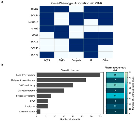
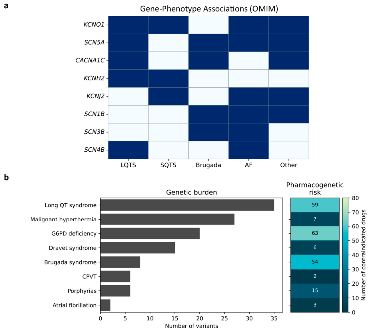
As shown in Figure 2a, several genes are associated with multiple clinical syndromes, highlighting the complexity of genotype–phenotype relationships underlying pharmacogenetic risk. Among all detected conditions, LQTS was the most frequent, observed in 35 individuals (29.41% of genotype-positive subjects), followed by MH (27 individuals, 22.69%) and G6PDD (20 individuals, 16.81%). Less frequent conditions included Dravet syndrome (DS; 15 individuals, 12.61%), BrS (8 individuals, 6.72%), porphyrias (6 individuals, 5.04%), catecholaminergic polymorphic ventricular tachycardia (CPVT; 6 individuals, 5.04%), and monogenic atrial fibrillation (AF; 2 individuals, 1.68%) (Figure 2b).
Figure 2.
Genetic and clinical heterogeneity of pharmacogenetically significant syndromes. (a) Overlap of hereditary arrhythmogenic syndromes at the gene level, demonstrating pleiotropy and the potential for a single gene to be associated with multiple phenotypes. Dark blue indicates the presence of a reported gene–phenotype association, whereas light blue indicates no established association. (b) Comparison of the number of patients with syndromes and the corresponding number of drugs associated with an increased risk of adverse drug reactions. Notes: LQTS—long QT syndrome, SQTS—short QT syndrome, AF—atrial fibrillation, CPVT—catecholaminergic polymorphic ventricular tachycardia.
Gene-level analysis revealed KCNQ1 as the primary contributor to LQTS, harboring the largest number of associated variants (24 variants, 18 unique). KCNH2 (n = 6, 5 unique) and SCN5A (n = 3) also significantly contributed to cardiac pharmacogenetic risk, reflecting their pleiotropic involvement in multiple arrhythmogenic syndromes. Additionally, single variants with corresponding carriers were detected in TRDN, CACNA1C, and KCNE1. RYR2 was the principal gene underlying CPVT; however, only one carrier was identified, whereas TECRL, TRDN, and CASQ2 variants collectively affected four additional individuals. Variants in HMBS, UROS, UROD, and CPOX causing porphyria were mostly singular occurrences, although UROS (NM_000375.3):c.63+1G>A was observed in three individuals; its clinical significance was lower due to carrier status compared to, for instance, CPOX(NM_000097.7):c.991C>T (p.Arg331Trp), which is associated with autosomal-dominant coproporphyria. Among individuals predisposed to MH, RYR1 variants predominated (9 unique variants; 14 carriers), whereas STAC3 variants were fewer in number (2 unique) but present in a similar number of carriers (13 individuals). Hemizygous G6PD variant carriers accounted for 35%, with no homozygous women identified. BrS in this cohort was associated exclusively with SCN5A variants.
Figure 2b summarizes, for each syndrome, both the number of affected patients and the corresponding number of drugs associated with increased ADR risk that should be avoided. Notably, LQTS and BrS were associated with the highest number of contraindicated drugs, emphasizing their particular relevance in the context of preventive pharmacogenomic screening.
For several recurrent variants, the following penetrance estimates were obtained from the literature: the RYR2 p.Arg420Trp variant associated with CPVT showed a penetrance of 25% [16]; the SCN1A p.Ala1669Val variant demonstrated complete penetrance in the reported family [17]; the CACNA1C p.Arg858His variant exhibited a penetrance of 50–60% [18]; both CASQ2 variants identified in our cohort were 100% penetrant (consistent with autosomal recessive inheritance); the G6PD p.Ser188Phe variant was found in 859 hemizygous males and 23 homozygous females in gnomAD v4.1.1, indicating low clinical penetrance; the KCNE1 p.Arg98Trp variant had an ECG penetrance (QTc > 460 ms) below 10% [19]; KCNH2 variants in our cohort were fully penetrant in the families where they were described; KCNQ1 variants consistently showed reduced penetrance, with some carriers presenting only ECG abnormalities without symptoms [20,21,22,23]; and a woman carrying the SCN5A p.Glu428Lys variant (classified as VUS in our study) had no QT prolongation [24].
3. Discussion
Among the 608 entries in the FDA table “Pharmacogenomic Biomarkers in Drug Labeling”, G6PDD (n = 41) and MH (n = 4) were predominantly represented [25], suggesting that the current pharmacogenetic regulatory framework is insufficiently adapted to account for risks associated with rare monogenic conditions. Nevertheless, out of 48 genes listed, 16 had documented associations in PharmGKB, as accessed via VarSome (retrieved 13 December 2025) [26]; KCNH2 ranked second after G6PD in terms of the number of associated drugs, while some single reported associations did not involve the primary phenotype (e.g., montelukast use in asthma and ABCC9 variants [27]). Importantly, carriers of variants in these genes may be required to avoid or use with extreme caution at least 179 drugs and other substances spanning multiple pharmacological classes, including analgesics and nonsteroidal anti-inflammatory drugs, antimalarial agents, antimicrobials (antibiotics, antifungals, and antiprotozoals), antiarrhythmic drugs, local and general anesthetics, anticonvulsants, psychotropic medications, diuretics, antidiabetic agents (particularly sulfonylureas), immunosuppressants, prokinetic agents, oncological and hematological therapies, antidotes, antigout medications, sympathomimetic agents, vitamins and vitamin-like compounds, as well as industrial and chemical substances with pharmacological effects. Notably, the studied cohort also comprised individuals harboring single alleles in genes with recessive inheritance, which, while not necessarily associated with overt disease, has important implications for genetic counseling and therapeutic decision-making.
Inherited arrhythmias represent a significant, albeit relatively rare, cause of sudden cardiac death (SCD). In the general population, SCD incidence ranges from approximately 1 per 100,000 in adolescents to 1 per 1000 among individuals aged 45–75, with a substantial proportion of sudden unexplained deaths in young adults and children (~10–15%) attributable to primary arrhythmogenic channelopathies in the absence of structural heart disease, such as LQTS, CPVT, and BrS [28]. CPVT is a less commonly recognized syndrome in predictive pharmacogenetics, most frequently caused by RYR2 variants. It manifests in children and adolescents during physical exertion or emotional stress, despite structurally normal hearts [29]. Its prevalence in Europe is estimated at approximately 1:10,000, and untreated disease carries a high risk of SCD, reaching up to 31% by age 30. A critical clinical feature of CPVT is that standard resuscitative and pharmacologic interventions, particularly catecholamine administration, may exacerbate arrhythmogenicity and lead to fatal outcomes [30].
Another condition often overlooked in pharmacogenomics is AF. GenCC classifies three genes as “Strong” (KCNQ1, GJA5, MYL4) and three as “Moderate” (KCNA5, NPPA, SCN2B) for monogenic AF. AF is one of the most prevalent arrhythmias, with a lifetime risk of 1:3–5 after age 45 and a global prevalence increase from 33.5 million to 59 million cases between 2010 and 2019 [31]. In our cohort, only SCN5A(NM_000335.5):c.1282G>A (p.Glu428Lys) (“Supportive”) and KCNA5(NM_002234.4):c.143A>G (p.Glu48Gly) variants were identified in two individuals, underscoring the limited representation of monogenic AF components detectable via WES. Despite the large number of drugs that can precipitate AF, only three are explicitly flagged for patients with pre-existing AF: acetylcholine, adenosine, and ibrutinib [32].
Although the classical neonatal estimate of LQTS prevalence (~1:2000) remains widely cited [33], population-based studies in Norway suggest that 1 in 100 individuals carries a pathogenic variant causing LQTS [34]. Limited Russian data also indicate a higher rate of QT interval prolongation (1.96%) detected among 2140 psychiatric patients [35]. The high population frequency of LQTS-associated variants combined with low clinical penetrance creates a substantial hidden-risk group in which life-threatening arrhythmias may be triggered by routine medications [36]. Notably, in 15–20% of individuals with a phenotypic LQTS presentation, no causative variant is identified; however, their clinical risk is comparable to genotype-positive patients [37]. In our study, this nosological group was the most frequently identified, representing 0.52% of the total cohort. Additionally, no patients with short QT syndrome (SQTS) were observed, likely reflecting the extremely low prevalence of this condition, with only approximately 43 clinically confirmed cases worldwide as of January 2025 [38].
BrS has an estimated global prevalence of approximately 1 in 2000 individuals, comparable to that of LQTS, with substantially higher rates reported in Southeast Asia—up to 35.5 per 1000 overall and up to 17.7 per 1000 in Thailand [39]. Although associations with BrS have been proposed for 22 genes to date, only SCN1B and SCN5A have achieved a “Definitive” level of evidence according to GenCC [40]. In BrS cohorts published between 2002 and 2022, genetic testing was performed in an average of 59% of patients, with an overall diagnostic yield of 26%. Both testing rates and diagnostic yield varied markedly across regions, ranging from 10% in Hong Kong to 100% in selected centers in China, Japan, France, and Spain, while diagnostic yield ranged from 10% in Japan to 67% in Belgium [41].
At the same time, it is important to distinguish true BrS from the Brugada ECG pattern, which may occur transiently and be induced by fever, medications, or electrolyte disturbances, without clinical manifestations of the disease. The Brugada pattern is considerably more common than overt BrS and is often detected in individuals without a family history of SCD, substantially complicating the interpretation of genetic findings and clinical risk assessment [42]. For example, a Brugada ECG pattern has been described in a patient with hypertrophic cardiomyopathy carrying pathogenic variants in MYBP3 and MYH7 [43]. Contemporary clinical observations and experimental data further indicate a close relationship between BrS and arrhythmogenic right ventricular cardiomyopathy/dysplasia (ARVC/D). In some patients, features of BrS and ARVC/D may coexist, combining characteristic Brugada ECG patterns with structural abnormalities of the right ventricle that meet diagnostic criteria for ARVC/D. In our study, no clinically significant PKP2 variants were identified; however, this gene was included based on its reported overlap with ARVC/D type 9 and its “Limited” evidence of association with BrS according to GenCC [44,45].
Variants in RYR1 are identified in the majority of patients with MH, accounting for approximately 50–60% of cases. Notably, pathogenic RYR1 variants are far more common in the general population (approximately 1 in 800 individuals), whereas a clinical MH crisis occurs in only 1 per 10,000–150,000 anesthetic exposures. Nevertheless, in the absence of timely treatment, MH-related mortality may reach up to 80% [46]. In our cohort, the number of carriers of RYR1 and STAC3 variants was approximately comparable when accounting for carrier status in all STAC3-positive individuals; however, the number of unique RYR1 variants exceeded that of STAC3 by a factor of 4.5. Patients with STAC3 variants frequently require surgical interventions due to multiple congenital anomalies, necessitating heightened vigilance during anesthesia. Given the autosomal recessive inheritance pattern, parents of affected individuals may be asymptomatic carriers [47]. The prevalence of STAC3-associated MH is difficult to estimate but appears to be extremely low based on the limited available literature [48].
Although the penetrance of RYR1 mutations is incomplete (estimated at 40.6% overall, with significantly higher penetrance in males [50% vs. 29.7% in females]) [49], the 2025 European Malignant Hyperthermia Group (EMHG) guidelines have introduced the diagnostic category MH genotype (MHG). According to these guidelines, any individual carrying a P or LP RYR1 variant is considered at increased risk of developing MH under anaesthesia and should be managed accordingly, regardless of penetrance estimates. Furthermore, the EMHG now provides a consensus definition of a clinical MH event and emphasises that functional characterization of variants remains key for pathogenicity classification [50].
DS is a rare epileptic encephalopathy with an incidence of approximately 2.2–6.5 cases per 100,000 individuals and a prevalence of 1.2–6.5 per 100,000 population (up to 0.0065%). In our cohort, the prevalence of genotype-positive DS was 0.22%, exceeding global estimates. Despite its rarity, DS is associated with high mortality (approximately 15.8 per 1000 patient-years, primarily due to sudden unexpected death in epilepsy [SUDEP]) and an earlier age at death [51]. It is well established that sodium channel blockers, particularly carbamazepine, oxcarbazepine, and lamotrigine, may exacerbate seizures and worsen cognitive outcomes in DS. Other antiepileptic drugs, including vigabatrin, rufinamide, and phenobarbital, may also increase seizure frequency in some patients [52]. The economic burden of DS is substantial, with costs estimated to be 1.5-fold higher than those for drug-resistant epilepsy and fivefold higher than for epilepsy in remission; mean annual costs have reached up to €29,872 in a multicenter German study [53].
Porphyrias constitute a group of rare disorders with prevalence varying widely by clinical subtype. In Europe, the most common forms—porphyria cutanea tarda, acute intermittent porphyria, and erythropoietic protoporphyria—occur at approximate frequencies of 1:10,000, 1:20,000, and 1:50,000–75,000, respectively. A decline in symptomatic acute hepatic porphyrias alongside increased detection of erythropoietic protoporphyria has been observed, likely reflecting improved diagnostics and reduced exposure to triggering factors [54]. In children, porphyrias often mimic common pediatric conditions, and clinical manifestations are frequently precipitated by medications and other external factors. Certain antiepileptic drugs (e.g., carbamazepine, phenytoin, valproate) are known to worsen disease severity. Given the overlap of clinical and biochemical features among porphyria subtypes, molecular confirmation is crucial for selecting safe therapies and identifying asymptomatic carriers within families [55].
G6PDD remains a major global public health concern. By 2021, its prevalence had reached 443 million cases worldwide, representing an increase of more than 80% compared with 1990, with the greatest burden observed in South Asia. The condition is more common in males, particularly in childhood and older age, and exhibits pronounced regional variability, underscoring the need for expanded screening programs and targeted public health interventions, especially in low- and middle-income countries [56]. In our cohort, G6PDD ranked third in frequency, with the important consideration that 13 women were identified as carriers.
4. Materials and Methods
4.1. Sample Collection
The study included 6739 anonymized WES datasets generated between 2020 and 2025. The cohort represents an unselected population-based sample, including neonates, pediatric and adult patients, as well as individuals undergoing carrier screening or preconception testing. Samples were primarily collected in Moscow hospitals (including multidisciplinary clinical centers and federal research institutions), with some patients referred from other regions. The cohort comprised 3462 females (51.4%) and 3277 males (48.6%). Although clinical and phenotypic data were available for a subset of samples, they were not incorporated into the present analysis, which was performed on anonymized exome data only.
4.2. DNA Sequencing and Bioinformatic Data Processing
DNA libraries were prepared from 500 ng of genomic DNA from peripheral blood samples using the MGIEasy Universal DNA Library Prep Set (MGI Tech, Shenzhen, China). DNA fragmentation was performed via ultrasonication (Covaris S-220, Covaris, Inc., Woburn, MA, USA) to an average fragment size of 250 bp. Exome enrichment was conducted using Agilent SureSelect Human All Exon v6/v7/v8 probes [57], and library concentrations were measured with Qubit Flex using the dsDNA HS Assay Kit (Invitrogen, Waltham, MA, USA). Library quality and fragment size distribution were assessed with Bioanalyzer 2100 (Agilent Technologies, Santa Clara, CA, USA). Libraries were circularized and sequenced in paired-end mode (PE100) on the DNBSEQ-G400 platform (MGI Tech), achieving an average coverage of 100×.
Raw sequencing reads (FastQ) were generated using basecallLite software (ver. 1.0.7.84) (MGI Tech) and quality-checked with FastQC v0.12.1 [58]. Low-quality bases and adapter sequences were trimmed using BBMap v38.96 [59]. Reads were aligned to the human reference genome GRCh38 with bwa-mem2 v2.2.1 [60], and BAM files were processed with SAMtools v1.9 [61]. Duplicate marking and enrichment metrics were obtained using Picard v2.22.4 [62]. Variant calling was performed using bcftools v1.9 [63] and DeepVariant v1.5.0 [64], followed by annotation with VEP v113 [65]. Potential sample relatedness was assessed during cohort-level quality control using pairwise PI_HAT estimates (threshold = 0.35). Finally, MultiQC v1.16 [66] was used for comprehensive quality control of the sequencing and analysis outputs.
4.3. Clinical Interpretation
Clinical significance of all identified variants, which had been reported at least once as pathogenic (P) or likely pathogenic (LP) in ClinVar, along with variants occasionally classified as of uncertain significance (VUS), was assessed according to ACMG/AMP guidelines [67], using the Varsome online platform [26] as an ACMG calculator, supplemented by a literature-based phenotype filter. Variants with an initial ACMG-based classification of VUS could be upgraded to LP if at least two independent publications reported a clear association with the relevant phenotype. Variants lacking any phenotype-supporting publication could be designated as LP at most, even if their ACMG score reached the P level. Variants that remained VUS after this assessment were included in Table S1 only if they had been reported at least once as P/LP in ClinVar; they were retained because, for some syndromes (notably MH/RYR1), many potentially actionable variants remain VUS due to the inherent difficulty of functional validation. In cases where a single rsID corresponded to multiple alternative substitutions at the same genomic position, all annotated variants were additionally evaluated independently using the ACMG/AMP classification framework. Variants were additionally reviewed manually in IGV [68]. The analysis focused on genes associated with inherited arrhythmia syndromes (including long and short QT syndromes, BrS, Timothy syndrome, Andersen–Tawil syndrome, AF, and CPVT), MH, G6PDD, porphyrias, and DS. Gene–disease associations were primarily determined based on data from OMIM [69], with additional validation of the level of evidence for gene–disease relationships using the GenCC database (not lower than the “Limited” level of evidence) [70]. As an exception, the TRPM4 gene was included because loss-of-function (LOF) variants in this gene have been reported to cause BrS [71]. In the interpretation of RYR1 variants associated with MH, we applied not only the standard ACMG/AMP criteria but also the disease-specific recommendations of the ClinGen Malignant Hyperthermia Susceptibility (MHS) Variant Curation Expert Panel, developed specifically for RYR1. These criteria account for gene-specific features of the available evidence, including modified application of the PS1, PM5, and PS4 rules, as well as the high prevalence of VUS [72]. For the interpretation of G6PD variants, we additionally followed the ACMG/AMP-based framework proposed by Geck et al., which incorporates functional data (activity in red blood cells and model systems), population frequency thresholds (PM2 < 0.001), and the use of PS4_Moderate (reports of variants in unrelated individuals) and PM1 (location in critical functional domains: NADP-binding, substrate-binding, or dimerization domain) [73].
It was taken into account that several genes are involved in the pathogenesis of multiple clinical syndromes, reflecting the overlapping nature of molecular mechanisms and increasing their clinical and pharmacogenetic relevance. The custom gene panel comprised 48 genes: ABCC9, ALAD, ALAS2, ANK2, CACNA1C, CACNA1S, CACNB2, CALM1, CALM2, CALM3, CASQ2, CAV3, CPOX, FECH, G6PD, GJA5, GPD1L, HCN4, HMBS, KCNA5, KCND3, KCNE1, KCNE2, KCNE3, KCNH2, KCNJ2, KCNJ5, KCNQ1, MYL4, NPPA, NUP155, PKP2, PPOX, RYR1, RYR2, SCN1A, SCN1B, SCN2B, SCN3B, SCN4B, SCN5A, SNTA1, STAC3, TECRL, TRDN, TRPM4, UROD, UROS. Among these, 32 genes were associated exclusively with autosomal-dominant syndromes under study, 8 exclusively with autosomal-recessive conditions, 6 with both autosomal-dominant and autosomal-recessive inheritance, and 2 genes were located on the X chromosome.
4.4. Population Frequency Comparisons
For population frequency comparisons, allele frequencies in the study cohort were compared with reference populations from gnomAD v4.1.1 NFE and RUSeq population clusters (Eur.Rus, Caucasus, and East Russia) using two-sided Fisher’s exact test. Multiple-testing correction was performed using the Bonferroni method separately for autosomal and X-linked analyses due to differences in inheritance patterns and allele counting strategies. Odds ratios (ORs) were calculated using 2 × 2 contingency tables. For rare variants containing zero counts in one or more cells, the Haldane–Anscombe correction (+0.5 added to each cell) was applied to avoid infinite OR estimates.
5. Conclusions
For the first time in a large cohort of 6739 individuals recruited in the Russian population, we identified clinically relevant variant findings associated with ADRs in 1.77% of individuals. LQTS and inherited arrhythmias were the most frequently identified conditions, which, in the absence of adequate clinical awareness, may lead to SCD, followed by MH, which is of particular relevance in the surgical and anesthetic setting. Incorporation of extended analyses of monogenic syndromes into standard pharmacogenomic testing panels has the potential to improve pharmacotherapy safety, provided that the identified variants are interpreted with attention to variant-specific penetrance, clinical validity, and the patient’s overall clinical context (e.g., family history, planned procedures, potential triggers). Further studies are needed to establish the clinical utility of this approach.
6. Limitations of Our Study
We focused on variants previously reported as P or LP in ClinVar, excluding those annotated as benign, which likely led to an underestimation of the true population prevalence of causative variants. This is particularly relevant for variants with high ACMG classification scores that have not yet been described in clinical practice. It should be noted that LOF variants predicted to be disease-causing, even when identified for the first time in a given individual, are important for personalized therapy and for assessing the risk of severe ADRs, although they require further functional and clinical validation. In addition, we did not include genes with insufficiently robust evidence for disease association, even in cases where convincing animal models have been established, such as ZFHX3 in the context of AF [74].
The presence of a pathogenic variant does not invariably translate into a clinical phenotype: some identified variants may exhibit incomplete penetrance or require additional genetic or environmental factors for disease manifestation, as observed in susceptibility to MH. Moreover, because anonymized exome datasets lacked clinical, longitudinal, and drug-exposure data, we could not determine whether variant carriers had manifested the corresponding syndromes or experienced ADRs. Thus, the reported prevalence reflects carrier status for P/LP variants rather than the clinical prevalence of disease. This limits the direct translation of carrier status into an absolute probability of ADRs but does not exclude an increased genetic susceptibility.
Although our cohort was relatively large (n = 6739), it may not fully capture the ethnic and regional diversity of the Russian population; therefore, the observed frequencies require validation in independent cohorts.
Finally, WES has inherent limitations in detecting copy number variants and noncoding regions that may contribute to ADR risk, as well as other methodological constraints typical of next-generation sequencing approaches.
Supplementary Materials
The following supporting information can be downloaded at: https://www.mdpi.com/article/10.3390/ijms27114851/s1.
Author Contributions
Conceptualization, A.A.B.; methodology, A.A.B.; software, V.V.C., I.A.V., O.N.S., Z.A.R. and M.I.K.; formal analysis, A.A.B.; investigation, A.O.S., A.F.S. and V.A.B.; resources, D.O.K.; data curation, V.V.C. and M.I.K.; supervision, D.O.K.; writing—original draft preparation, A.A.B.; writing—review and editing, D.O.K.; visualization, A.A.B. All authors have read and agreed to the published version of the manuscript.
Funding
The work was supported by the Ministry of Science and Higher Education of the Russian Federation (Strategic Academic Leadership Program “Priority 2030”, agreement No. 075-15-2025-200/GTR-G).
Institutional Review Board Statement
This study was conducted in accordance with the Declaration of Helsinki, and approval was obtained from the Local Research Ethics Committee of Russian National Medical University (Protocol No. 241, from 26 June 2024), and all participants provided written informed consent prior to data collection.
Informed Consent Statement
Informed consent was obtained from all subjects involved in this study.
Data Availability Statement
The sequence data are generated from patient samples and therefore are only available under restricted access.
Conflicts of Interest
The authors declare no conflicts of interest. The funders had no role in the design of the study; in the collection, analyses, or interpretation of data; in the writing of the manuscript; or in the decision to publish the results.
References
- Potter, E.; Reyes, M.; Naples, J.; Dal Pan, G. FDA Adverse Event Reporting System (FAERS) Essentials: A Guide to Understanding, Applying, and Interpreting Adverse Event Data Reported to FAERS. Clin. Pharmacol. Ther. 2025, 118, 567–582. [Google Scholar] [CrossRef]
- Beijer, H.J.M.; De Blaey, C.J. Hospitalisations caused by adverse drug reactions (ADR): A meta-analysis of observational studies. Pharmacol. World Sci. 2002, 24, 46–54. [Google Scholar] [CrossRef]
- Komagamine, J. Prevalence of Urgent Hospitalizations Caused by Adverse Drug Reactions: A Cross-Sectional Study. Sci. Rep. 2024, 14, 6058. [Google Scholar] [CrossRef]
- Zhai, J.; Yan, H.; Zhang, J.; Yan, H.; Ma, J.; Zhang, S. A Comprehensive Analysis of Adverse Drug Reactions in 2020–2023: Case Studies. Front. Pharmacol. 2025, 16, 1628347. [Google Scholar] [CrossRef]
- Montastruc, J.; Lafaurie, M.; De Canecaude, C.; Durrieu, G.; Sommet, A.; Montastruc, F.; Bagheri, H. Fatal Adverse Drug Reactions: A Worldwide Perspective in the World Health Organization Pharmacovigilance Database. Br. J. Clin. Pharmacol. 2021, 87, 4334–4340. [Google Scholar] [CrossRef]
- Yang, R.; Yin, N.; Zhang, Y.; Faiola, F. Visualization of FDA Adverse Drug Reaction Reports: Development and Usability Study of the VisDrugs Web Server. JMIR Form. Res. 2025, 9, e71519. [Google Scholar] [CrossRef]
- De La Torre, B.G.; Albericio, F. The Pharmaceutical Industry in 2024: An Analysis of the FDA Drug Approvals from the Perspective of Molecules. Molecules 2025, 30, 482. [Google Scholar] [CrossRef]
- McInnes, G.; Lavertu, A.; Sangkuhl, K.; Klein, T.E.; Whirl-Carrillo, M.; Altman, R.B. Pharmacogenetics at Scale: An Analysis of the UK Biobank. Clin. Pharmacol. Ther. 2021, 109, 1528–1537. [Google Scholar] [CrossRef]
- Sangkuhl, K.; Whirl-Carrillo, M.; Whaley, R.M.; Woon, M.; Lavertu, A.; Altman, R.B.; Carter, L.; Verma, A.; Ritchie, M.D.; Klein, T.E. Pharmacogenomics Clinical Annotation Tool (Pharm CAT). Clin. Pharmacol. Ther. 2020, 107, 203–210. [Google Scholar] [CrossRef]
- Micaglio, E.; Locati, E.T.; Monasky, M.M.; Romani, F.; Heilbron, F.; Pappone, C. Role of Pharmacogenetics in Adverse Drug Reactions: An Update towards Personalized Medicine. Front. Pharmacol. 2021, 12, 651720. [Google Scholar] [CrossRef] [PubMed]
- Nakano, M.; Funayama, M.; Takata, T.; Wakisaka, R.; Koyama, G.; Koreki, A.; Ishida, T.; Uchida, H.; Mimura, M. Caution for Psychiatrists: Malignant Hyperthermia Risks with the Anesthetic Agent Succinylcholine (Suxamethonium) during Electroconvulsive Therapy. BMC Psychiatry 2024, 24, 411. [Google Scholar] [CrossRef] [PubMed]
- Streeten, E.A.; See, V.Y.; Jeng, L.B.J.; Maloney, K.A.; Lynch, M.; Glazer, A.M.; Yang, T.; Roden, D.; Pollin, T.I.; Daue, M.; et al. KCNQ1 and Long QT Syndrome in 1/45 Amish: The Road From Identification to Implementation of Culturally Appropriate Precision Medicine. Circ. Genom. Precis. Med. 2020, 13, e003133. [Google Scholar] [CrossRef] [PubMed]
- CredibleMeds: QTdrugs List//AZCERT, Inc.—Last Updated: 7 November 2025. Available online: https://crediblemeds.org (accessed on 13 December 2025).
- BrugadaDrugs.org. Safe Drug Use and the Brugada Syndrome. Available online: https://www.brugadadrugs.org/ (accessed on 14 December 2025).
- Gonsalves, S.G.; Dirksen, R.T.; Sangkuhl, K.; Pulk, R.; Alvarellos, M.; Vo, T.; Hikino, K.; Roden, D.; Klein, T.E.; Poler, S.M.; et al. Clinical Pharmacogenetics Implementation Consortium (CPIC) Guideline for the Use of Potent Volatile Anesthetic Agents and Succinylcholine in the Context of RYR 1 or CACNA 1S Genotypes. Clin. Pharmacol. Ther. 2019, 105, 1338–1344, Correction in Clin. Pharmacol. Ther. 2019, 106, 1408. [Google Scholar] [CrossRef]
- Bauce, B.; Rampazzo, A.; Basso, C.; Bagattin, A.; Daliento, L.; Tiso, N.; Turrini, P.; Thiene, G.; Danieli, G.A.; Nava, A. Screening for Ryanodine Receptor Type 2 Mutations in Families with Effort-Induced Polymorphic Ventricular Arrhythmias and Sudden Death. J. Am. Coll. Cardiol. 2002, 40, 341–349. [Google Scholar] [CrossRef]
- Cestèle, S.; Harper, A.J.; Marra, S.; Leroudier, N.; Rusconi, R.; Dhifallah, S.; Horrocks, I.A.; Kirkham, F.J.; Morgan, I.M.R.; Singh, J.; et al. Elicited Repetitive Daily Blindness Associated With Gain-of-Function SCN1A Variants and Responsiveness to Sodium Channel Blockers. Neurol. Genet. 2026, 12, e200352. [Google Scholar] [CrossRef]
- Gardner, R.J.M.; Crozier, I.G.; Binfield, A.L.; Love, D.R.; Lehnert, K.; Gibson, K.; Lintott, C.J.; Snell, R.G.; Jacobsen, J.C.; Jones, P.P.; et al. Penetrance and Expressivity of the R858H CACNA1C Variant in a Five-generation Pedigree Segregating an Arrhythmogenic Channelopathy. Mol. Genet. Genom. Med. 2019, 7, e00476. [Google Scholar] [CrossRef]
- Roberts, J.D.; Asaki, S.Y.; Mazzanti, A.; Bos, J.M.; Tuleta, I.; Muir, A.R.; Crotti, L.; Krahn, A.D.; Kutyifa, V.; Shoemaker, M.B.; et al. An International Multicenter Evaluation of Type 5 Long QT Syndrome: A Low Penetrant Primary Arrhythmic Condition. Circulation 2020, 141, 429–439. [Google Scholar] [CrossRef]
- Bdier, A.Y.; Al-Ghamdi, S.; Verma, P.K.; Dagriri, K.; Alshehri, B.; Jiman, O.A.; Ahmed, S.E.; Wilde, A.A.M.; Bhuiyan, Z.A.; Al-Aama, J.Y. Autosomal Recessive Long QT Syndrome, Type 1 in Eight Families from Saudi Arabia. Mol. Genet. Genom. Med. 2017, 5, 592–601. [Google Scholar] [CrossRef]
- Zhou, H.; Lai, W.; Zhu, W.; Xie, J.; Liu, X.; Shen, Y.; Yuan, P.; Liu, Y.; Cao, Q.; He, W.; et al. Genotype-Based Clinical Manifestation and Treatment of Chinese Long QT Syndrome Patients with KCNQ1 Mutations—R380S and W305L. Cardiol. Young 2016, 26, 754–763. [Google Scholar] [CrossRef]
- Chen, S.; Zhang, L.; Bryant, R.; Vincent, G.; Flippin, M.; Lee, J.; Brown, E.; Zimmerman, F.; Rozich, R.; Szafranski, P.; et al. KCNQ1 Mutations in Patients with a Family History of Lethal Cardiac Arrhythmias and Sudden Death. Clin. Genet. 2003, 63, 273–282. [Google Scholar] [CrossRef]
- Ereiz, J.; Cvitkušić Lukenda, K.; Vučić, D.; Bitunjac, I.; Silović, J.; Miškić, B. Familial Long QT Syndrome: A Case Report. Cardiol. Croat. 2023, 19, 126. [Google Scholar] [CrossRef]
- Wu, J.; Mizusawa, Y.; Ohno, S.; Ding, W.-G.; Higaki, T.; Wang, Q.; Kohjitani, H.; Makiyama, T.; Itoh, H.; Toyoda, F.; et al. A hERG Mutation E1039X Produced a Synergistic Lesion on IKs Together with KCNQ1-R174C Mutation in a LQTS Family with Three Compound Mutations. Sci. Rep. 2018, 8, 3129. [Google Scholar] [CrossRef] [PubMed]
- Table of Pharmacogenomic Biomarkers in Drug Labeling//U.S. Food and Drug Administration (FDA).—Last Updated: 23 September 2024. Available online: https://www.fda.gov/drugs/science-and-research-drugs/table-pharmacogenomic-biomarkers-drug-labeling (accessed on 13 December 2025).
- Kopanos, C.; Tsiolkas, V.; Kouris, A.; Chapple, C.E.; Albarca Aguilera, M.; Meyer, R.; Massouras, A. VarSome: The Human Genomic Variant Search Engine. Bioinformatics 2019, 35, 1978–1980. [Google Scholar] [CrossRef]
- Hirvensalo, P.; Tornio, A.; Neuvonen, M.; Tapaninen, T.; Paile-Hyvärinen, M.; Kärjä, V.; Männistö, V.T.; Pihlajamäki, J.; Backman, J.T.; Niemi, M. Comprehensive Pharmacogenomic Study Reveals an Important Role of UGT1A3 in Montelukast Pharmacokinetics. Clin. Pharmacol. Ther. 2018, 104, 158–168. [Google Scholar] [CrossRef] [PubMed]
- Gollob, M.; Spears, D. Genetics of Inherited Primary Arrhythmia Disorders. Appl. Clin. Genet. 2015, 8, 215–233. [Google Scholar] [CrossRef]
- Schneider, L.; Begovic, M.; Zhou, X.; Hamdani, N.; Akin, I.; El-Battrawy, I. Catecholaminergic Polymorphic Ventricular Tachycardia: Advancing From Molecular Insights to Preclinical Models. J. Am. Heart Assoc. 2025, 14, e038308. [Google Scholar] [CrossRef]
- Bellamy, D.; Nuthall, G.; Dalziel, S.; Skinner, J.R. Catecholaminergic Polymorphic Ventricular Tachycardia: The Cardiac Arrest Where Epinephrine Is Contraindicated*. Pediatr. Crit. Care Med. 2019, 20, 262–268. [Google Scholar] [CrossRef]
- Linz, D.; Gawalko, M.; Betz, K.; Hendriks, J.M.; Lip, G.Y.H.; Vinter, N.; Guo, Y.; Johnsen, S. Atrial Fibrillation: Epidemiology, Screening and Digital Health. Lancet Reg. Health-Eur. 2024, 37, 100786. [Google Scholar] [CrossRef]
- Tamargo, J.; Villacastín, J.; Caballero, R.; Delpón, E. Drug-Induced Atrial Fibrillation. A Narrative Review of a Forgotten Adverse Effect. Pharmacol. Res. 2024, 200, 107077. [Google Scholar] [CrossRef]
- Schwartz, P.J.; Stramba-Badiale, M.; Crotti, L.; Pedrazzini, M.; Besana, A.; Bosi, G.; Gabbarini, F.; Goulene, K.; Insolia, R.; Mannarino, S.; et al. Prevalence of the Congenital Long-QT Syndrome. Circulation 2009, 120, 1761–1767. [Google Scholar] [CrossRef]
- Berge, K.E.; Haugaa, K.H.; Früh, A.; Anfinsen, O.-G.; Gjesdal, K.; Siem, G.; Øyen, N.; Greve, G.; Carlsson, A.; Rognum, T.O.; et al. Molecular Genetic Analysis of Long QT Syndrome in Norway Indicating a High Prevalence of Heterozygous Mutation Carriers. Scand. J. Clin. Lab. Investig. 2008, 68, 362–368. [Google Scholar] [CrossRef]
- Malin, D.I.; Bulatova, D.R.; Shchustova, L.Y.; Gordeeva, R.P. Prevalence and risk Factors for long QT interval Syndrome in Patients of the City Psychiatric hospital. Mod. Ther. Ment. Disord. 2023, 3, 50–56. [Google Scholar] [CrossRef]
- Zaklyazminskaya, E.V.; Abriel, H. Prevalence of Significant Genetic Variants in Congenital Long QT Syndrome Is Largely Underestimated. Front. Pharmacol. 2012, 3, 72. [Google Scholar] [CrossRef]
- Shimamoto, K.; Dagradi, F.; Ohno, S.; Spazzolini, C.; Crotti, L.; Giovenzana, F.L.F.; Musu, G.; Pedrazzini, M.; Kusano, K.; Takegami, M.; et al. Clinical Features, Long-Term Prognosis, and Clinical Management of Genotype-Negative Long QT Syndrome Patients. JACC Clin. Electrophysiol. 2024, 10, 2584–2596. [Google Scholar] [CrossRef]
- Boulmpou, A.; Giannopoulos, A.; Papadopoulos, C.; Giannopoulos, G.; Papagiannis, I.; Zormpas, G.; Keivanidou, A.; Fidani, L.; Vassilikos, V. The Uncommon Phenomenon of Short QT Syndrome: A Scoping Review of the Literature. J. Pers. Med. 2025, 15, 105. [Google Scholar] [CrossRef]
- Vutthikraivit, W.; Rattanawong, P.; Putthapiban, P.; Sukhumthammarat, W.; Vathesatogkit, P.; Ngarmukos, T.; Thakkinstian, A. Worldwide Prevalence of Brugada Syndrome: A Systematic Review and Meta-Analysis. Acta Cardiol. Sin. 2018, 34, 267–277, Erratum in Acta Cardiol. Sin. 2019, 35, 192. [Google Scholar] [CrossRef]
- Xu, T.; Wang, S.; Wang, J.; Xing, J. Brugada Syndrome Update. Front. Physiol. 2025, 15, 1520008. [Google Scholar] [CrossRef]
- Chung, C.T.; Bazoukis, G.; Matusik, P.T.; Zaklyazminskaya, E.; Radford, D.; Rajan, R.; Liu, T.; Mok, N.S.; Lee, S.; Tse, G. Differences in the Rate and Yield of Genetic Testing in Patients With Brugada Syndrome: A Systematic Review and Analysis of Cohort Studies. Ann. Clin. Cardiol. 2023, 5, 11–16. [Google Scholar] [CrossRef]
- Adytia, G.J.; Sutanto, H. Brugada Phenocopy vs. Brugada Syndrome: Delineating the Differences for Optimal Diagnosis and Management. Curr. Probl. Cardiol. 2024, 49, 102566. [Google Scholar] [CrossRef]
- Leelaviwat, N.; Benjanuwattra, J.; Vutthikraivit, W.; Kewcharoen, J.; Trongtorsak, A.; Abdelnabi, M.; Del Rio-Pertuz, G.; Mekraksakit, P.; Thangjui, S.; Sethi, P. Genotypic Overlap Between Brugada Syndrome and Hypertrophic Cardiomyopathy or Brugada Phenocopy? J. Am. Coll. Cardiol. 2023, 81, 3870. [Google Scholar] [CrossRef]
- Jeong, J.H.; Lee, H.S.; Choi, Y.Y.; Kim, Y.G.; Shim, J.; Hwang, J.H.; Yun, S.G.; Cho, Y.J.; Kim, Y.-H.; Choi, J.-I. Clinical Role of Genetic Testing for the Brugada Syndrome Overlapping with Arrhythmogenic Cardiomyopathy. Int. J. Arrhythm. 2024, 25, 12. [Google Scholar] [CrossRef]
- Kataoka, S.; Serizawa, N.; Kitamura, K.; Suzuki, A.; Suzuki, T.; Shiga, T.; Shoda, M.; Hagiwara, N. An Overlap of Brugada Syndrome and Arrhythmogenic Right Ventricular Cardiomyopathy/Dysplasia. J. Arrhythm. 2016, 32, 70–73. [Google Scholar] [CrossRef]
- Saha, A.K.; Pinyavat, T. Genetic Testing for Malignant Hyperthermia Susceptibility—Threading the Needle in the Haystack. Genes 2025, 16, 1281. [Google Scholar] [CrossRef]
- Silva, M.S.; Nakamura, R.; Arjona, M.R.; Monaco, T.P.C.D.; Malito, M.L.; Sampaio, T.O.; Oliveira, S.L.D.; Magalhães, J.S.D.A.; Machado-Costa, M.C.; Silva, H.C.A. STAC3 Gene Congenital Myopathy and Malignant Hyperthermia: A Crossroads between Neurology and Anesthesia. Arq. Neuropsiquiatr. 2025, 83, 001–006. [Google Scholar] [CrossRef]
- Miller, D.M.; Daly, C.; Aboelsaod, E.M.; Gardner, L.; Hobson, S.J.; Riasat, K.; Shepherd, S.; Robinson, R.L.; Bilmen, J.G.; Gupta, P.K.; et al. Genetic Epidemiology of Malignant Hyperthermia in the UK. Br. J. Anaesth. 2018, 121, 944–952. [Google Scholar] [CrossRef]
- Ibarra Moreno, C.A.; Hu, S.; Kraeva, N.; Schuster, F.; Johannsen, S.; Rueffert, H.; Klingler, W.; Heytens, L.; Riazi, S. An Assessment of Penetrance and Clinical Expression of Malignant Hyperthermia in Individuals Carrying Diagnostic Ryanodine Receptor 1 Gene Mutations. Anesthesiology 2019, 131, 983–991. [Google Scholar] [CrossRef]
- Rüffert, H.; Gillies, R.; Hopkins, P.M.; Glahn, K.P.E.; Johannsen, S.; Kamsteeg, E.-J.; Hellblom, A.; Stowell, K.; Bilmen, J.; Klincova, M.; et al. European Malignant Hyperthermia Group 2025 Guidelines for the Investigation of Malignant Hyperthermia Susceptibility. Br. J. Anaesth. 2026, 136, 653–661. [Google Scholar] [CrossRef]
- Sullivan, J.; Benítez, A.; Roth, J.; Andrews, J.S.; Shah, D.; Butcher, E.; Jones, A.; Cross, J.H. A Systematic Literature Review on the Global Epidemiology of Dravet Syndrome and Lennox–Gastaut Syndrome: Prevalence, Incidence, Diagnosis, and Mortality. Epilepsia 2024, 65, 1240–1263. [Google Scholar] [CrossRef]
- Gao, C.; Pielas, M.; Jiao, F.; Mei, D.; Wang, X.; Kotulska, K.; Jozwiak, S. Epilepsy in Dravet Syndrome—Current and Future Therapeutic Opportunities. J. Clin. Med. 2023, 12, 2532. [Google Scholar] [CrossRef]
- Strzelczyk, A.; Kalski, M.; Bast, T.; Wiemer-Kruel, A.; Bettendorf, U.; Kay, L.; Kieslich, M.; Kluger, G.; Kurlemann, G.; Mayer, T.; et al. Burden-of-Illness and Cost-Driving Factors in Dravet Syndrome Patients and Carers: A Prospective, Multicenter Study from Germany. Eur. J. Paediatr. Neurol. 2019, 23, 392–403. [Google Scholar] [CrossRef]
- Ramanujam, V.S.; Anderson, K.E. Porphyria Diagnostics—Part 1: A Brief Overview of the Porphyrias. Curr. Protoc. Hum. Genet. 2015, 86, 17–20. [Google Scholar] [CrossRef] [PubMed]
- Chakraborty, A.; Muranjan, M.; Karande, S.; Kharkar, V. Porphyrias: Uncommon Disorders Masquerading as Common Childhood Diseases. J. Postgrad. Med. 2023, 69, 164–171. [Google Scholar] [CrossRef]
- Yu, Z.; Xiong, Q.; Wang, Z.; Li, L.; Niu, T. Global, Regional, and National Burden of Glucose-6-Phosphate Dehydrogenase (G6PD) Deficiency from 1990 to 2021: A Systematic Analysis of the Global Burden of Disease Study 2021. Front. Genet. 2025, 16, 1593728. [Google Scholar] [CrossRef]
- Belova, V.; Pavlova, A.; Afasizhev, R.; Moskalenko, V.; Korzhanova, M.; Krivoy, A.; Cheranev, V.; Nikashin, B.; Bulusheva, I.; Rebrikov, D.; et al. System Analysis of the Sequencing Quality of Human Whole Exome Samples on BGI NGS Platform. Sci. Rep. 2022, 12, 609. [Google Scholar] [CrossRef]
- Andrews, S. FastQC: A Quality Control Tool for High Throughput Sequence Data. 2010. Available online: http://www.bioinformatics.babraham.ac.uk/projects/fastqc/ (accessed on 20 November 2025).
- Joint Genome Institute. Available online: https://jgi.doe.gov/data-and-tools/software-tools/bbtools/bb-tools-user-guide/bbduk-guide/ (accessed on 20 April 2025).
- Li, H.; Durbin, R. Fast and Accurate Short Read Alignment with Burrows–Wheeler Transform. Bioinformatics 2009, 25, 1754–1760. [Google Scholar] [CrossRef]
- Li, H.; Handsaker, B.; Wysoker, A.; Fennell, T.; Ruan, J.; Homer, N.; Marth, G.; Abecasis, G.; Durbin, R. 1000 Genome Project Data Processing Subgroup The Sequence Alignment/Map Format and SAMtools. Bioinformatics 2009, 25, 2078–2079. [Google Scholar] [CrossRef]
- Picard Toolkit, version 2.22.4; Broad Institute: Cambridge, MA, USA, 2019. Available online: https://broadinstitute.github.io/picard/ (accessed on 20 April 2025).
- Li, H. A Statistical Framework for SNP Calling, Mutation Discovery, Association Mapping and Population Genetical Parameter Estimation from Sequencing Data. Bioinformatics 2011, 27, 2987–2993. [Google Scholar] [CrossRef] [PubMed]
- Poplin, R.; Chang, P.-C.; Alexander, D.; Schwartz, S.; Colthurst, T.; Ku, A.; Newburger, D.; Dijamco, J.; Nguyen, N.; Afshar, P.T.; et al. A Universal SNP and Small-Indel Variant Caller Using Deep Neural Networks. Nat. Biotechnol. 2018, 36, 983–987. [Google Scholar] [CrossRef]
- McLaren, W.; Gil, L.; Hunt, S.E.; Riat, H.S.; Ritchie, G.R.S.; Thormann, A.; Flicek, P.; Cunningham, F. The Ensembl Variant Effect Predictor. Genome Biol. 2016, 17, 122. [Google Scholar] [CrossRef]
- Ewels, P.; Magnusson, M.; Lundin, S.; Käller, M. MultiQC: Summarize Analysis Results for Multiple Tools and Samples in a Single Report. Bioinformatics 2016, 32, 3047–3048. [Google Scholar] [CrossRef] [PubMed]
- Richards, S.; Aziz, N.; Bale, S.; Bick, D.; Das, S.; Gastier-Foster, J.; Grody, W.W.; Hegde, M.; Lyon, E.; Spector, E.; et al. Standards and Guidelines for the Interpretation of Sequence Variants: A Joint Consensus Recommendation of the American College of Medical Genetics and Genomics and the Association for Molecular Pathology. Genet. Med. 2015, 17, 405–424. [Google Scholar] [CrossRef] [PubMed]
- Robinson, J.T.; Thorvaldsdóttir, H.; Winckler, W.; Guttman, M.; Lander, E.S.; Getz, G.; Mesirov, J.P. Integrative Genomics Viewer. Nat. Biotechnol. 2011, 29, 24–26. [Google Scholar] [CrossRef] [PubMed]
- Online Mendelian Inheritance in Man, OMIM®. McKusick-Nathans Institute of Genetic Medicine, Johns Hopkins University (Baltimore, MD). Available online: https://omim.org/ (accessed on 13 December 2025).
- DiStefano, M.T.; Goehringer, S.; Babb, L.; Alkuraya, F.S.; Amberger, J.; Amin, M.; Austin-Tse, C.; Balzotti, M.; Berg, J.S.; Birney, E.; et al. The Gene Curation Coalition: A Global Effort to Harmonize Gene–Disease Evidence Resources. Genet. Med. 2022, 24, 1732–1742. [Google Scholar] [CrossRef] [PubMed]
- Liu, H.; Chatel, S.; Simard, C.; Syam, N.; Salle, L.; Probst, V.; Morel, J.; Millat, G.; Lopez, M.; Abriel, H.; et al. Molecular Genetics and Functional Anomalies in a Series of 248 Brugada Cases with 11 Mutations in the TRPM4 Channel. PLoS ONE 2013, 8, e54131. [Google Scholar] [CrossRef]
- Johnston, J.J.; Dirksen, R.T.; Girard, T.; Hopkins, P.M.; Kraeva, N.; Ognoon, M.; Radenbaugh, K.B.; Riazi, S.; Robinson, R.L.; Saddic, L.A., III; et al. Updated Variant Curation Expert Panel Criteria and Pathogenicity Classifications for 251 Variants for RYR1-Related Malignant Hyperthermia Susceptibility. Hum. Mol. Genet. 2022, 31, 4087–4093. [Google Scholar] [CrossRef]
- Geck, R.C.; Powell, N.R.; Dunham, M.J. Functional Interpretation, Cataloging, and Analysis of 1,341 Glucose-6-Phosphate Dehydrogenase Variants. Am. J. Hum. Genet. 2023, 110, 228–239. [Google Scholar] [CrossRef]
- Jameson, H.S.; Hanley, A.; Hill, M.C.; Xiao, L.; Ye, J.; Bapat, A.; Ronzier, E.; Hall, A.W.; Hucker, W.J.; Clauss, S.; et al. Loss of the Atrial Fibrillation-Related Gene, Zfhx3, Results in Atrial Dilation and Arrhythmias. Circ. Res. 2023, 133, 313–329. [Google Scholar] [CrossRef]

Disclaimer/Publisher’s Note: The statements, opinions and data contained in all publications are solely those of the individual author(s) and contributor(s) and not of MDPI and/or the editor(s). MDPI and/or the editor(s) disclaim responsibility for any injury to people or property resulting from any ideas, methods, instructions or products referred to in the content. |
© 2026 by the authors. Licensee MDPI, Basel, Switzerland. This article is an open access article distributed under the terms and conditions of the Creative Commons Attribution (CC BY) license.