


Abstract
Radioguided surgery has become a key component of contemporary breast cancer care, supporting less invasive approaches while maintaining oncologic safety. This narrative review summarizes current practice and recent developments in radioguided breast and axillary surgery, from established molecular imaging workflows to emerging three-dimensional and intraoperative technologies. Modern breast cancer management is increasingly shaped by tumor biology and the widespread use of neoadjuvant systemic therapy, which is transforming surgical decision-making and driving a shift toward personalized, patient-tailored pathways. In this context, radioguided techniques help maintain procedural accuracy despite therapy-induced changes in breast and nodal anatomy, enabling reliable lesion localization and targeted management of the axilla. We discuss sentinel lymph node strategies and de-escalation concepts, including targeted axillary dissection (TAD) after neoadjuvant therapy using marked nodes and selective removal approaches. We also review localization methods, including radioactive seed–based techniques, and the expanding role of molecular imaging–guided surgery to support intraoperative decision-making. Particular attention is paid to technologies aimed at improving surgical precision and margin assessment, including portable/freehand SPECT concepts and intraoperative PET/CT-based specimen imaging for immediate evaluation of excised tissue. Finally, we highlight how artificial intelligence and digital tools may enable workflow optimization, navigation, image interpretation, and decision support, accelerating the transition toward individualized treatment. Overall, integrating molecular information with real-time 3D guidance can help tailor breast and axillary management to each patient while reducing morbidity.
1. Introduction
Breast cancer management has undergone substantial evolution, with surgical strategies increasingly aiming to balance oncologic effectiveness with preservation of patients’ quality of life. The rising incidence of breast cancer and widespread early detection have further increased the demand for minimally invasive surgical approaches, which are associated with fewer postoperative complications, shorter hospital stays, and reduced healthcare costs [1,2,3].
In parallel, molecular imaging technologies are being used more frequently across the breast cancer pathway, including diagnosis, staging, treatment guidance, and follow-up. This expansion has been supported by hybrid imaging modalities such as SPECT/CT, PET/CT, PET/MRI, and dedicated breast PET systems (dbPET), enabling more precise anatomical–functional correlation and improved procedural planning [2,4].
Within this therapeutic landscape, radioguided surgery has gained a central role, ranging from radioactive seed localization (RSL) and radioguided occult lesion localization (ROLL) to combined approaches for lesion and sentinel node targeting (SNOLL), as well as techniques for identifying pathologic nodes and guiding selective axillary surgery (e.g., MARI and targeted axillary dissection, TAD) [1,4]. These image-guided applications provide a platform for technological innovation and for more individualized surgical planning in breast cancer.
Radioguided surgery has become firmly established in clinical practice through sentinel lymph node biopsy (SLNB), enabling surgical de-escalation—particularly in axillary management—while maintaining staging accuracy in appropriately selected patients [5,6,7,8]. Importantly, current trends in breast and axillary surgery are increasingly influenced by tumor biology and by the widespread use of neoadjuvant systemic therapy, which can alter breast and nodal anatomy and make intraoperative localization and decision-making more challenging. In this setting, radioguided and molecular imaging–guided approaches can provide additional certainty and support personalized, patient-tailored treatment pathways [5,6,7,8].
This narrative review focuses on radioguided breast and axillary surgery in the context of molecular imaging and the emergence of devices capable of providing near–real-time three-dimensional intraoperative information, moving the field toward 3D navigation and minimally invasive precision surgery. We highlight current clinical applications, emerging technologies, and future directions that may facilitate personalized management across different clinical scenarios in breast cancer [1,2,3].
Despite the widespread adoption of radioguided techniques in breast cancer surgery, several clinically relevant gaps and areas of uncertainty persist. Axillary management remains highly variable across institutions, particularly in patients treated with neoadjuvant systemic therapy, where the optimal balance between oncologic safety and surgical de-escalation is still evolving [4,9]. Differences in guideline interpretation, access to hybrid imaging and marking technologies, and local expertise contribute to heterogeneous real-world practice. In addition, while intraoperative verification strategies are increasingly recognized as important, there is no consensus on how emerging molecular and imaging-based tools should be integrated into routine workflows [2,10].
At the same time, rapid technological convergence is reshaping the surgical landscape. Advances in molecular imaging, intraoperative navigation, three-dimensional guidance, specimen-based verification, and artificial intelligence are occurring in parallel, often addressed separately in the literatura [2,4,10]. Existing reviews frequently focus on isolated techniques or single steps of the surgical pathway, without providing an integrated, workflow-oriented perspective that connects preoperative imaging, intraoperative guidance, verification, and decision-making—particularly in the context of personalized and response-adapted breast cancer treatment.
Beyond nuclear and radioguided techniques, recent imaging-driven and AI-assisted diagnostic approaches—such as hyperspectral imaging and other emerging modalities—illustrate the broader technological convergence shaping personalized breast cancer care, although their direct intraoperative applicability remains limited [11,12].
In this setting, the present narrative review aims to provide an updated and integrative synthesis of radioguided breast and axillary surgery, explicitly linking molecular imaging, targeted axillary strategies, intraoperative navigation, and emerging digital tools. By framing these technologies within clinically meaningful scenarios, levels of evidence, and implementation considerations, this review seeks to support personalized surgical decision-making while highlighting both opportunities and current limitations in modern breast cancer care.
This article was conducted as a narrative review of the literature. Relevant publications were identified through searches of major biomedical databases, including PubMed and Scopus, with a focus on studies published over the last decade, complemented by seminal earlier works where appropriate. The review prioritizes clinically relevant evidence, including guideline documents, prospective and retrospective clinical studies, key feasibility and translational reports, and authoritative reviews in the fields of breast surgery, nuclear medicine, and molecular imaging. Emphasis was placed on literature addressing radioguided breast and axillary surgery, targeted axillary strategies, intraoperative imaging and navigation, specimen verification, and emerging digital and artificial intelligence–based tools, with the aim of providing an integrated, workflow-oriented perspective.
2. Evolution and Scope of Image-Guided and Radioguided Surgery
This section outlines the historical evolution and conceptual scope of image-guided and radioguided surgery, providing the foundation for current breast and axillary applications.
2.1. Evolution of Image-Guided Surgery (1950s–Present)
The evolution of image-guided surgery has been driven by the progressive integration of imaging modalities into intraoperative decision-making. Early milestones include the introduction of fluorescence-based angiography in the 1950s and the use of intraoperative X-ray devices and ultrasound in the 1960s, initially in cardiac surgery and neurosurgery. Intraoperative ultrasound expanded into general surgery during the 1970s and became a widely adopted tool for real-time anatomical guidance [3,13].
From the 1980s onward, nuclear medicine–based approaches gained relevance, as radiopharmaceuticals began to be used for the identification of lymph nodes and tumor targets, enabling intraoperative localization beyond what could be achieved with palpation or conventional imaging alone [2,3]. In parallel, other intraoperative targeting concepts have been explored (e.g., magnetic tracers, optoacoustic imaging, and Raman spectroscopy), reflecting a broader trend toward functional and molecular guidance in the operating room [2,3]. Since the 1990s, digital navigation systems based on computed tomography (CT) and magnetic resonance imaging (MRI) have further expanded the scope of surgical guidance across multiple specialties, paving the way for modern hybrid and three-dimensional workflows [2,3].
Overall, these developments have shifted the field from purely anatomical guidance toward techniques that integrate functional, molecular, and spatial information, aiming to increase precision while supporting less invasive surgical strategies—an evolution that is particularly relevant in breast cancer, where both axillary management and breast-conserving surgery benefit from accurate targeting and verification [2,3].
2.2. Definition and Core Principles of Radioguided Surgery
Radioguided surgery can be defined as a set of surgical techniques that use real-time radiation detection in the operating room to localize and guide the excision of tissues or structures that have been labeled with a radiotracer or radioactive source. In practice, a radiopharmaceutical (or a sealed radioactive source, such as a seed) is administered or placed before surgery to mark a target of interest—such as a sentinel lymph node, a suspicious lymph node, or a non-palpable lesion—allowing intraoperative detection with dedicated devices (e.g., handheld gamma probes and/or portable imaging systems) [1,13,14].
Several core principles underlie radioguided workflows: (i) target definition and labeling using a tracer/source with suitable physical and biological properties; (ii) preoperative mapping and planning, often supported by planar imaging and hybrid modalities (e.g., SPECT/CT), to describe target location and potential drainage patterns; (iii) intraoperative detection and navigation, providing audio-visual feedback to guide the approach and confirm target removal; and (iv) verification steps, including ex vivo assessment of the specimen or surgical bed, to reduce the risk of missed targets and to support procedural completeness [2,14,15]. These principles have enabled radioguided surgery to expand from sentinel node procedures to broader applications such as selective axillary strategies (e.g., marked-node retrieval within targeted axillary dissection) and radioguided localization in breast-conserving surgery [2,14,15].
3. Molecular and Hybrid Imaging as the Preoperative Backbone
This section reviews the role of molecular and hybrid imaging modalities as the preoperative backbone of radioguided breast and axillary surgery, highlighting their contribution to surgical planning and personalization.
3.1. SPECT/CT in Breast Cancer: Sentinel Node Mapping, Complex Drainage, and Surgical Planning
Preoperative lymphatic mapping is a cornerstone of radioguided breast surgery, particularly for sentinel lymph node biopsy (SLNB). Planar lymphoscintigraphy provides initial functional information; however, hybrid SPECT/CT improves anatomical localization and increases confidence when drainage is atypical or when targets are located in anatomically complex regions [15,16,17]. By combining the functional distribution of radiocolloids with CT-based morphology, SPECT/CT can better delineate sentinel nodes in relation to critical structures and may reveal additional nodes not clearly separated on planar imaging, especially in patients with high background activity near the injection site, obesity, or altered drainage pathways after prior surgery or radiotherapy [15,16,17].
In clinical practice, SPECT/CT supports individualized surgical planning by clarifying the number and location of sentinel nodes (axillary and extra-axillary), facilitating incision planning, and potentially reducing operative time and uncertainty. It is also particularly valuable when internal mammary chain drainage is suspected, where accurate localization may influence staging and adjuvant treatment decisions in selected patients [4,15].
3.2. PET/CT and PET/MRI: Where They Add Value and Key Limitations
PET/CT, most commonly with 18F-FDG, is widely used for staging and response assessment in breast cancer, particularly in locally advanced disease and in specific biological subtypes where metabolic imaging provides complementary information to conventional imaging [2,4]. PET-based approaches offer the advantage of whole-body assessment and can guide systemic treatment decisions; however, standard whole-body PET/CT has limited spatial resolution for small lesions and may be suboptimal for detecting very small foci or certain low-grade histologies [2,4].
PET/MRI combines metabolic information with the superior soft-tissue contrast of MRI and has been explored in scenarios where detailed local assessment is required. Nevertheless, cost, limited availability, workflow complexity, and heterogeneous evidence across clinical indications remain barriers to broad implementation. Importantly, for surgical decision-making at the breast and axillary level, PET-based information is most impactful when it can be translated into actionable localization strategies—either through radioguided targeting, hybrid approaches, or intraoperative specimen assessment workflows (addressed later in this review) [2,4].
3.3. Dedicated Breast PET (dbPET): Role and Evidence
Dedicated breast PET systems (dbPET) have been developed to overcome spatial resolution limitations of whole-body PET/CT by enabling high-resolution, breast-focused imaging. These systems can improve detection of small lesions and may provide additional information in dense breasts or in cases where conventional imaging yields equivocal findings [4,18]. Early clinical studies suggest that dbPET may show higher sensitivity for subcentimeter tumors compared with whole-body PET/CT, potentially aiding local assessment and characterization in selected settings [4,18].
Despite these advantages, dbPET currently has limited availability, and standardized indications are still evolving. Further comparative studies are needed to clarify its incremental value over established modalities (mammography, ultrasound, MRI, and standard PET/CT), and to define how dbPET findings should be integrated into surgical planning and personalized treatment pathways. Nevertheless, dbPET illustrates the broader trend toward higher-resolution molecular imaging that, when coupled with radioguided or intraoperative technologies, may support more precise and individualized breast cancer surgery [4,18].
4. Axillary Management in Breast Cancer
This section focuses on contemporary axillary management in breast cancer, emphasizing surgical de-escalation strategies, targeted approaches, and the impact of neoadjuvant systemic therapy on decision-making.
4.1. From ALND to SLNB and Surgical De-Escalation
Axillary management in breast cancer has changed profoundly over the past decades, moving from routine axillary lymph node dissection (ALND) toward less invasive approaches that preserve staging accuracy while reducing morbidity. Sentinel lymph node biopsy (SLNB), introduced in the 1990s, became the standard for clinically node-negative patients and has consistently demonstrated lower rates of lymphedema, shoulder dysfunction, and sensory morbidity compared with ALND, without compromising oncologic outcomes in appropriately selected patients [6,8].
Landmark randomized trials and long-term follow-up studies have supported further de-escalation of axillary surgery in selected settings. Together with modern systemic therapy and radiotherapy, evidence from trials such as ACOSOG Z0011 and IBCSG 23-01, as well as multicenter experiences, has contributed to a paradigm in which completion ALND may be omitted in subsets of patients with limited sentinel-node involvement, provided that multidisciplinary management is optimized [7,19,20]. Consequently, the “best” axillary procedure is increasingly determined by clinical stage, tumor biology, planned systemic therapy, and radiotherapy fields—highlighting the need for personalized, guideline-informed decision-making [5,6].
4.2. Neoadjuvant Systemic Therapy and Post-NACT Staging Challenges
The expanding use of neoadjuvant systemic therapy (NACT) has further transformed axillary surgery. NACT is now frequently considered not only in locally advanced disease but also in earlier stages for specific biological subtypes, with the dual aim of improving systemic control and enabling surgical downstaging. As a result, the axillary status at diagnosis and the response to therapy must be integrated into surgical planning [5,6].
However, NACT can induce fibrosis and alter lymphatic drainage patterns, which may affect SLNB performance when used as a post-NACT staging tool—particularly in patients who are clinically node-positive at diagnosis and convert to ycN0. Early prospective studies highlighted concerns about detection rates and false-negative rates when SLNB is performed after NACT in this subgroup, prompting the development of strategies to improve accuracy [5].
In this evolving landscape, axillary management is no longer “one-size-fits-all.” Instead, it requires a workflow that aligns imaging, marking, surgery, and pathology. A multidisciplinary approach—radiology, nuclear medicine, breast/oncologic surgery, radiation oncology, and pathology—is essential to ensure that the intended targets are identified and retrieved, and that the pathological information obtained is meaningful for subsequent treatment decisions [2,5,6].
4.3. Targeted Approaches After NACT: MARI, TAD, RISAS, and Marking Strategies
To address the limitations of post-NACT SLNB in initially node-positive patients, targeted strategies have been developed to ensure retrieval of the originally involved node. A key concept is marking the biopsy-proven positive node before NACT and selectively removing it at surgery, either alone (targeted lymph node biopsy, TLNB) or in combination with SLNB [5,21,22].
One established approach is the MARI procedure (marking the axillary node with a radioactive iodine-125 seed), in which an I-125 seed is placed in a biopsy-proven positive node before NACT and the marked node is selectively removed after therapy using intraoperative detection. This strategy aims to reduce uncertainty arising from altered lymphatic drainage by anchoring the surgical target to the disease-proven node [5,21,22].
A broader framework is targeted axillary dissection (TAD), which typically combines removal of the marked node (clip/seed/other marker) with SLNB to improve staging accuracy and reduce false-negative rates compared with SLNB alone in selected patients after NACT [21,22]. Variations in marking methods (clips with intraoperative localization, radioactive seeds, magnetic markers, radar/reflector systems) reflect local resources and expertise, but all require a well-defined workflow and close coordination between radiology, nuclear medicine, surgery, and pathology [21,22].
Prospective and multicenter experiences have supported the diagnostic performance of targeted approaches. For example, approaches combining marked-node removal with SLNB (including strategies such as RISAS) have reported favorable identification and false-negative rates in selected post-NACT populations, supporting their role as less invasive alternatives to routine ALND when guideline criteria are met and adequate multidisciplinary infrastructure is in place [5,22]. Importantly, patient selection remains central: the extent of nodal disease at presentation, imaging response, number of suspicious nodes, and planned radiotherapy influence whether targeted strategies can safely replace ALND in individual cases [5,22].
From a practical standpoint, appropriate patient selection is central to the safe implementation of targeted axillary strategies after neoadjuvant systemic therapy. Key factors include the initial nodal burden at diagnosis (e.g., limited versus extensive nodal involvement), radiologic response of the axilla to therapy, the number of biopsy-proven positive nodes identified and marked before treatment, and the feasibility of reliably retrieving all intended targets at surgery. In addition, planned radiotherapy fields and the overall multidisciplinary treatment strategy influence whether targeted approaches can replace completion axillary lymph node dissection in individual patients.
In clinical practice, targeted axillary dissection is most commonly considered in patients who present with limited node-positive disease at diagnosis and convert to ycN0 after neoadjuvant therapy, provided that the originally involved node has been successfully marked and can be retrieved. Combining removal of the marked node with sentinel lymph node biopsy improves staging confidence and reduces false-negative rates compared with sentinel node biopsy alone. Conversely, patients with a high initial nodal burden, poor radiologic response, or inability to reliably retrieve marked targets may still benefit from more extensive axillary surgery. Thus, rather than a one-size-fits-all solution, targeted strategies should be applied within a structured, multidisciplinary workflow aligned with current guidelines and institutional expertise.
4.4. Tracers, Injection Approaches, and the Role of SPECT/CT
For SLNB, the most commonly used radiotracers are 99mTc-labeled colloids (e.g., sulfur colloid in the United States and 99mTc-nanocolloids in Europe). Injection technique varies (peritumoral, intratumoral, intradermal, subareolar/periareolar), and different approaches may influence visualization of extra-axillary drainage. Many centers favor superficial injections for practicality and rapid lymphatic visualization, while deeper injections may increase identification of alternative drainage pathways in selected higher-risk situations [1,4,16].
Because axillary decision-making is increasingly individualized, preoperative imaging is not simply “confirmatory”—it is part of the workflow that supports accurate surgery. Hybrid SPECT/CT can improve localization of sentinel nodes, clarify Berg level distribution, and identify additional nodes that may be missed on planar imaging. This is particularly valuable in patients with prior breast/axillary procedures, ipsilateral recurrence, obesity, or complex drainage patterns, where accurate mapping can influence both surgical approach and radiotherapy planning [4,16].
4.5. Intraoperative Detection Tools: Probes, Portable Gamma Cameras, and Workflow Integration
Intraoperatively, radioguided procedures rely on real-time detection, most commonly using a handheld gamma probe optimized for low- to medium-energy photons. Portable gamma cameras can provide 2D images that complement the auditory/numeric feedback of probes and may facilitate faster localization, assessment from different angles, and verification of target removal—particularly when targets are deep, multiple, or close to the injection site [14,17,23,24].
Crucially, the benefits of these technologies depend on workflow integration. Successful axillary personalization requires (i) reliable preoperative identification and/or marking of the intended targets, (ii) clear intraoperative localization strategies, (iii) standardized specimen handling and pathological assessment (including confirmation that the marked node and sentinel nodes have been retrieved), and (iv) coordinated postoperative decision-making. When these steps are aligned, radioguided and targeted approaches can support de-escalation while maintaining oncologic confidence—helping tailor axillary management to each patient in the modern era of biology-driven and response-adapted breast cancer therapy [14,17,23,24].
5. Breast Surgery: Localization and Margin Assessment
This section addresses radioguided localization techniques in breast-conserving surgery and the ongoing challenge of margin assessment, highlighting both established workflows and emerging solutions.
5.1. Localization Techniques: ROLL, RSL, SNOLL—Strengths and Limitations
Breast-conserving surgery (BCS) is a standard approach for many patients with early-stage breast cancer and is commonly combined with radiotherapy, achieving oncologic outcomes comparable to mastectomy in appropriately selected cases while offering advantages in cosmetic results and quality of life [25].
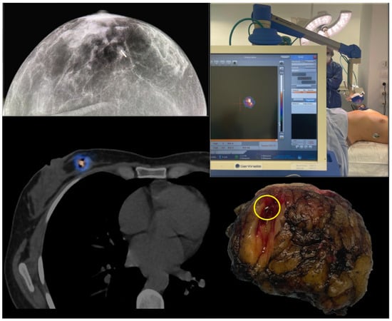
As screening and imaging improve, an increasing proportion of breast cancers present as non-palpable lesions, making accurate localization essential for successful BCS. Several radioguided strategies have been developed to translate preoperative imaging information into precise intraoperative targeting. Radioguided occult lesion localization (ROLL) uses intralesional or perilesional injection of a radiotracer to guide excision with a gamma probe. Radioactive seed localization (RSL) uses a sealed radioactive source (commonly I-125) placed under imaging guidance to mark the lesion and facilitate flexible scheduling and reliable intraoperative detection (Figure 1). Sentinel node and occult lesion localization (SNOLL) combines breast lesion targeting with sentinel node mapping, allowing coordinated management of breast and axilla within a unified radioguided workflow [1,4].
Figure 1.
Radioactive seed localization (RSL) using an I-125 seed for a non-palpable breast tumor. Upper left: mammographic image demonstrating the radioactive seed positioned within the target lesion. Lower left: multimodal SPECT/CT image showing radiotracer uptake from the seed and its precise anatomical localization on CT. Upper right: intraoperative localization of the radioactive seed using a portable gamma camera over the patient in the operating room. Lower right: macroscopic view of the surgical specimen after lumpectomy, confirming the presence of the radioactive seed within the excised tumor.
In ROLL-based workflows, 99mTc-labeled macroaggregated albumin (99mTc-MAA) can be used as a robust local tracer for lesion marking, providing a well-defined intraoperative signal that supports targeted excision in non-palpable disease and complements conventional specimen radiography/ultrasound depending on the clinical setting [26].
Beyond single-point targeting, I-125 seeds can also be deployed strategically to support more individualized resections, for example by marking not only the lesion center but also selected borders or anatomical landmarks to guide tailored excision planes in challenging cases. This “multi-marker” concept may be particularly helpful after neoadjuvant therapy or when oncoplastic planning requires precise spatial control. In addition, I-125 seeds generally produce minimal or no MRI artifacts compared with some alternative localization materials, which may facilitate integration with MRI-based planning in selected workflows [1].
Across techniques, radioguided localization can provide high targeting accuracy and intraoperative confidence, particularly when closely integrated with radiology and nuclear medicine workflows. Practical differences include logistics (timing of placement/injection), regulatory requirements for sealed sources, equipment availability, and institutional familiarity. Importantly, optimal selection is context-dependent, and patient-tailored approaches may consider tumor location, imaging phenotype, breast density, planned neoadjuvant therapy, and the expected complexity of margin assessment [4].
5.2. The Margin Problem: Why Re-Excision Remains Common
Despite technical advances, achieving clear margins remains a major challenge in BCS. Positive margins—often defined as “ink on tumor”—are associated with increased risk of local recurrence, and re-excision is still frequently performed to obtain adequate clearance [27,28]. Reported reoperation rates vary widely between centers, reflecting differences in case-mix, surgical technique, pathological processing, and institutional thresholds for additional surgery [27,28].
A key limitation is the lack of a universally adopted intraoperative method that is simultaneously accurate, rapid, cost-effective, and easy to integrate into routine practice. Conventional approaches (e.g., specimen radiography, intraoperative ultrasound, frozen section, imprint cytology) may be helpful but have limitations related to sampling, time requirements, or performance in specific histologies (e.g., extensive in situ components) [27,28,29]. Delays and additional procedures can increase patient anxiety, postpone adjuvant therapy, worsen cosmetic outcomes, and increase healthcare costs [27,28,29].
This unmet need has stimulated interest in intraoperative and near-real-time technologies capable of providing spatially resolved assessment of the excised specimen, bridging the gap between preoperative imaging and definitive pathology. In the following sections, we review emerging 3D navigation tools (including freehand SPECT concepts) and intraoperative molecular imaging approaches (including PET-based specimen imaging and Cerenkov luminescence imaging) that aim to improve targeting and margin assessment, potentially reducing re-excision while supporting personalized breast-conserving strategies [2,27].
7. Intraoperative Molecular Imaging and Specimen Assessment
This section reviews intraoperative molecular imaging approaches for specimen assessment and verification, focusing on PET-based techniques and Cerenkov luminescence imaging.
7.1. Cerenkov Luminescence Imaging with 18F-FDG: Concept and Clinical Translation
Cerenkov luminescence imaging (CLI) is an optical technique that detects faint light emitted by charged particles generated during the decay of certain PET radionuclides, most notably 18F. When patients receive 18F-FDG preoperatively, excised tissue can be imaged with compact optical systems to visualize radiotracer distribution at high spatial resolution. This approach effectively bridges molecular imaging and intraoperative assessment by providing a rapid, specimen-focused readout that can complement conventional specimen radiography and pathology workflows [33].
Clinical feasibility studies in breast-conserving surgery have demonstrated that CLI can detect increased signal in tumor-containing specimens and may provide useful information on margin proximity, with encouraging concordance versus definitive histopathology for margin distance assessment in selected cohorts [33]. Practical strengths include compact equipment, fast acquisition, and the ability to leverage widely available PET tracers. However, limitations remain, including sensitivity challenges for small in situ components, potential signal attenuation with depth, and the influence of intraoperative factors (e.g., cautery-related artifacts) on image quality. As such, CLI is best viewed as an enabling technology with clear translational appeal, but requiring further standardization and outcome-focused validation before broad adoption [33].
At present, Cerenkov luminescence imaging should be considered a translational and investigational technique. While first-in-human and early clinical studies support its feasibility and potential utility for intraoperative margin assessment, evidence remains limited and heterogeneous. Further standardization and outcome-driven validation are required before CLI can be recommended beyond specialized centers and research-oriented settings.
7.2. Specimen PET/CT (Micro-PET/CT and High-Resolution Approaches): Evidence and Workflow
While CLI provides an optical readout, specimen PET/CT approaches aim to generate a three-dimensional molecular map of the excised specimen, potentially offering a more comprehensive assessment of tracer uptake across all margins. High-resolution specimen imaging systems—including micro-PET/CT and dedicated specimen PET/CT workflows—have been explored to evaluate whether 18F-FDG uptake patterns can identify close/positive margins intraoperatively and thereby reduce re-excision rates [34,35].
Conceptually, the workflow is: (i) radiotracer administration (commonly 18F-FDG) prior to surgery; (ii) standard tumor excision; (iii) rapid specimen transport to the imaging device; (iv) acquisition and reconstruction of 3D PET/CT images; and (v) interpretation using visual assessment and/or quantitative thresholds to identify suspicious uptake near a margin that may trigger immediate targeted re-excision [34,35].
Early studies suggest that specimen PET/CT can reliably visualize tracer uptake in tumor tissue and enable volumetric margin assessment, although performance depends on tumor biology, tracer-to-background ratio, image resolution, and the chosen decision thresholds. A recurring practical challenge is detecting non-invasive disease components (e.g., DCIS) and certain histologies with lower or more heterogeneous FDG uptake, which may reduce sensitivity for margin involvement despite otherwise strong performance for invasive components [34,35]. Nevertheless, these approaches are appealing because they offer whole-specimen 3D assessment without the sampling limitations inherent to frozen section or imprint cytology, and because they align with a workflow paradigm centered on real-time verification.
With regard to clinical maturity, intraoperative or specimen-based PET/CT imaging remains an early-phase or investigational approach. Current evidence is derived from pilot studies and limited clinical series, demonstrating technical feasibility and promising correlations with histopathology, but without definitive proof of impact on re-excision rates or long-term outcomes. Broader clinical adoption will depend on prospective validation, standardized interpretation criteria, and demonstration of cost-effectiveness within routine surgical workflows.
7.3. Strengths, Pitfalls, and Implementation Considerations
Intraoperative molecular specimen imaging is a promising strategy to support breast-conserving surgery, but its clinical impact depends on implementation details and careful patient selection. Key strengths include: (i) molecular specificity, reflecting viable tumor metabolism (or, in the future, receptor-targeted tracers); (ii) 3D margin awareness across the entire specimen; and (iii) compatibility with verification-driven workflows aimed at reducing reoperations.
However, several pitfalls must be addressed. Histology and phenotype matter: FDG uptake may be lower in low-grade tumors, certain lobular carcinomas, and in situ components, and treatment effects after neoadjuvant therapy can further alter uptake patterns, potentially affecting interpretability. Logistics and timing are also central—radiotracer administration, operating room coordination, specimen handling, imaging acquisition time, and rapid communication of results must fit within an efficient surgical pathway. Radiation safety and workflow governance require clear protocols that involve nuclear medicine, surgery, radiology, medical physics, and pathology, particularly when handling PET radionuclides and when imaging is performed close to the operating room environment [34,35].
Looking forward, the concept of “molecular specimen assessment” may evolve beyond FDG. As targeted radiopharmaceuticals expand and hybrid intraoperative technologies mature, specimen imaging could become an integral component of personalized surgical oncology—helping tailor the extent of resection to tumor biology, response to therapy, and patient-specific risk, while minimizing overtreatment and reinterventions [36,37].
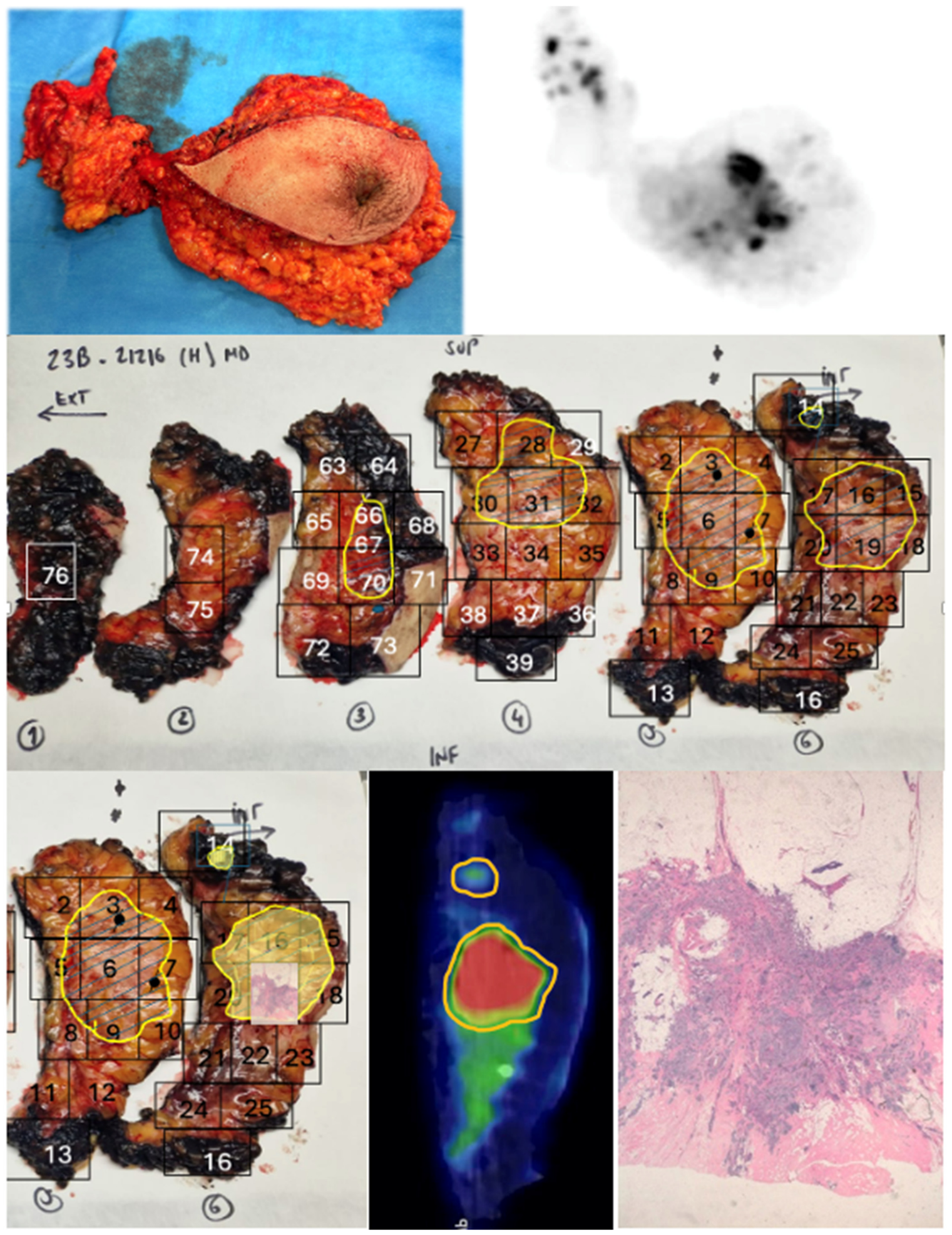
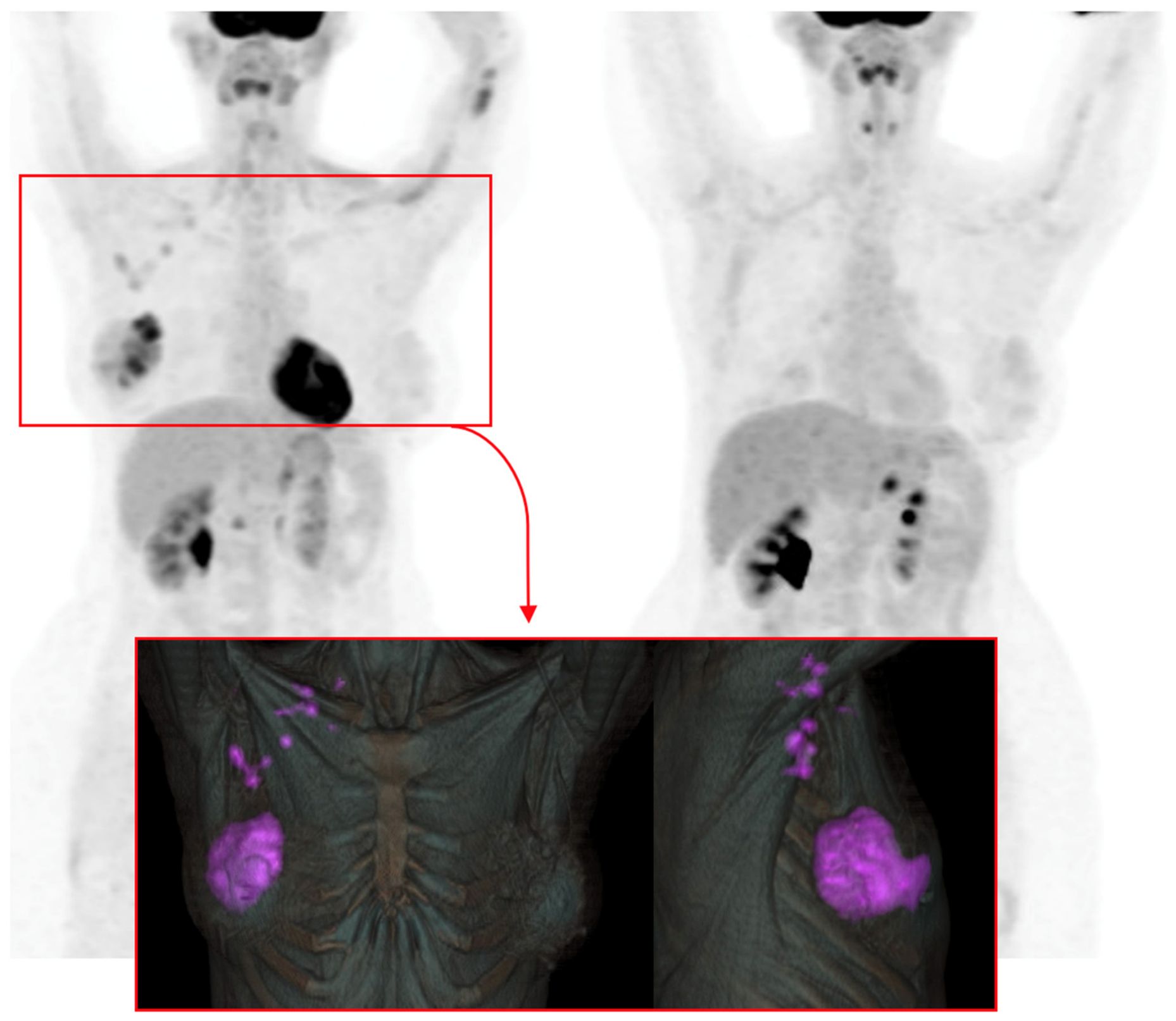
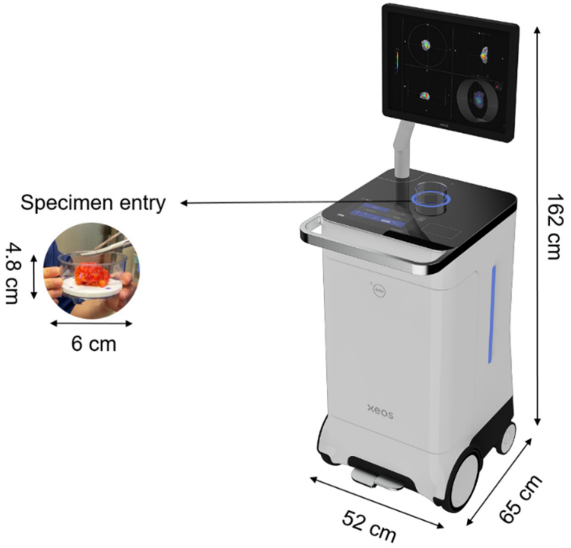
Beyond preoperative staging, molecular imaging is increasingly moving into the operating room as a tool for real-time or near–real-time verification of surgical outcomes, including ex vivo assessment of resection specimens (“back-table” verification). A workflow-based view of precision surgery highlights this verification step as essential to close the loop between preoperative imaging, intraoperative guidance, and pathology, ultimately enabling more consistent, patient-tailored decision-making. In this context, intraoperative and specimen-focused approaches (e.g., Cerenkov luminescence imaging and high-resolution specimen PET/CT) can complement conventional techniques by providing functional information linked to radiotracer uptake and by supporting margin-oriented surgical decisions (Figure 4, Figure 5 and Figure 6).
Figure 4.
Molecularly guided breast surgery using 18F-FDG in a combined mastectomy and axillary lymph node dissection performed through a single surgical approach. This case represents the first reported experience of molecular surgery of this type in Spain. (Upper left): anterior macroscopic view of the en bloc surgical specimen, including breast and axillary contents. (Upper right): ex vivo PET/CT assessment demonstrating heterogeneous and multifocal radiotracer uptake within the breast specimen, as well as multiple focal FDG-avid lesions in the axillary component, corresponding to pathologically involved lymph nodes (n = 10). (Middle panels): macroscopic sectioning of the specimen with marked pathological segments, shown in coronal orientation and matched to corresponding PET/CT slices. (Lower panels): comparison between macroscopic histopathologic sections and ex vivo molecular imaging, demonstrating mirror-image correspondence of the primary lesion and a superior peripheral focus, with full concordance between PET findings and histology. The mirror-image correspondence reflects clear concordance between the ex vivo molecular (PET/CT) findings of the specimen and the corresponding histopathological sections, confirming spatial agreement between metabolic activity and tumor tissue. Histological section stained with haematoxylin and eosin (H&E); approximate magnification ×20.
Figure 5.
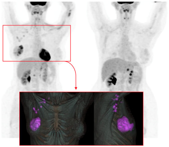
Preoperative and postoperative 18F-FDG PET/CT imaging of the patient undergoing molecularly guided surgery shown in Figure 4. Upper left: preoperative PET/CT demonstrating FDG-avid lesions in the breast and axilla, displayed with three-dimensional volumetric reconstruction to support surgical planning and target definition. Lower panels: Volumetric segmentation was generated using an artificial intelligence–based algorithm, anterior and lateral views highlighting areas of increased glucose metabolism (shown in purple), corresponding to the primary breast lesion and axillary involvement. Upper right: postoperative PET/CT follow-up performed three months after surgery, demonstrating no evidence of residual or recurrent metabolically active disease.
Figure 6.
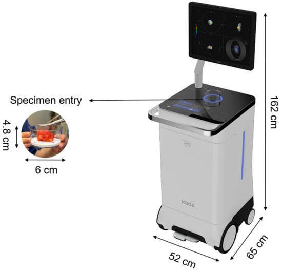
AURA 10 mobile PET/CT system for intraoperative and specimen imaging. This figure illustrates the first commercially available portable PET/CT system introduced in Europe. The system consists of a shielded main body housing the integrated PET and CT components, designed for ex vivo molecular and anatomical assessment of surgical specimens. A high-resolution touchscreen monitor is mounted on a short articulated arm connected to the main shielded unit, allowing intuitive and fully tactile interaction by the physician. The central circular imaging chamber accommodates the surgical specimen for evaluation. The mobile design, including visible system dimensions and integrated wheels, facilitates easy positioning and maneuverability within the operating room. An example of specimen positioning during imaging is also shown.
8. Artificial Intelligence and Digital Tools
This section explores the role of artificial intelligence and digital tools as enabling technologies across the radioguided surgical pathway, from planning to intraoperative decision support.
8.1. AI as an Enabling Layer Across the Radioguided Surgical Pathway
Artificial intelligence (AI) is increasingly becoming an enabling layer across the entire radioguided and molecular surgery pathway, supporting computer-assisted diagnosis, preoperative planning, intraoperative navigation, real-time detection, and specimen verification. A workflow-based view of precision surgery positions AI as a key component to integrate multimodal data streams (hybrid imaging, tracking, intraoperative signals, pathology feedback) and to close the loop between planning, execution, and verification—an approach that aligns with the goal of personalized, patient-tailored surgical decision-making. Recent perspectives within nuclear medicine and radioguided surgery also emphasize continuous innovation, multidisciplinary collaboration, and the need to translate technological advances into clinically meaningful endpoints [10,38].
These developments are consistent with recent imaging-focused studies exploring computer-assisted and multimodal diagnostic strategies in breast cancer, underscoring the expanding role of advanced image analysis and AI-driven tools in personalized care [11,12].
8.2. Practical AI Use-Cases Relevant to Breast Radioguided and Molecular Surgery
In breast cancer, AI-enabled tools can be mapped to clinically relevant tasks at multiple stages of care. First, AI may improve segmentation and quantification on SPECT/CT and PET/CT, facilitating target definition (lesions, sentinel nodes, marked nodes) and supporting more consistent surgical planning. Second, AI-driven image registration and fusion can help align preoperative hybrid imaging with intraoperative coordinates, enabling 3D navigation concepts and augmented visualization to guide targeted approaches in both the breast and axilla. Third, AI may support quality control and hotspot detection in intraoperative imaging (including freehand/portable 3D systems), potentially reducing operator dependence by standardizing acquisition and interpretation. Finally, AI-assisted interpretation of intraoperative or specimen-based molecular signals may contribute to margin-oriented decision support, prioritizing directed shaving or immediate re-excision in selected scenarios and strengthening verification-driven workflows.
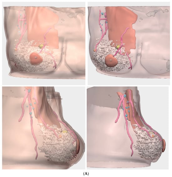
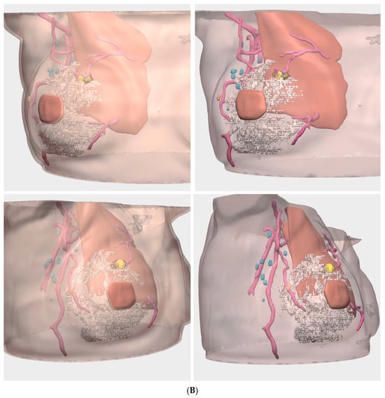
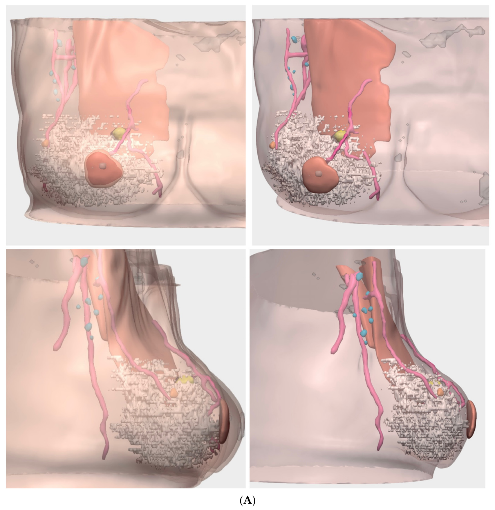
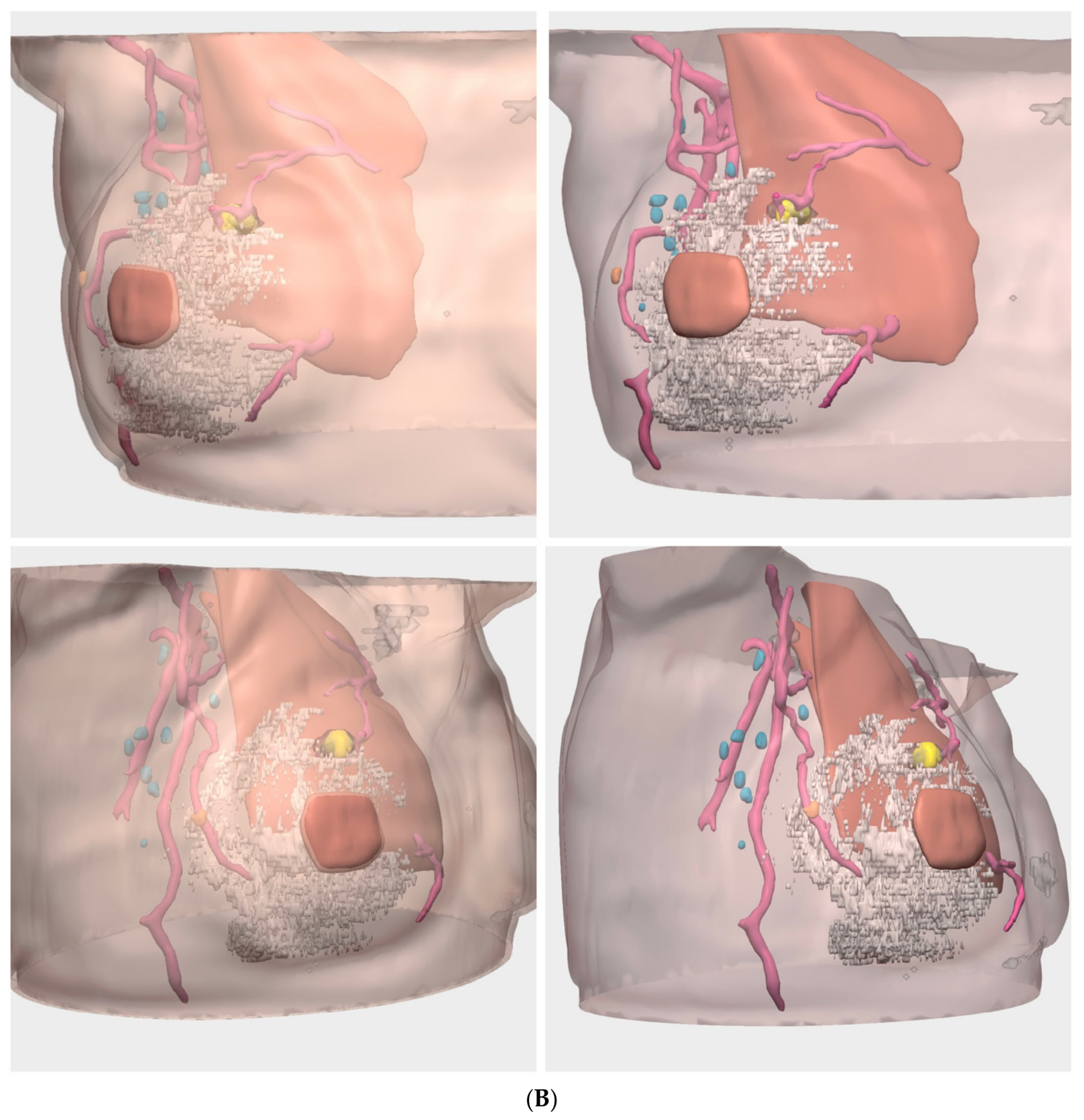
A clinically relevant example of artificial intelligence–supported integration is the use of convolutional neural network–based radiomics and segmentation applied to multimodal 18F-FDG PET/CT imaging for three-dimensional breast and axillary modeling. Unlike most AI approaches in breast imaging, which are predominantly based on magnetic resonance imaging acquired in the prone position, this strategy enables patient-specific anatomical reconstruction in both prone and supine positions. Supine PET/CT acquisition allows representation of breast anatomy in a configuration that is directly reproducible in the operating room, thereby improving spatial correspondence between preoperative imaging, surgical navigation, and intraoperative decision-making. By combining prone and supine datasets, this approach illustrates how AI-assisted multimodal imaging can bridge the gap between diagnostic imaging and surgically relevant anatomy, enhancing personalization without replacing clinical judgment (Figure 7A,B).
Figure 7.
(A) Artificial intelligence–based radiomic segmentation and three-dimensional reconstruction of 18F-FDG PET/CT acquired in the prone position. Upper panels show anterior views with and without skin rendering, while lower panels show lateral views with and without skin. The primary breast tumor is segmented in yellow, axillary lymph nodes in blue, and vascular structures in red, together with visualization of surrounding muscle and normal glandular tissue. The reconstruction demonstrates a metabolically active lesion located in the upper inner quadrant of the breast, in close proximity to a vascular structure, with an apparent safety margin from the pectoral muscle and the skin surface. (B) Artificial intelligence–based radiomic segmentation and three-dimensional reconstruction of 18F-FDG PET/CT acquired in the supine position. Upper panels display anterior views with and without skin rendering, while lower panels show lateral views with and without skin, mirroring the visualization shown in (A). The primary tumor is segmented in yellow, axillary lymph nodes in blue, and vascular structures in red, together with surrounding muscle and normal glandular tissue. In the supine, anatomically reproducible position, the reconstruction reveals relevant spatial changes compared with the prone configuration, including increased distance to the nipple–areolar complex, closer spatial relationship with vascular structures, and lateral breast displacement rather than inferior gravitational deformation. This representation reflects surgical positioning and facilitates improved correspondence with intraoperative anatomy.
8.3. Barriers to Implementation: Validation, Bias, and Clinical Integration
Despite rapid progress, clinical adoption requires robust validation, including multicenter generalization, attention to dataset shift and bias, and transparent reporting of performance across patient subgroups and tumor phenotypes. Interoperability with surgical navigation systems and hospital IT infrastructure, along with prospective evaluation using clinically meaningful outcomes (e.g., reduction in re-excision rates, improved staging accuracy after neoadjuvant therapy, decreased morbidity through axillary de-escalation), will be essential [10,38]. Successful implementation is inherently multidisciplinary: nuclear medicine, radiology, surgery, pathology, medical physics, and data science must align to ensure that AI outputs remain interpretable, actionable, and safe within real-world workflows—particularly when decisions affect the extent of resection or the omission of more invasive axillary procedures [10,38].
From a clinical readiness standpoint, most AI-supported tools in radioguided and molecular breast surgery are currently at an early adoption stage. Evidence is predominantly derived from retrospective analyses, pilot implementations, or technical validation studies, with limited prospective evaluation using clinically meaningful endpoints. Consequently, AI should currently be viewed as an enabling layer that supports, rather than replaces, expert multidisciplinary decision-making, pending robust clinical validation and regulatory alignment.
9. Conclusions and Future Directions
This section summarizes key concepts and discusses future directions for personalized radioguided and molecular breast cancer surgery.
From an outcome-oriented perspective, the clinical value of radioguided and molecular imaging–supported surgery lies not only in technical feasibility, but in its potential impact on patient-centered endpoints. Across breast and axillary procedures, these approaches aim to reduce re-excision rates, minimize surgical morbidity—particularly lymphedema through axillary de-escalation—and support more accurate staging and treatment adaptation. Intraoperative verification and specimen-based assessment may help avoid delayed reoperations, shorten time to adjuvant therapy, and improve workflow efficiency. Ultimately, linking technological innovation to measurable clinical outcomes is essential to justify adoption and to ensure that personalization translates into tangible benefits for patients and healthcare systems.
Breast cancer surgery is undergoing continuous transformation, driven by advances in tumor biology, systemic therapy (including the expanding use of neoadjuvant treatment), and a growing emphasis on de-escalation strategies that preserve oncologic safety while improving quality of life. In parallel, nuclear medicine is evolving rapidly—both technologically and pharmacologically—creating new opportunities to support more precise localization, navigation, and verification in the operating room. Together, these trends generate new clinical needs and reinforce the importance of flexible, patient-centered decision-making across diverse scenarios [10,38].
Guidelines provide an essential evidence-based framework, yet real-world implementation must account for local resources and expertise: not all centers have identical access to hybrid imaging, intraoperative navigation tools, or specimen-imaging technologies. In this context, the most impactful pathway forward is a multidisciplinary and personalized workflow, integrating radiology, nuclear medicine, surgery, pathology (and, when relevant, radiation oncology and medical physics) to select the best strategy for each patient. The overarching goal is to deliver less invasive, more accurate surgery, reduce unnecessary morbidity (particularly in the axilla), and improve the overall patient experience without compromising oncologic outcomes [6,38].
Looking ahead, several converging directions may further reshape radioguided and molecular breast surgery. First, the maturation of intraoperative 3D navigation (including freehand/portable SPECT concepts) and verification-driven workflows may increase reproducibility and confidence in complex cases, supporting personalized de-escalation strategies. Second, intraoperative molecular specimen assessment—particularly PET-based specimen imaging—has the potential to become a disruptive tool for margin-oriented decision-making, enabling more consistent real-time verification and potentially reducing re-excision rates in breast-conserving surgery.
Finally, the future is likely to be strongly influenced by the expanding portfolio of molecularly targeted radiopharmaceuticals. Beyond 18F-FDG, tracers aligned with tumor phenotype (e.g., immuno-PET approaches such as 89Zr-trastuzumab for HER2 expression, or 18F-fluoroestradiol for estrogen receptor imaging) may enable more selective “molecular guidance” in the operating room—supporting lesion characterization, response-adapted targeting after neoadjuvant therapy, and potentially tracer-driven personalization of both breast and axillary procedures [36,37]. As these agents and enabling technologies mature, the integration of molecular imaging, intraoperative navigation, specimen verification, and AI-assisted decision support may define the next era of precision breast cancer surgery: more personalized, less aggressive, and ultimately more aligned with the needs and outcomes that matter most to each patient [2,9,10,38].
Author Contributions
Conceptualization, J.O.C. and M.T.; methodology, J.O.C.; investigation, J.O.C., J.S.S., C.C.A., R.D.E., L.F.L., C.S.B., D.C.S., E.B.V., E.M.S. and V.L.F.; resources, E.B.V., E.M.S. and V.L.F.; writing—original draft preparation, J.O.C.; writing—review and editing, M.T., B.B., S.V.S. and J.M.C.A.; supervision, S.V.S. and J.M.C.A.; project administration, J.O.C. and M.T. All authors have read and agreed to the published version of the manuscript.
Funding
This research received no external funding.
Institutional Review Board Statement
Not applicable.
Informed Consent Statement
Not applicable.
Data Availability Statement
The raw data supporting the conclusions of this article will be made available by the authors on request.
Conflicts of Interest
The authors declare no conflicts of interest.
References
- Vidal-Sicart, S.; Valdés Olmos, R.A. New devices in radioguided surgery. Clin. Transl. Imaging 2023, 11, 545–557. [Google Scholar] [CrossRef]
- Boekestijn, I.; van Oosterom, M.N.; Dell’Oglio, P.; van Velden, F.H.P.; Pool, M.; Maurer, T.; Rietbergen, D.D.D.; Buckle, T.; van Leeuwen, F.W.B. The current status and future prospects for molecular imaging-guided precision surgery. Cancer Imaging 2022, 22, 48. [Google Scholar] [CrossRef] [PubMed]
- Buckle, T.; Schilling, C.; Maurer, T.; Vidal-Sicart, S. Image-guided surgery: From classical techniques to novel aspects and approaches. Q. J. Nucl. Med. Mol. Imaging 2021, 65, 187–189. [Google Scholar] [CrossRef] [PubMed]
- Berneta, L.; Piñero, A.; Martínez, M.; Vidal-Sicart, S.; Algara, M.; Palomares, E. Consensus of the Spanish Society of Breast Senology and Pathology (SESPM) on Selective Biopsy of the Sentinel Node (BSGC) and axillary management in breast cancer. Rev. Senol. Patol. Mamar. 2022, 35, 243–259. [Google Scholar]
- Perrone, E.; Collarino, A.; Pereira Arias-Bouda, L.M.; den Hartog, W.; Garganese, G.; Van der Hage, J.A.; Di Giuda, D.; Giammarile, F. Molecular imaging to guide breast cancer surgery. Clin. Transl. Imaging 2023, 11, 521–531. [Google Scholar] [CrossRef]
- Galimberti, V.; Cole, B.F.; Zurrida, S.; Viale, G.; Luini, A.; Veronesi, P.; Baratella, P.; Chifu, C.; Sargenti, M.; Intra, M.; et al. Axillary dissection versus no axillary dissection in patients with sentinel-node micrometastasis (IBCSG 23-01): A phase 3 randomised controlled trial. Lancet Oncol. 2013, 14, 297–305. [Google Scholar] [CrossRef] [PubMed]
- Fisher, B.; Jeong, J.H.; Anderson, S.; Bryant, J.; Fisher, E.R.; Wolmark, N. Twenty-five-year follow-up of a randomized trial comparing radical mastectomy, total mastectomy, and total mastectomy followed by radiation. N. Engl. J. Med. 2002, 347, 567–575. [Google Scholar] [PubMed]
- Solá, M.; Alberro, J.A.; Fraile, M.; Santesteban, P.; Ramos, M.; Fabregas, R.; Moral, A.; Ballester, B.; Vidal, S. Complete axillary lymph node dissection versus clinical follow-up in breast cancer patients with sentinel node micrometastases: Final results from the multicentric clinical trial AATRM 048/13/2000. Ann. Surg. Oncol. 2013, 20, 120–127. [Google Scholar] [PubMed]
- Loibl, S.; André, F.; Bachelot, T.; Barrios, C.H.; Bergh, J.; Burstein, H.J.; Cardoso, M.J.; Carey, L.A.; Dawood, S.; Del Mastro, L.; et al. ESMO Clinical Practice Guideline: Early breast cancer diagnosis, treatment and follow-up. Ann. Oncol. 2024, 35, 159–182. [Google Scholar]
- Vidal-Sicart, S.; Goñi, E.; Cebrecos, I.; Rioja, M.E.; Perissinotti, A.; Sampol, C.; Vidal, O.; Saavedra-Pérez, D.; Ferrer, A.; Martí, C.; et al. Cirugía radioguiada y cirugía molecular. Rev. Esp. Med. Nucl. Imagen Mol. 2024, 43, 39–54. [Google Scholar] [CrossRef]
- Leung, J.-H.; Karmakar, R.; Mukundan, A.; Thongsit, P.; Chen, M.-M.; Chang, W.-Y.; Wang, H.-C. Systematic Meta-Analysis of Computer-Aided Detection of Breast Cancer Using Hyperspectral Imaging. Bioengineering 2024, 11, 1060. [Google Scholar] [CrossRef] [PubMed]
- Hsu, H.; Lee, K.-H.; Karmakar, R.; Mukundan, A.; Attar, R.S.; Liu, P.-H.; Wang, H.-C. From Innovation to Application: Can Emerging Imaging Techniques Transform Breast Cancer Diagnosis? Diagnostics 2025, 15, 2718. [Google Scholar] [CrossRef] [PubMed]
- Povoski, S.P. The history of radioguided surgery: Early historical milestones and the development of later innovative clinical applications. In Radioguided Surgery; Herrmann, K., Nieweg, O.E., Povoski, S.P., Eds.; Springer International: Berlin/Heidelberg, Germany, 2016; pp. 3–12. [Google Scholar]
- Povoski, S.P.; Neff, R.L.; Mojzisik, C.M.; O’Malley, D.M.; Hinkle, G.H.; Hall, N.C.; Murrey, A.D.; Knopp, M.V.; Martin, E.W. A comprehensive overview of radioguided surgery using gamma detection probe technology. World J. Surg. Oncol. 2009, 7, 11. [Google Scholar] [CrossRef] [PubMed]
- Vidal-Sicart, S.; Rioja, E.M.; Paredes, P.; Keshtgar, M.R.; Olmos, R.A.V. Contribution of perioperative imaging to radioguided surgery. Q. J. Nucl. Med. Mol. Imaging 2014, 58, 140–160. [Google Scholar] [PubMed]
- Banys-Paluchowski, M.; de Boniface, J. Axillary staging in node-positive breast cancer converting to node negativity through neoadjuvant chemotherapy: Current evidence and perspectives. Scand. J. Surg. 2023, 112, 117–125. [Google Scholar] [CrossRef] [PubMed]
- Vidal-Sicart, S.; Vermeeren, L.; Solà, O.; Paredes, P.; Valdés-Olmos, R.A. The use of a portable gamma camera for preoperative lymphatic mapping: A comparison with a conventional gamma camera. Eur. J. Nucl. Med. Mol. Imaging 2011, 38, 636–641. [Google Scholar] [PubMed]
- van Loevezijn, A.A.; van der Noordaa, M.E.M.; Stokkel, M.P.M.; van Werkhoven, E.D.; Groen, E.J.; Loo, C.E.; Elkhuizen, P.H.M.; Sonke, G.S.; Russell, N.S.; van Duijnhoven, F.H.; et al. Three-year follow-up of de-escalated axillary treatment after neoadjuvant systemic therapy in clinically node-positive breast cancer: The MARI-protocol. Breast Cancer Res. Treat. 2022, 193, 37–48. [Google Scholar] [CrossRef] [PubMed]
- Giuliano, A.E.; Ballman, K.V.; McCall, L.; Beitsch, P.D.; Brennan, M.B.; Kelemen, P.R.; Ollila, D.W.; Hansen, N.M.; Whitworth, P.W.; Blumencranz, P.W.; et al. Effect of axillary dissection vs no axillary dissection on 10-year overall survival among women with invasive breast cancer and sentinel node metastasis: The ACOSOG Z0011 randomized clinical trial. JAMA 2017, 318, 918–926. [Google Scholar] [CrossRef] [PubMed]
- Morton, D.L. Lymphoscintigraphy and sentinel node localization in breast cancer. J. Nucl. Med. 1996, 37, 1006–1015. [Google Scholar] [CrossRef] [PubMed]
- Simons, J.M.; van Nijnatten, T.J.A.; van der Pol, C.C.; van Diest, P.J.; Jager, A.; van Klaveren, D. Diagnostic accuracy of radioactive iodine seed placement in the axilla with sentinel lymph node biopsy after neoadjuvant chemotherapy in node-positive breast cancer. JAMA Surg. 2022, 157, 991–999. [Google Scholar] [CrossRef] [PubMed]
- Gómez, A.d.A.; Ferrer, F.V.; Rebolleda, J.F.; Jurado, R.S.; Errando, J.P.; Leiva, V.B.; Bataller, L.C.; García, J.Á.G.; Comas, A.G.-V.; Diana, C.A.F. Comparative study of the sensitivity of MAMMI-PET in the diagnosis of breast cancer with regard to other imaging tests. Rev. Senol. Patol. Mamar. 2023, 36, 100414. [Google Scholar] [CrossRef]
- Heller, S.; Zanzonico, P. Nuclear probes and intraoperative gamma cameras. Semin. Nucl. Med. 2011, 41, 166–181. [Google Scholar] [CrossRef] [PubMed]
- Siman, W.; Kappadath, S.C. Performance characteristics of a new pixelated portable gamma camera. Med. Phys. 2012, 39, 3435–3444. [Google Scholar] [CrossRef] [PubMed]
- Krag, D.N.; Anderson, S.J.; Julian, T.B.; Brown, A.M.; Harlow, S.P.; Costantino, J.P.; Ashikaga, T.; Weaver, D.L.; Mamounas, E.P.; Jalovec, L.M.; et al. Sentinel node resection compared with conventional axillary lymph node dissection in clinically node-negative patients with breast cancer: NSABP B-32 randomised phase 3 trial. Lancet Oncol. 2010, 11, 927–933. [Google Scholar] [PubMed]
- Orozco-Cortés, J.; Romero, Á.B.; Garrigos, G.; Estellés, N.; Mut, T.; Reyes; Martínez, E.; Medina, R.; Peláez, S.; Abreu, P.; et al. Implementation of the use of SPECT-portable for evaluation of surgical margins in breast cancer with indication of ROLL: First results. Rev. Esp. Med. Nucl. Imagen Mol. (Engl. Ed.) 2023, 42, 147–155. [Google Scholar] [CrossRef] [PubMed]
- St John, E.R.; Al-Khudairi, R.; Ashrafian, H.; Athanasiou, T.; Takats, Z.; Hadjiminas, D.J.; Darzi, A.; Leff, D.R. Diagnostic accuracy of intraoperative techniques for margin assessment in breast cancer surgery: A meta-analysis. Ann. Surg. 2017, 265, 300–310. [Google Scholar] [CrossRef] [PubMed]
- Butler-Henderson, K.; Lee, A.H.; Price, R.I.; Waring, K. Intraoperative assessment of margins in breast-conserving therapy: A systematic review. Breast 2014, 23, 112–119. [Google Scholar] [PubMed]
- Tang, R.; Buckley, J.M.; Fernandez, L.; Coopey, S.; Aftreth, O.; Michaelson, J.; Saksena, M.; Lei, L.; Specht, M.; Gadd, M.; et al. Micro-computed tomography (Micro-CT): A novel approach for intraoperative breast cancer specimen imaging. Breast Cancer Res. Treat. 2013, 139, 311–316. [Google Scholar] [PubMed]
- Bluemel, C.; Matthies, P.; Herrmann, K.; Povoski, S.P. 3D scintigraphic imaging and navigation in radioguided surgery: Freehand SPECT technology and its clinical applications. Expert Rev. Med. Devices 2016, 13, 339–351. [Google Scholar] [CrossRef] [PubMed]
- Declipse® SPECT; SurgicEye: Munich, Germany; Available online: http://www.surgiceye.com/m/declipseSPECT/openSurgery.html (accessed on 10 November 2015).
- Hurtado, Á.E.; Orozco-Cortés, J.; Ibarra, P.C.; González, U.L.; Romero, Á.B.; de la Cruz, M.N.; Gelo, C.S.; Calabuig, L.C.; Sánchez, P.A.; Dólera, T.M.; et al. Concordance between freehand SPECT and conventional scintigraphy for sentinel lymph node detection in breast cancer. Rev. Esp. Med. Nucl. Imagen Mol. (Engl. Ed.) 2024, 43, 79–83. [Google Scholar] [CrossRef] [PubMed]
- Grootendorst, M.R.; Cariati, M.; Pinder, S.E.; Kothari, A.; Douek, M.; Kovacs, T.; Hamed, H.; Pawa, A.; Nimmo, F.; Owen, J.; et al. Intraoperative assessment of tumor resection margins in breast-conserving surgery using 18F-FDG Cerenkov luminescence imaging: A first-in-human feasibility study. J. Nucl. Med. 2017, 58, 891–898. [Google Scholar] [CrossRef] [PubMed]
- Göker, M.; Marcinkowski, R.; Van Bockstal, M.; Keereman, V.; Van Holen, R.; Van Dorpe, J.; Vandenberghe, S.; Brans, B.; Depypere, H.; Broecke, R.V.D. 18F-FDG micro-PET/CT for intra-operative margin assessment during breast-conserving surgery. Acta Chir. Belg. 2020, 120, 366–374. [Google Scholar] [CrossRef] [PubMed]
- De Crem, A.S.; Tummers, P.; Depypere, H.; Braems, G.; Salihi, R.; Vergauwen, G.; Cisternino, G.; Van de Vijver, K.; De Visschere, P.; De Man, K.; et al. Intraoperative margin assessment using specimen PET-CT (BIMAP). npj Breast Cancer 2025, 11, 101. [Google Scholar] [CrossRef] [PubMed]
- Mankoff, D.; Balogová, S.; Dunnwald, L.; Dehdashti, F.; DeVries, E.; Evangelista, L.; Van Kruchten, M.; Vaz, S.C.; Fowler, A.; Linden, H.; et al. SNMMI Procedure Standard/EANM Practice Guideline for 18F-Fluoroestradiol PET imaging of patients with estrogen receptor–positive breast cancer. J. Nucl. Med. 2024, 65, 221–229. [Google Scholar] [PubMed]
- Bensch, F.; Brouwers, A.H.; Hooge, M.N.L.-D.; de Jong, J.R.; van der Vegt, B.; Sleijfer, S.; de Vries, E.G.E.; Schröder, C.P. 89Zr-trastuzumab PET supports clinical decision making in breast cancer patients when HER2 status cannot be determined by standard work-up. Eur. J. Nucl. Med. Mol. Imaging 2018, 45, 2300–2306. [Google Scholar] [CrossRef] [PubMed]
- Cavinato, L.; Kirienko, M.; Papp, L.; Shi, K.; Visvikis, D.; Chiti, A.; Sollini, M. Digital twin system in computational nuclear oncology: Roadmap, opportunities and challenges. EANM J. 2025, 1, 100007. [Google Scholar] [CrossRef]
Disclaimer/Publisher’s Note: The statements, opinions and data contained in all publications are solely those of the individual author(s) and contributor(s) and not of MDPI and/or the editor(s). MDPI and/or the editor(s) disclaim responsibility for any injury to people or property resulting from any ideas, methods, instructions or products referred to in the content. |
© 2026 by the authors. Licensee MDPI, Basel, Switzerland. This article is an open access article distributed under the terms and conditions of the Creative Commons Attribution (CC BY) license.