


Abstract
Background/Objectives: Serum protein electrophoresis (SPE) is a widely used laboratory test for the detection and monitoring of monoclonal gammopathies, including multiple myeloma (MM). Although SPE is usually recommended in the presence of specific clinical or laboratory abnormalities, monoclonal gammopathies may occasionally develop rapidly and without typical symptoms. This case report aims to emphasize the diagnostic value of SPE in identifying an unexpected and fast-evolving monoclonal gammopathy. Methods: We report the clinical and laboratory eight-month follow-up of a 58-year-old male who initially underwent SPE for unrelated clinical conditions. Serial SPE analyses were performed using capillary zone electrophoresis. When abnormalities emerged, immunotyping and serum free light chain (FLC) assays were conducted. The diagnostic workup was completed with bone marrow aspiration, flow cytometry, and imaging studies according to current international diagnostic criteria. Results: The initial SPE (November 2023) showed a normal protein profile. After eight months, follow-up SPE revealed a prominent monoclonal spike in the gamma region (2.9 g/dL), associated with increased total serum proteins (91 g/L; range 64–82 g/L), elevated IgA levels (20.0 g/L; range 0.4–3.5 g/L), and a markedly abnormal κ/λ FLC ratio (54.00; range 0.31–1.56). Bone marrow analysis demonstrated >18% plasma cell infiltration, confirming the diagnosis of IgA-κ MM. The patient underwent standard therapy followed by autologous stem cell transplantation, achieving disease remission. Conclusions: This case highlights that clinically relevant monoclonal gammopathies may arise rapidly in the absence of classical diagnostic features. Routine SPE represents a cost-effective and accessible screening tool that can identify subtle protein abnormalities, prompting the timely use of more specific and invasive diagnostic procedures for aggressive plasma cell disorders.
1. Introduction
Serum protein electrophoresis (SPE) is a low-cost and simple laboratory test used in clinical practice to identify patients with monoclonal proteins, Multiple Myeloma (MM), immunoglobulin light-chain (AL) amyloidosis, lymphoid malignant diseases, and other serum protein disorders [1].
SPE is performed using capillary zone electrophoresis (CZE), a technique that separates proteins within narrow fused-silica capillaries under a high-voltage electric field. In CZE, analytes migrate through the capillary according to their charge-to-size ratio, and separation is further influenced by electroosmotic flow. Detection is carried out by continuous UV absorbance, producing a high-resolution electropherogram [2]. In CZE, human serum is exposed to an electrical current, which separates the proteins based on size and electrical charge into six fractions: albumin, alpha-1 (α1), alpha-2 (α2), beta-1 (β1), beta-2 (β2) and gamma (γ) [3]. CZE can differentiate healthy SPE patterns from disease conditions, such as the appearance of a monoclonal peak or band in the γ-region. In fact, interpretation of the SPE mainly focuses on the γ-region, which primarily contains immunoglobulins IgG, IgA, and IgM, since changes in this area are often associated with serious diseases. An increase in γ-globulins can produce different electrophoretic patterns. For example, monoclonal gammopathies like MM show a sharp and narrow peak (M-spike) in the γ-region. Conversely, an elevated and broad γ-region is typical of polyclonal gammopathies, which can occur in conditions such as chronic infections, autoimmune and connective tissue diseases [4,5], liver cirrhosis, Hodgkin’s disease, chronic lymphocytic leukemia, or amyloidosis. The γ band is decreased in agammaglobulinemia and hypogammaglobulinemia syndromes [6]. While various conditions can lead to an increase in the γ-region on serum protein electrophoresis, those that produce a distinct, spike-like peak within the γ-globulin zone are of clinical significance [7]. These are known as monoclonal gammopathies, a heterogeneous group of disorders characterized by clonal proliferation of plasma cells or B lymphocytes, resulting in the abnormal production of a monoclonal immunoglobulin (M protein) or free light chains (FLCs), typically detectable in the blood, urine, or both [8], and often indicates the presence of MM. To characterize the monoclonal nature of the peak, when an abnormal γ-region peak is detected, immunotyping (IT) is performed by overlaying antisera against heavy (IgG, IgA, IgM) and light (κ, λ) chains. The combination of SPE and IT thus provides both a screening and confirmatory approach in diagnosing a wide range of monoclonal gammopathies.
MM, which represents approximately 10–15% of all hematologic malignancies [9], is a clonal plasma cell neoplasm characterized by excessive production of monoclonal immunoglobulins (M proteins), usually exceeding 3 g/dL, along with ≥10% infiltration of clonal plasma cells in the bone marrow. Clinically, MM presents with a spectrum of symptoms, most notably bone pain, anemia, renal dysfunction, and hypercalcemia [10,11]. The M protein can act as a key tumor marker in MM and related monoclonal gammopathies, reflecting the underlying clonal plasma cell proliferation and production of a uniform immunoglobulin [12].
As the disease progresses, the clonal proliferation of malignant plasma cells within the bone marrow disrupts normal haematopoiesis and immunoglobulin production, leading to a range of clinical manifestations, including osteolytic lesions, focal bone resorption, and an increased risk of osteopenia, osteoporosis, and pathological fractures [13]. Despite significant advances in therapeutic strategies over the past two decades that have notably improved patient outcomes, MM remains an incurable malignancy, with a median overall survival of approximately 6 years following diagnosis [14]. Therefore, early and accurate diagnosis is critical to optimizing patient management. In this context, clinical laboratories play an essential role through the implementation of SPE and IT, which are fundamental in the early detection of monoclonal proteins and confirming diagnosis. This case presents an unusual and rarely documented rapid progression from a normal electrophoretic profile to overt MM within eight months, emphasizing the diagnostic relevance of routine SPE even in apparently asymptomatic patients. To our knowledge, such an acute transformation confirmed by serial laboratory and imaging findings has not been previously reported in the literature.
2. Materials and Methods
This study was conducted between November 2023 and July 2024. The patient was admitted to San Pio Hospital in Benevento.
The diagnostic workup for the patient included serial serum protein electrophoresis (SPE), immunotyping (IT), bone marrow aspiration, flow cytometry, and imaging studies (PET and MRI), according to current international diagnostic criteria.
Serum Protein Electrophoresis and Immunotyping
Serum protein electrophoresis (SPE) was performed using capillary zone electrophoresis (CZE) on the Capillarys 3 Octa system (Sebia, Lisses, France). This technique separates serum proteins within fused-silica capillaries under a high-voltage electric field based on their charge-to-size ratio, with migration further influenced by electroosmotic flow.
Blood samples were collected in serum tubes, allowed to clot, and centrifuged at 3000 rpm for 10 min. Prior to analysis, serum was visually inspected for hemolysis, lipemia, and icterus. Separation was carried out in fused-silica capillaries (internal diameter 25–75 μm) using an alkaline buffer (pH ~8.6–10.0) with an applied voltage. Proteins were detected by continuous ultraviolet (UV) absorbance at a wavelength of 200–214 nm, generating a high-resolution electropherogram.
Under these analytical conditions, serum proteins were resolved into six major fractions: albumin, alpha-1 (α1), alpha-2 (α2), beta-1 (β1), beta-2 (β2), and gamma (γ). Electropherograms were processed using Phoresis Core software (version 9.32; Sebia, Lisses, France , which automatically integrated peak areas and provided quantitative results expressed as relative percentages and absolute concentrations (g/dL or g/L). IT was performed using the same system with antisera against IgG, IgA, IgM, κ, and λ chains to confirm monoclonal components.
Analytical performance, including precision and reproducibility, was verified according to internal laboratory quality control procedures, and each run included internal quality controls at both normal and pathological levels. CZE allows for the reliable detection of monoclonal components, which typically appear as narrow peaks in the γ-region. Consequently, clinical interpretation focused primarily on this region, which contains immunoglobulins (IgG, IgA, and IgM).
3. Results
In November 2023, a 58-year-old male patient was undergoing clinical follow-up for recurrent prostatitis. The patient’s past medical history was otherwise unremarkable, with no family history of hematological malignancies or significant comorbidities. At that time, he was not taking any long-term medications, except for occasional treatments for prostatic symptoms. As part of this evaluation, the clinician requested laboratory tests to rule out comorbidities and gain a more comprehensive clinical picture. Baseline laboratory values, including a complete blood count (CBC), renal function, and calcium levels, were all within reference ranges (Table 1).
Table 1.
Clinical and laboratory parameters at baseline (November 2023) and follow-up (July 2024).
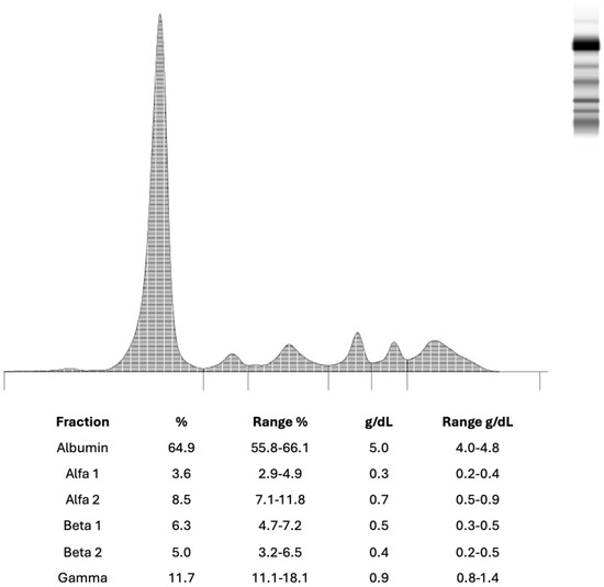
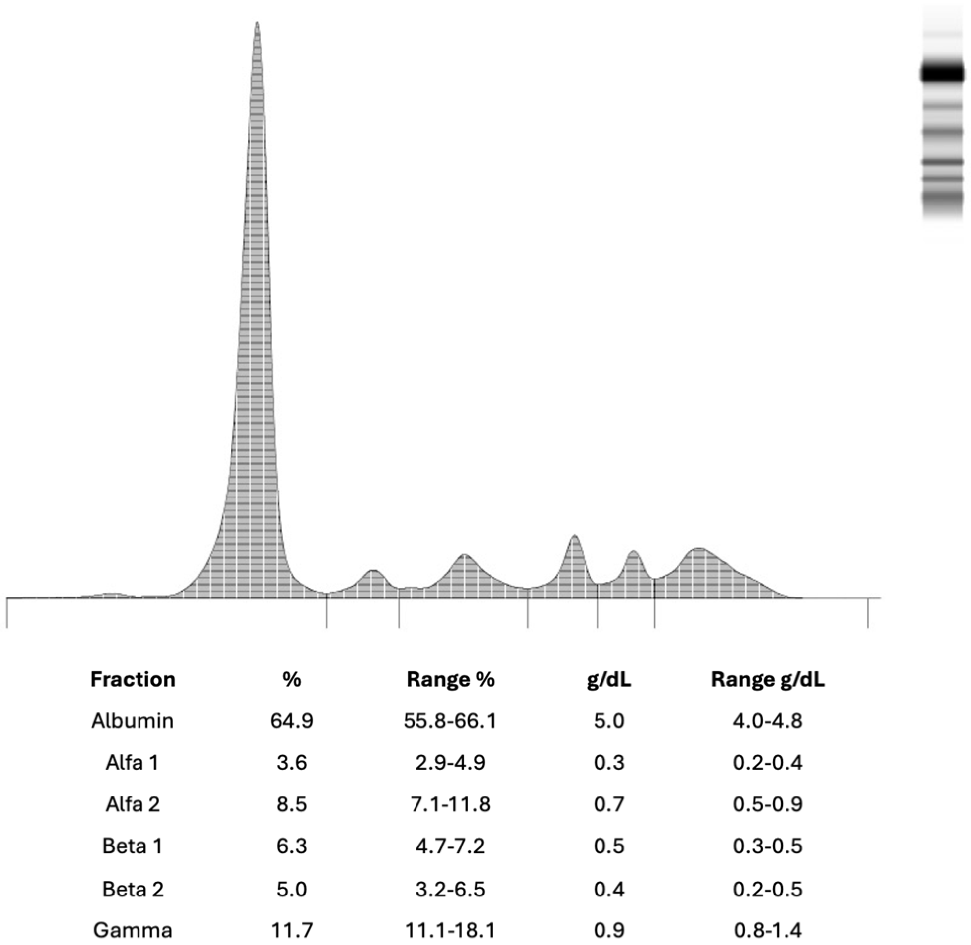
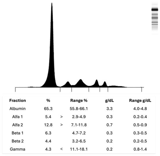
Although the patient did not exhibit CRAB (hypercalcaemia, renal involvement, anaemia, and bone lesions) criteria, serum protein electrophoresis (SPE) was ordered due to mild but persistent asthenia of unclear origin. The baseline SPE (November 2023) revealed a normal protein profile showed a normal protein profile (Figure 1), with a physiological distribution of serum proteins across all major fractions and no monoclonal spikes.
Figure 1.
Normal serum protein electrophoresis pattern in a 58–year-old male patient from November 2023.
The patient showed no significant abnormalities in the SPE, except for an elevated prostate-specific antigen (PSA) of 11 μg/L (reference range: <4.0) and a free PSA of 2.52 μg/L, corresponding to a free/total PSA ratio of approximately 23%, which falls within the intermediate risk range for prostate cancer.
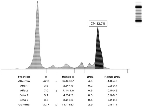
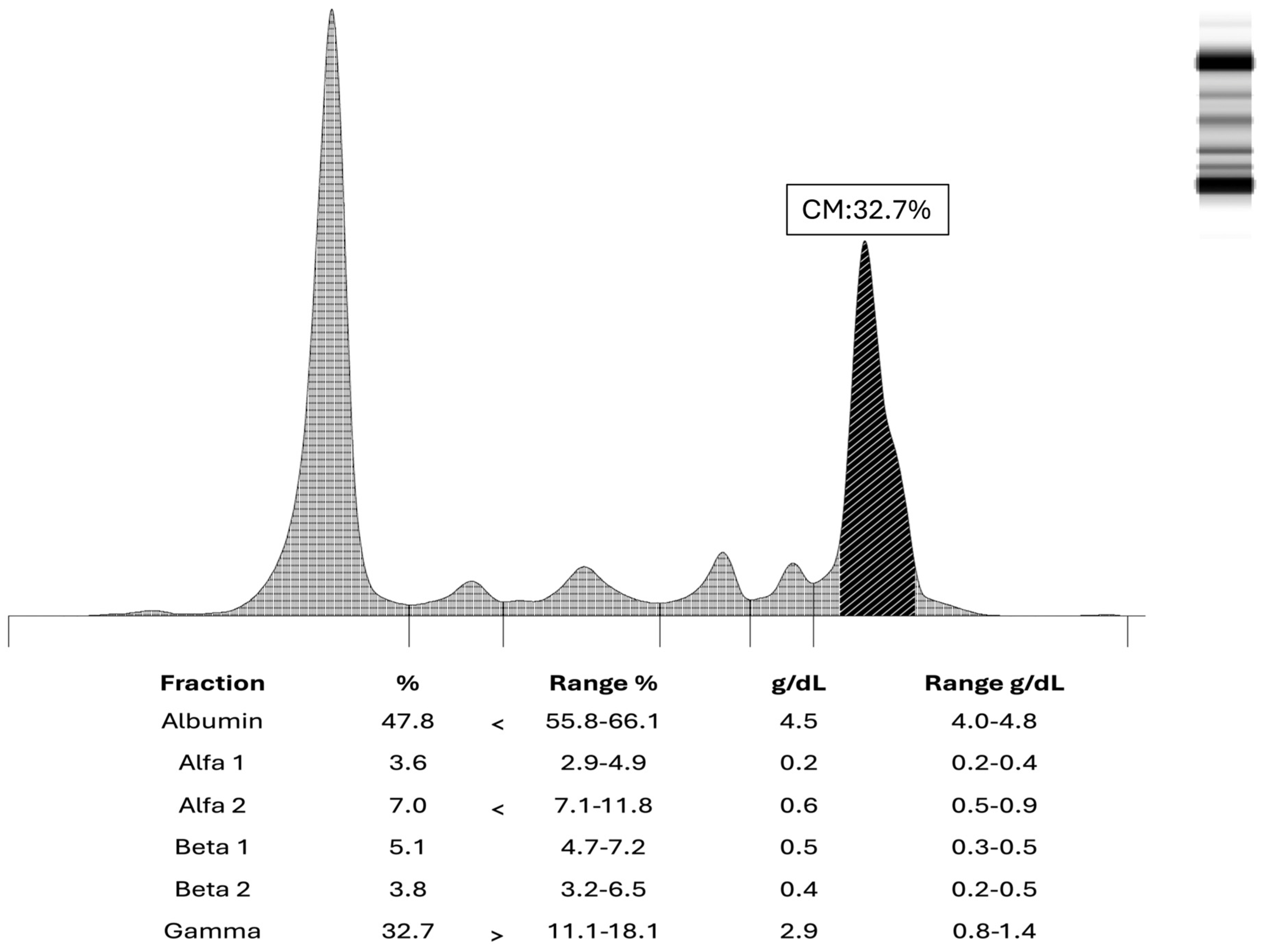
The patient returned for follow-up eight months later, in July 2024. While prostatitis was again confirmed (PSA 9.83 μg/L; free PSA 2.24 μg/L), clinicians noted a significantly altered SPE pattern compared to the previous test (Figure 2). This follow-up analysis revealed a prominent monoclonal band in the gamma region (γ-globulin fraction: 32%; reference range: 11.1–18.8%) and an inverted albumin/gamma ratio of 0.99, strongly suggestive of a monoclonal gammopathy. These findings were accompanied by a notable increase in total serum proteins (91 g/L) and a stable but slightly increased serum calcium level (2.47 mmol/L), as detailed in Table 1.
Figure 2.
Pathological serum protein electrophoresis pattern in a 58-year-old male patient from July 2024.
Serum protein electrophoresis showed a monoclonal spike in the gamma region, indicative of a monoclonal gammopathy. No alterations were observed in the other fractions (albumin, alpha and beta).
Detailed laboratory investigations in July 2024 confirmed a markedly increased serum IgA (20.0 g/L; reference range: 0.4–3.5 g/L) and raised serum κ (kappa) light chains (7.76 g/L; reference range: 1.7–3.7 g/L). Haemoglobin (138 g/L; reference range: 120–170 g/L), serum calcium (2.47 mmol/L; reference range: 2.12–2.52 mmol/L) and creatinine levels (79.6 μmol/L; reference range: 53–106 μmol/L) remained stable and consistent with those detected in November 2023.
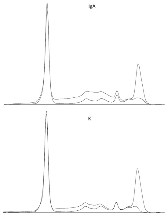
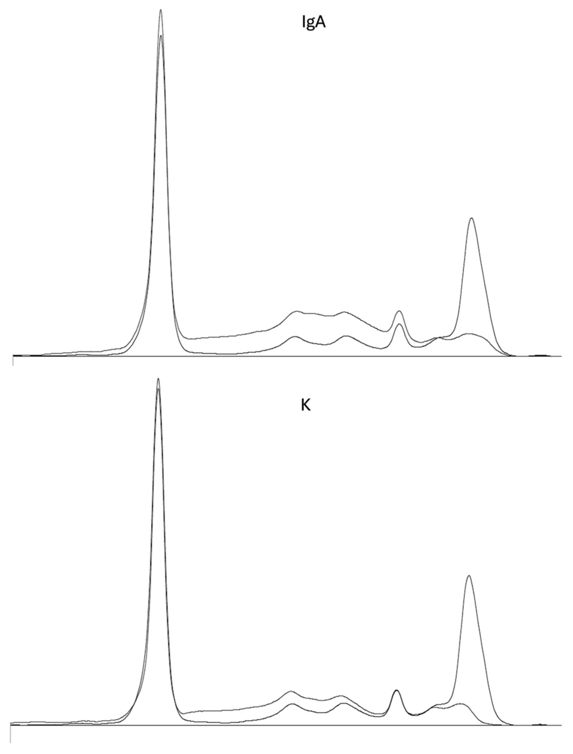
Immunotyping confirmed the nature of the monoclonal component as an IgA-κ type (Figure 3).
Figure 3.
Immunotyping (IT) electropherogram of the patient’s serum (July 2024). The IT pattern revealed a distinct monoclonal band in the gamma region, which reacts specifically with anti-IgA (IgA) and anti-kappa (K) antibodies.
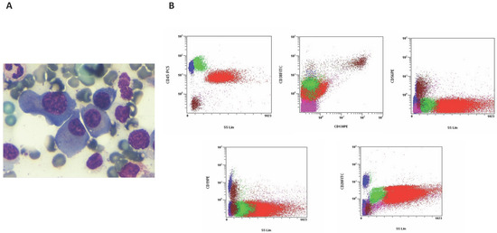
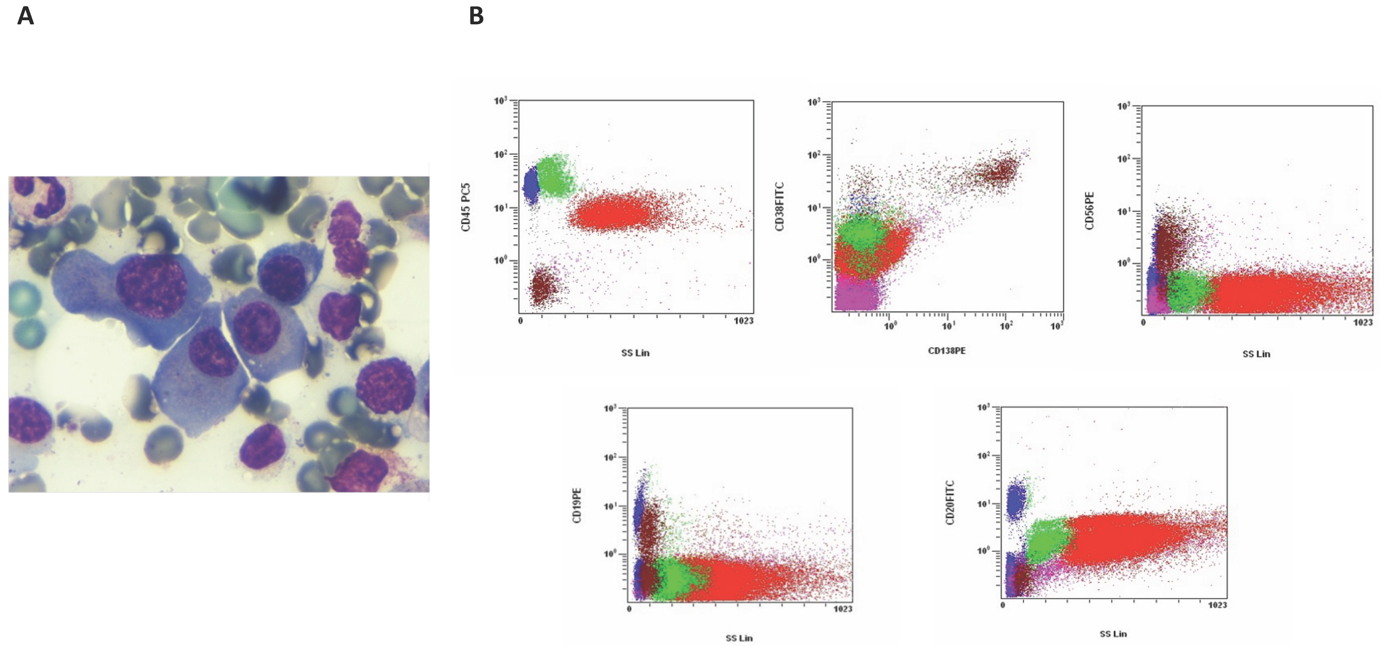
Serum free light chain analysis revealed a κ concentration of 0.584 g/L (reference range: 0.00670–0.02240) and a λ (lambda) concentration of 0.0108 g/L (reference range: 0.00830–0.02700), resulting in a markedly elevated κ/λ ratio of 54.00 (reference range: 0.31–1.56). To confirm the diagnosis, a bone marrow analysis was performed. Morphological examination of the bone marrow aspirate revealed over 18% of plasma cell infiltration (Figure 4A). Flow cytometry identified a pathological plasma cell population with the following immunophenotype: CD45−, CD38+, CD138+, CD56+, CD19+, CD28+, CD20−, CD33+, CD117+ and CD13− (Figure 4B).
Figure 4.
Bone marrow findings in the patient from July 2024. (A) Bone marrow aspirate shows a marked plasma cell infiltration (18%), indicative of a plasma cell disorder (May–Grünwald–Giemsa stain, 1000×). (B) Flow cytometry dot plots illustrating the gating strategy and immunophenotypic profile of plasma cells, characterized by CD45−, CD38+, CD56+, CD19+, and CD20− expression.
Imaging studies (PET and MRI) further supported the diagnosis of MM, in accordance with current diagnostic guidelines [10]. After the diagnosis of multiple myeloma, the patient underwent standard induction therapy followed by autologous stem cell transplantation, in accordance with current international treatment guidelines. Following this treatment, the disease entered remission, with persistent hypogammaglobulinemia, as shown by SPE results reported (Figure 5).
Figure 5.
Serum protein electrophoresis (SPE) following autologous stem cell transplantation in a patient with multiple myeloma.
The analysis shows disease remission with persistent hypogammaglobulinemia after standard induction therapy and autologous stem cell transplantation, according to international treatment guidelines.
4. Discussion
Serum protein electrophoresis (SPE) is a crucial screening tool for detecting monoclonal gammopathies and other serum protein abnormalities [15]. It plays a significant role in the detection and monitoring of these conditions; its application in clinical diagnostics is also continually evolving with the newest Artificial Intelligence (AI) support [16]. Moreover, an emerging diagnostic approach, such as MALDI-TOF Mass spectrometry, has recently been evaluated to optimize the detection and identification of M protein. MALDI-TOF Mass spectrometry demonstrated 92% concordance with immunofixation electrophoresis (IFE) to identify immunoglobulin subtypes, even in cases where classical IFE results are ambiguous or negative [17].
At his initial presentation in November 2023, a 58-year-old male patient had unremarkable laboratory findings, except for elevated PSA levels, which were consistent with recurrent prostatitis. Notably, serum protein profiles, haemoglobin, calcium, and creatinine were within reference ranges, emphasizing that monoclonal gammopathies can emerge rapidly and silently in the absence of clinical symptoms [18]. At follow-up within eight months following the initial evaluation, the patient demonstrated a striking alteration in serum protein pattern, including a distinct monoclonal spike in the γ-region. Additionally, the albumin-to-gamma ratio decreased, in association with elevated total protein levels, and at the same time increased serum IgA and serum κ light chains. Identification of IgA-κ monoclonal component by IT, together with bone marrow biopsy analysis and imaging studies, confirmed the diagnosis of MM. This form of MM, characterized by the IgA-κ subtype, can often lead to significant renal and skeletal complications. Moreover, as observed in this report, it may present with a highly variable clinical course, ranging from an indolent to a more aggressive disease form [19].
This case highlights the asymptomatic or subtle disease progression of MM, even in patients being evaluated for unrelated conditions. The classical clinical features of MM, commonly summarized by the CRAB acronym (hypercalcaemia, renal involvement, anaemia and bone lesions) [20], were not observed in this patient, except for bone lesions identified only through imaging analysis.
The rapid evolution observed in this case highlights the clinical variability of MM and shows that significant monoclonal gammopathies can develop in patients without hematological features. Although some monoclonal gammopathies progress to malignancies at lower rates [21], our report may suggest that rapid progression is also possible, emphasizing the importance of careful clinical and laboratory follow-up when unexpected findings occur. Current guidelines recommend SPE in the evaluation of patients with unexplained anemia, bone pain, renal dysfunction, or hypercalcemia (CRAB criteria) [22]. However, this case highlights that significant gammopathies can emerge without these classic symptoms, emphasizing the usefulness of clinical vigilance. While SPE may not detect plasma cell dyscrasias earlier than high-sensitivity methods like flow cytometry or imaging, its clinical significance is rooted in its broad availability and low cost. In this case, the routine repetition of SPE served as the critical diagnostic trigger, justifying the subsequent use of more expensive and specific investigations like bone marrow aspiration and PET-MRI. Thus, SPE acts as a fundamental ‘gatekeeper’ in the diagnostic algorithm of Multiple Myeloma. The rapid progression from an undetectable monoclonal component to MM in less than 1 year, as observed in this patient, also suggests potential underlying biological aggressiveness, as well-documented for the IgA myelomas [23].
This case underscores the pivotal role of serum protein electrophoresis in the detection, diagnosis, and management of MM. It highlights the necessity of continuous collaboration between clinical laboratories and healthcare providers to ensure that subtle laboratory abnormalities are recognized, appropriately investigated, and translated into timely clinical interventions. Though a large study (iStopMM) was recently published [24], additional epidemiological investigations are still needed, specifically to better characterize and highlight rare or atypical cases such as the one presented here.
Ongoing research and refined screening protocols could enhance the early detection of aggressive forms of MM, especially in asymptomatic individuals [10].
5. Conclusions
This case study demonstrates the importance of SPE as a routine diagnostic test, particularly for the detection of MM and other gammopathies. Despite the absence of evident clinical symptoms, the patient showed a rapid progression from an undetectable monoclonal gammopathy to symptomatic MM within months, underscoring the potential aggressiveness of certain plasma cell disorders, particularly those involving IgA isotypes. We acknowledge the apparent lack of correlation between the first and second evaluations regarding the FLC and immunotyping results. However, at the time of the initial assessment, the electrophoretic pattern was interpreted as within normal limits, with no features suggestive of a monoclonal component. Consequently, there was no indication to proceed with immediate follow-up testing, such as immunotyping or serum free light chain (FLC) analysis.
Further research into integrating advanced diagnostic techniques with traditional electrophoretic methods is essential to improve diagnostic precision and patient outcomes. Nonetheless, SPE remains a rapid, informative, and reliable test in clinical practice.
In conclusion, this case indicates that these diseases, although rare, can develop quickly, highlighting the potential of SPE as a reliable and accessible primary screening tool. Developing integrated diagnostic strategies and conducting comprehensive further epidemiological studies are essential for better understanding and managing monoclonal gammopathies.
Author Contributions
Conceptualization, S.I., M.S. and P.C.; methodology, A.C.; software, Y.P.; validation, L.M.P., P.C.; formal analysis, M.S.; investigation, P.C.; resources, A.C.; data curation, M.S.; writing—original draft preparation, P.C.; writing—review and editing, P.C., L.M.P. and M.S.; visualization, F.F.; supervision, P.C.; project administration, P.C.; funding acquisition, L.M.P. All authors have read and agreed to the published version of the manuscript.
Funding
This research received no external funding.
Institutional Review Board Statement
Ethical review and approval were not required for this study in accordance with the San Pio Hospital Benevento guidelines and the CARE guidelines for case reports, as it involves a single patient case and the data were collected during routine clinical practice.
Informed Consent Statement
Written informed consent has been obtained from the patient to publish this paper.
Data Availability Statement
The original contributions presented in this study are included in the article. Further inquiries can be directed to the corresponding author.
Conflicts of Interest
The authors declare no conflicts of interest.
Abbreviations
The following abbreviations are used in this manuscript:
| CRAB | Hypercalcaemia, renal involvement, anaemia and bone lesions |
| CZE | Capillary zone electrophoresis |
| FLC | Free light chain |
| IFE | Immunofixation electrophoresis |
| IT | Immunotyping |
| MM | Multiple myeloma |
| SPE | Serum protein electrophoresis |
References
- Vavricka, S.R.; Burri, E.; Beglinger, C.; Degen, L.; Manz, M. Serum protein electrophoresis: An underused but very useful test. Digestion 2009, 79, 203–210. [Google Scholar] [CrossRef]
- Henskens, Y.; de Winter, J.; Pekelharing, M.; Ponjee, G. Detection and identification of monoclonal gammopathies by capillary electrophoresis. Clin. Chem. 1998, 44, 1184–1190. [Google Scholar] [CrossRef]
- Capillary Zone Electrophoresis in the Evaluation of Serum Protein Abnormalities | American Journal of Clinical Pathology | Oxford Academic, (n.d.). Available online: https://academic.oup.com/ajcp/article-abstract/110/2/248/1758983?redirectedFrom=PDF&login=false (accessed on 28 September 2025).
- Zhu, S.; Li, W.; Lin, M.; Li, T. Serum Protein Electrophoresis and Immunofixation Electrophoresis Detection in Multiple Myeloma. J. Coll. Physicians Surg. Pak. 2021, 31, 864–867. [Google Scholar] [CrossRef]
- Janz, S. Waldenström macroglobulinemia: Clinical and immunological aspects, natural history, cell of origin, and emerging mouse models. Int. Sch. Res. Not. 2013, 2013, 815325. [Google Scholar] [CrossRef]
- Seltzer, G.; Baron, S.; Toporek, M. Idiopathic hypogammaglobulinemia and agammaglobulinemia; review of the literature and report of a case. N. Engl. J. Med. 1955, 252, 252–255. [Google Scholar] [CrossRef] [PubMed]
- Prisi, S.; Khurana, V.; Saijpaul, R.; Verma, R.; Chandra, L.; Koner, B.C. Unraveling the Possibilities of Monoclonal Protein Migration, Identification, and Characterization in SPEP on Capillary Zone Electrophoresis. J. Lab. Physicians 2022, 14, 505–510. [Google Scholar] [CrossRef] [PubMed]
- Chopra, G.S.; Gupta, P.K.; Mishra, D.K. Evaluation of Suspected Monoclonal Gammopathies: Experience in a Tertiary Care Hospital. Med. J. Armed Forces India 2006, 62, 134–137. [Google Scholar] [CrossRef] [PubMed]
- Rajkumar, S.V. Multiple myeloma: 2022 update on diagnosis, risk stratification, and management. Am. J. Hematol. 2022, 97, 1086–1107. [Google Scholar] [CrossRef]
- Rajkumar, S.V.; Dimopoulos, M.A.; Palumbo, A.; Blade, J.; Merlini, G.; Mateos, M.-V.; Kumar, S.; Hillengass, J.; Kastritis, E.; Richardson, P.; et al. International Myeloma Working Group updated criteria for the diagnosis of multiple myeloma. Lancet Oncol. 2014, 15, e538–e548. [Google Scholar] [CrossRef]
- Rajkumar, S.V. Updated Diagnostic Criteria and Staging System for Multiple Myeloma. Am. Soc. Clin. Oncol. Educ. Book 2016, 35, e418–e423. [Google Scholar] [CrossRef] [PubMed]
- Plano, F.; Corsale, A.M.; Gigliotta, E.; Camarda, G.; Vullo, C.; Di Simone, M.; Azgomi, M.S.; Speciale, M.; Carlisi, M.; Caccamo, N.; et al. Monoclonal Gammopathies and the Bone Marrow Microenvironment: From Bench to Bedside and Then Back Again. Hematol. Rep. 2023, 15, 23–49. [Google Scholar] [CrossRef] [PubMed]
- Sandal, R.; Mishra, K.; Jandial, A.; Khadwal, A.; Malhotra, P. Multiple myeloma and pepperpot skull. QJM Int. J. Med. 2018, 111, 917. [Google Scholar] [CrossRef]
- Gau, Y.-C.; Yeh, T.-J.; Hsu, C.-M.; Hsiao, S.Y.; Hsiao, H.-H. Pathogenesis and Treatment of Myeloma-Related Bone Disease. Int. J. Mol. Sci. 2022, 23, 3112. [Google Scholar] [CrossRef]
- Genzen, J.R.; Murray, D.L.; Abel, G.; Meng, Q.H.; Baltaro, R.J.; Rhoads, D.D.; Delgado, J.C.; Souers, R.J.; Bashleben, C.; Keren, D.F.; et al. Screening and Diagnosis of Monoclonal Gammopathies: An International Survey of Laboratory Practice. Arch. Pathol. Lab. Med. 2018, 142, 507–515. [Google Scholar] [CrossRef]
- Shen, R.; Hu, Y.; Wu, Q.; Zhu, J.; Xu, W.; Pan, B.; Shao, W.; Wang, B.; Guo, W. Achieving a New Artificial Intelligence System for Serum Protein Electrophoresis to Recognize M-Spikes. ACS Omega 2025, 10, 5770–5777. [Google Scholar] [CrossRef]
- Lee, J.; Choi, J.H.; Kim, E.-H.; Im, J.; Hwang, H.; Yang, S.; Lee, J.H.; Lee, K.; Song, J.; Park, S.; et al. Detecting M-Protein via Mass Spectrometry and Affinity Beads: Enrichment With Mixed Kappa-Lambda Beads Enables Prompt Application in Clinical Laboratories. Ann. Lab. Med. 2024, 44, 518–528. [Google Scholar] [CrossRef]
- Sive, J.; Cuthill, K.; Hunter, H.; Kazmi, M.; Pratt, G.; Smith, D. British Society of Haematology, Guidelines on the diagnosis, investigation and initial treatment of myeloma: A British Society for Haematology/UK Myeloma Forum Guideline. Br. J. Haematol. 2021, 193, 245–268. [Google Scholar] [CrossRef]
- Chea, M.; Rigolot, L.; Canali, A.; Vergez, F. Minimal Residual Disease in Acute Myeloid Leukemia: Old and New Concepts. Int. J. Mol. Sci. 2024, 25, 2150. [Google Scholar] [CrossRef]
- Padala, S.A.; Barsouk, A.; Barsouk, A.; Rawla, P.; Vakiti, A.; Kolhe, R.; Kota, V.; Ajebo, G.H. Epidemiology, Staging, and Management of Multiple Myeloma. Med. Sci. 2021, 9, 3. [Google Scholar] [CrossRef] [PubMed]
- Kyle, R.A.; Therneau, T.M.; Rajkumar, S.V.; Offord, J.R.; Larson, D.R.; Plevak, M.F.; Melton, L.J. A long-term study of prognosis in monoclonal gammopathy of undetermined significance. N. Engl. J. Med. 2002, 346, 564–569. [Google Scholar] [CrossRef] [PubMed]
- Rajkumar, S.V.; Kumar, S. Multiple myeloma current treatment algorithms. Blood Cancer J. 2020, 10, 94. [Google Scholar] [CrossRef] [PubMed]
- Kyle, R.A.; Durie, B.G.M.; Landgren, O.; Blade, J. Monoclonal gammopathy of undetermined significance (MGUS) and smoldering (asymptomatic) multiple myeloma: IMWG consensus perspectives risk factors for progression and guidelines for monitoring and management. Leukemia 2021, 24, 1121–1127. [Google Scholar] [CrossRef] [PubMed]
- Rögnvaldsson, S.; Love, T.J.; Thorsteinsdottir, S.; Reed, E.R.; Óskarsson, J.Þ.; Pétursdóttir, Í.; Sigurðardóttir, G.Á.; Viðarsson, B.; Önundarson, P.T.; Agnarsson, B.A.; et al. Kristinsson, Iceland screens, treats, or prevents multiple myeloma (iStopMM): A population-based screening study for monoclonal gammopathy of undetermined significance and randomized controlled trial of follow-up strategies. Blood Cancer J. 2021, 11, 94. [Google Scholar] [CrossRef] [PubMed]

Disclaimer/Publisher’s Note: The statements, opinions and data contained in all publications are solely those of the individual author(s) and contributor(s) and not of MDPI and/or the editor(s). MDPI and/or the editor(s) disclaim responsibility for any injury to people or property resulting from any ideas, methods, instructions or products referred to in the content. |
© 2026 by the authors. Licensee MDPI, Basel, Switzerland. This article is an open access article distributed under the terms and conditions of the Creative Commons Attribution (CC BY) license.