


Abstract
The increasing production and accumulation of plastic waste, coupled with insufficient recycling practices, contribute to the growing presence of plastic in the environment. Nanoplastic particles are of particular concern, as they pose greater (health and environmental) risks and exhibit wider dispersion compared to macroplastics. The blood–brain barrier may be exposed to nanoplastics present in the blood, which could affect its functionality or even pass through and damage the central nervous system. This study examined the effects of polystyrene (PS) nanoparticles with different chemical surface modifications (pristine, carboxylated, aminated) and sizes (50 nm and 100 nm) on cells of the neurovascular unit (NVU): human brain endothelial cells, astrocytes, and pericytes. Results indicated that only high concentrations of nanoparticles (100 μg/mL and 300 μg/mL) applied for 48 h decreased cell viability and barrier integrity significantly. Specifically, 50 nm carboxylated PS particles reduced barrier integrity and altered tight junction gene expression substantially. Fluorescent labelling of the investigated particles enabled to confirm their uptake by all tested cell types of the NVU, but also highlighted that the labelling changes the particles’ properties. Furthermore, cell culture medium-dependent particle agglomeration and increase of size were inversely correlated with cellular internalisation, which has to be considered for future risk assessments.
1. Introduction
Plastic has become ubiquitous in the human environment and due to widespread and steadily increasing use, long degradation half-life in nature, and lagging behind of collection and recycling processes, it can be predicted that the exposure of basically every organism in earths’ biosphere towards plastic particles will continue to increase for the upcoming decades—even if production of plastics would be confined in the near future—with unforeseeable consequences. Notably, polystyrene (PS) constitutes a substantial proportion, comprising 7% of global plastic, approximately 0.67 billion metric tons [1]. Degradation of macroplastics in nature occurs via physical, chemical and biological processes, which ultimately break down plastic to nano-sized fragments, nanoplastics (NPs), of 1–1000 nm size [2]. Nanoparticles are prone to cellular uptake and, hence, may generally bear higher risks for human and animal health. Furthermore, whereas macroplastic can be collected manually or removed by e.g., mechanical means from nature, it is virtually impossible to engage protective measures against nanoparticles. However, the effects of nanoplastics on the cellular and molecular levels remain widely unclear, i.a., because of a lack of sensitive and specific detection methods, and challenges in standardising study designs, as reference materials representing the vast diversity of polymer types, additives and physical fragment shapes are not available [3]. Compared to reported plastic concentrations in human blood (1.6 μg/mL) [4], recent studies have demonstrated cytotoxicity of 25–100 nm PS-NPs only at much higher concentrations (30–300 µg/mL) in the human intestinal cell line, Caco-2 [5], in hepatocellular carcinoma cells, HepG2 [6], and in lung epithelial cells, A549 cells [7], and reported dose-dependent toxicity, impacts on gene expression and cell viability [5,6,7].
NP characteristics, particularly size and surface chemistry, influence the toxic potential. Smaller particles typically exhibit stronger effects, although data on human brain toxicity remains limited [8]. On the other hand, agglomeration could reduce their toxic potential, with smaller particles showing higher agglomeration propensity [9]. Additionally, surface chemistry plays an important role in cellular uptake and impact on cellular functions [10].
The blood–brain barrier (BBB) is the interface between blood and brain tissue. Its primary function is to keep the homeostasis in the brain parenchyma by functioning as a physical, transport and metabolic barrier [11]. Specific transcellular transport mechanisms regulate the passage of molecules, while tight junctions between brain endothelial cells (BECs) restrict the paracellular permeation of ions, pathogens, particles and substances [12]. Besides BECs, astrocytes, pericytes, neurons and microglia form a complex and dynamic structure, the so-called neurovascular unit (NVU). The basement membrane surrounds the BECs and embeds the pericytes, while astrocytic end-feet are basolaterally in contact with the capillaries [13].
To model the human BBB in vitro, numerous approaches have been developed, from cell lines such as the mostly used BBB line hCMEC/D3 [14], to cutting-edge techniques utilising human-induced pluripotent stem cell (hiPSC)-derived brain capillary endothelial-like cells (BCELCs), which are capable of forming physiologically relevant, very tight barriers. In this study, both BBB models were applied.
The abundance of plastic in the environment and its entry into organisms, including humans, is well-documented, but its effects on BBB integrity and function remain incompletely characterised. While the presence of plastic particles in human blood samples has been reported [4], and studies in a mouse model have shown that orally administered PS-NPs have been detected in the brain within 2 h after gavage [15], it is crucial to differentiate between particle presence and functional BBB modulation. Recent literature highlighted that factors such as particle size, surface chemistry, exposure dose, and the presence of a biomolecular corona significantly influence the interaction of MNPs with the BBB [16]. Importantly, many studies utilised fluorescently labelled particles or retrospective tissue analysed prone to contamination, dye leaching, and aggregation artefacts, calling for rigorous experimental controls and caution in interpreting particle presence as BBB translocation. This study addressed the potential toxicity of PS-NP on NVU cells, utilising human cerebral microvascular endothelial cells, human primary astrocytes and pericytes. The toxicity of PS-NPs with different chemical surface properties (pristine, carboxylated and aminated) and sizes (50 nm and 100 nm) on NVU cell lines was tested after 48 h of treatment. Cell viability assays identified carboxylated (negatively charged) 50 nm PS-NPs as the most toxic across the three assessed NVU cell types, whereas larger and differently modified particles affected cell viability only modestly at the highest concentrations applied.
Additionally, fluorescently labelled NPs were used to visualise cellular uptake in brain endothelial cells, astrocytes and pericytes after 48 h and revealed cell-type-dependent differences in NP uptake.
The effects of PS-NPs on BBB functionality were assessed by evaluating barrier integrity using two ThinCert® models, one based on hCMEC/D3 cells and the other one on hiPSC-BCELCs. Surprisingly, cells responded to NP treatments by up- or downregulation of transendothelial electrical resistance (TEER), depending on treatment duration (24, 48 h) and particle properties. Finally, potential changes in gene expression induced by NP exposure were analysed by high-throughput qPCR, revealing a number of differently expressed transcripts depending on the cell model and the applied NP types.
2. Materials and Methods
2.1. Nanoparticles
Six types of PS-NPs were used: Polybead® Microspheres 0.1 µm (#00876; Polysciences, Inc., Washington, PA, USA), Polybead® Carboxylate Microspheres 0.1 µm (#16688; Polysciences), Polybead® Amino Microspheres 0.1 µm (#16586; Polysciences), Polybead® Microspheres 0.05 µm (#08691; Polysciences), Polybead® Carboxylate Microspheres 0.05 µm (#15913; Polysciences), and DiagPoly amine polystyrene particles 0.05 µm (#DFO-L027; CD Bioparticles, Shirley, NY, USA). The particles were stored at 4 °C.
2.1.1. Particle Measurements
Aliquots of particles in 1.5 mL HPLC glass vials closed with parafilm and secured on a styrofoam holder underwent a ten-minute treatment in an ultrasound bath at 35 kHz without heat. Subsequently, to discern particle characteristics within distinct cell culture media, NPs were diluted to 100 μg/mL in deionised water and cell media, incubated for 48 h at 37 °C, 5% CO2, 95% air atmosphere, and 95% humidity. After incubation, samples were further diluted in deionised water immediately before measurement to achieve an optimal particle concentration of 50–200 particles per frame, which was maintained for all NTA measurements, including cell culture experiments, to ensure tracking accuracy and reproducibility. This dilution step was performed solely for analytic purposes and did not affect the original incubation concentrations. Measurements were conducted using a Nanoparticle Tracking Analyser (NTA; PMX-420-12F-R5, ZetaView® Quatt; Particle Metrix GmbH, Inning am Ammersee, Germany). Size measurements of 50 nm and 100 nm particles were conducted at 25 °C with a 520 nm laser wavelength, shutter set to 100, and a frame rate of 15. Sensitivity was set to 75 for 50 nm particles and 60 for 100 nm particles. For zeta potential, 100 nm NPs were assessed at 25 °C with a 488 nm laser, 60 sensitivity, shutter at 100, and frame rate of 15, while 50 nm particles were measured with a sensitivity of 75.
For NP-cell interaction experiments, labelled fluorescent particles were measured with a 640 nm laser and 660 nm filter cut-off at 25 °C using the following parameters: sensitivity set to 100, shutter at 32, and frame rate of 15. When adaptations were necessary due to a high fluorescent background, the shutter was raised to 100.
2.1.2. Ultrasonic Pre-Treatment and Sterilisation of Nanoparticles for Cell Interaction Studies
Prior to cell experiments, NP aliquots (with an additional 10%) underwent ultrasonic pre-treatment as described in Section 2.1.1. They were then transferred to sterile spectroscopic glass cuvettes placed under a laminar flow hood with 15 W UV lamps (distance specified in Figure S1) for a 30 min sterilisation.
2.1.3. Labelling of Nanopolystyrene with Atto665 Carboxylated Dye
For the uptake study, NP aliquots were sonicated for 10 min (see Section 2.1.1), then diluted to 1 mg/mL in deionised water and incubated with 6 µg/mL Atto665 carboxylated dye (#AD 665-21; Atto-TEC GmbH, Siegen, Germany) at RT on a shaker at 100 rpm for 2 h. The dispersions were then transferred to D-Tubes™ Dialyzer Maxi, MWCO 3.5 kDa (#71508-3; Millipore, Sigma-Aldrich®, St. Louis, MO, USA), placed in a styrofoam holder within a 5 L beaker containing 2 L of deionised water. The dialysis ran for 5 days with daily water change.
2.2. Cell Culture
All cells were cultured in tissue culture plates and flasks at 37 °C, 5% CO2, 95% air atmosphere, and 95% humidity. hCMEC/D3 cells (#SCC066, Merck Millipore, Darmstadt, Germany) were maintained in EBM-2 (#CC3156; Lonza, Basel, Switzerland) supplemented with 5% FBS (#F9665; Sigma-Aldrich), 100 U/mL Penicillin, 100 µg/mL Streptomycin (1% P/S; #P4333; Merck, Darmstadt, Germany), 10 mM HEPES (#H0887; Sigma-Aldrich), 5 μg/mL ascorbic acid (#A4544; Sigma; stock solution: 1 mg/mL ascorbic acid in EBM-2 medium), and 1 ng/mL human-basic fibroblast growth factor (hbFGF; #F0291; Sigma-Aldrich; stock solution: 200 ng/mL hbFGF in DPBS containing 0.1% BSA). Cells were plated on 0.5% gelatine-coated tissue flask (30 min at RT; #G7041; Sigma-Aldrich) and split weekly at a 1:3 ratio.
Human primary astrocytes (#SC-1800-5, Provita AG, Berlin, Germany) and pericytes (#SC-1200, Provita AG) were grown in astrocyte or pericyte medium (#SC-1801, #SC-1201; Provita AG) supplemented with 2% FBS (#0010; ScienCell, Carlsbad, CA, USA), 1% P/S, and 1% astrocyte or pericyte growth supplements (#1852, #1252; ScienCell). Cultures were maintained in cell culture flasks coated with 0.15% poly-L-lysine (1 h at 37 °C; #0413; ScienCell), split weekly, and seeded at 6.9 × 10³ cells/cm2.
The SBAD0201 cell line had been generated by IMI StemBANCC, and was kindly provided by Dr Zameel Cader, University of Oxford, and followed a neuro-endothelial co-differentiation protocol adapted from previous publications [17,18]. Human-induced pluripotent stem cells (hiPSC) were cultured on Matrigel-coated 6-well plates (#354230; Corning, Corning, NY, USA; #140675; NuncTM, Thermo Fisher Scientific, Waltham, MA, USA), passaged, and seeded at 7.5 × 10³ cells/cm2. After expansion in mTeSR-1 medium (#85850; Stemcell Technologies, Vancouver, BC, Canada), cells were treated for 6 days with unconditioned medium for neuro-endothelial co-differentiation. Cells were then cultured in human endothelial SFM (#11111-044; GibcoTM, Sigma-Aldrich) with additives. ThinCert® inserts (0.336 cm2/insert area, 0.4 µm pore size; #662160; Greiner Bio-One, Kremsmünster, Austria) were coated with collagen IV (0.4 mg/mL; #C5533; Sigma-Aldrich; the lyophilised powder was dissolved in 0.005% acetic acid at 4 °C overnight to 1 mg/mL collagen IV stock solution) and fibronectin (0.1 mg/mL; #F1141; Sigma) in 24-well plates (#662160; Greiner Bio-One) and incubated overnight at 37 °C, 5% CO2, 95% air atmosphere, and 95% humidity. Next, on day 8, inserts were washed twice with sterile filtered distilled H2O, and cells were detached and reseeded at 106 cells/cm2. To reduce NP agglomeration, the medium was changed to VascuLife® (#LM-0002; CellSystems, Troisdorf, Germany) with 1% Platelet-poor plasma-derived serum (PDS; BT-214-10; Bioquote Ltd., York, UK) on day 9. Cells were differentiated for particle experiments by day 10.
SH-SY5Y neuroblastoma cells were cultured in DMEM/F12 (1:1) (#21331-020; GibcoTM, Sigma-Aldrich) with 10% FBS, 1% P/S, 2 mM L-Glutamine (1% L-Glut; #25030-081; GibcoTM, Sigma-Aldrich). Cells were subcultivated at a 1:6–1:8 ratio before reaching confluence.
2.3. Cell Viability Measured with MTT
hCMEC/D3 cells were seeded at 4 × 10⁴ cells/cm2 in 0.5% gelatine-coated 96-well plates (#655180, Greiner Bio-One), while human primary astrocytes and pericytes were seeded at 6.9 × 10³ cells/cm2 on poly-L-lysine-coated plates (#0413; ScienCell). Media were changed on days 2 and 5 after seeding. On day 5, hCMEC/D3 cells received medium with 0.25% FBS, while astrocytes and pericytes continued with full maintenance medium. On day 6, UV-sterilised NPs in basal media (hCMEC/D3: from EBM-2 to VascuLife®, astrocytes/pericytes: respective media) were applied at 1, 3, 10, 30, 100, and 300 μg/mL, with medium (basal VascuLife®) and vehicle controls (basal VascuLife® with deionised water). Cells were incubated for 48 h at 37 °C, 5% CO2, 95% air atmosphere, and 95% humidity. An MTT assay (0.25 mg/mL of MTT in DPBS; #M2128, Sigma, #14190-094, Gibco) was used for cell viability assessment, with a 2 h incubation, followed by a wash with 200 µL ice-cold DPBS/well and lysis with 100 µL DMSO (#A994.2; Carl Roth, Karlsruhe, Germany). After 10 min incubation at 37 °C, 5% CO2, 95% air atmosphere, and 95% humidity, absorbance was measured at 570 nm on a plate reader (Multimode Plate Reader EnSpire 2300, Perkin Elmer, Waltham, MA, USA).
Prior to differentiation (day 0), SH-SY5Y cells were seeded at 104 cells/cm2 in 0.5% gelatine-coated 96-well plates. Differentiation occurred in two phases: on day 1, growth medium was replaced by “differentiation medium 1” (DMEM/F12 (1:1) basal medium with 15% FBS, 1% P/S, 1% L-Glut and 10 µM retinoic acid (RA; #722262, StemCell Technologies)), which was changed on day 4 again. In the second phase, on day 6, medium was changed to “differentiation medium 2” (DMEM/F12 (1:1) supplemented with 1% P/S, 1% L-Glut and 50 ng/mL brain-derived neurotrophic factor (BDNF; #450-02, PeproTech, Rocky Hill, NJ, US). On day 8, a second medium change with differentiation medium 2 was done. On day 10, differentiated cells underwent the above-described protocol: NP treatment in basal medium for 48 h, followed by MTT assay. Differentiation efficiency was confirmed in preliminary experiments by increased MAP2 expression. All NP exposures were evaluated relative to internal vehicle controls to ensure consistency across experiments.
2.4. Uptake Study of Fluorescently Labelled Nanoplastic
Cells were cultured on precoated 12 mm glass coverslips (gelatine for hCMEC/D3; poly-L-lysine for astrocytes and pericytes; #550.012-C; Lab Comercial, Barcelona, Spain) at seeding densities of 4 × 10⁴ cells/cm2 for hCMEC/D3 and 6.9 × 10³ cells/cm2 for astrocytes and pericytes. Media changes followed the protocol in Section 2.3. On day 6, UV-treated fluorescently labelled NPs were applied to cells at 10 µg/mL, with a 48 h incubation at 37 °C, 5% CO2, 95% air atmosphere, and 95% humidity, followed by fluorescent staining.
2.5. Fluorescent Staining
Cells were fixed with 4% paraformaldehyde (#158127, Sigma-Aldrich) in DPBS for 15 min, blocked with 0.5% fish gelatine (#G7041, Sigma) in DPBS (immunofluorescent, IF block), and stored overnight at 4 °C. They were then permeabilised with 0.5% Triton X-100 (#T8787, Sigma-Aldrich) in DPBS for 5 min, incubated in IF block for 15 min, and stained with Coralite® Plus 488-Phalloidin (1:2000, #PF00001; ProteinTech Group, Sankt Leon-Rot, Germany) in IF block for 1 h, protected from light. After three DPBS washes, 1 µg/mL DAPI (#6843.1, Carl Roth) in ultrapure water (Milli-Q®, MQ) was applied, followed by a final MQ water wash. Samples were mounted using quick-hardening EverBriteTM Mounting Medium (#23003, Biotium, Inc., Fremont, CA, USA). Images were acquired with a Zeiss LSM700 confocal microscope (Carl Zeiss AG, Jena, Germany; plan-apochromat 63×/1.40 oil objective, diode lasers 405, 555, 639; laser power: 1%, 2%, 1%, detector gain: 520, 550, 550) and processed with Fiji (Image J 1.54p, Java 21.0.7, NIH, USA).
2.6. Transendothelial Electrical Resistance Measurements
hCMEC/D3 cells were seeded at 4 × 10⁴ cells/cm2 on collagen IV-(0.04 mg/mL) and fibronectin (0.1 mg/mL)-coated ThinCert® inserts (0.336 cm2/insert area, 0.4 µm pore size) in 24-well plates, incubated at 37 °C for 4 h, and washed three times with PBS. Media changes followed Section 2.3. On day 6, the medium was refreshed with basal VascuLife®, and transendothelial electrical resistance (TEER) measurements were conducted after a 40 min incubation at RT, followed by sterilised NP application in basal VascuLife® (single types at several concentrations or multiple types at 100 µg/mL). Each setup included vehicle controls and blanks (medium only without cells). TEER was measured immediately post-NP application and again at 24 and 48 h.
The hiPSC-BCELCs followed a similar protocol: after differentiation on inserts (Section 2.2), cells were conditioned in VascuLife®, and initial TEER was measured. NP dispersions were applied, including an additional control with VascuLife® and 1% PDS. TEER measurements were taken immediately, at 24 and then at 48 h post-treatment.
For TEER analysis, average blank values without cells were subtracted from each data point, then multiplied by the insert membrane area. For TEER blank inserts during each experiment, the according experimental medium was used, since preliminary experiments assessing whether added NPs influence the TEER value did not result in significant differences. Biological replicates were averaged, and standard deviations were calculated. TEER values were expressed as a percentage relative to the vehicle control average.
2.7. High-Throughput Quantitative Real-Time PCR
For RNA isolation, the AllPrep DNA/RNA/Protein Mini kit (#80004; Qiagen, Venlo, The Netherlands) was used according to the manufacturer’s instructions. Cells were harvested in RLT buffer with 1% freshly added β-mercaptoethanol (#60-24-2; Sigma-Aldrich), and lysates from three to four inserts per treatment were pooled and frozen at −80 °C. As previously described in detail [19], RNA was purified, and its concentration and quality were assessed using the NanodropTM ND 2000 spectrophotometer (Thermo Scientific; PEQLAB Biotechnologie GmbH, Erlangen, Germany). Samples were stored at −80 °C until use.
For cDNA synthesis, 250 ng of RNA was converted to 20 µL cDNA using the High-Capacity Reverse Transcriptase Kit (#4368814; Applied BiosystemsTM, Thermo Scientific). To concentrate targets, pre-amplification was conducted with primers at tenfold concentration along with Qiagen Mastermix and HotStar Plus Taq Polymerase (#203603; Qiagen), as previously described [20,21,22]. High-throughput qPCR was performed on a BiomarkTM System (Fluidigm®) with pre-amplified cDNA in 96 sample × 96 target chips, targeting housekeeping genes and BBB-related genes (Table S1). Threshold cycle (Ct) values were normalised to the housekeeping gene PPIA, and relative quantification was done with the 2−ΔCt method. Differential gene expression, illustrated as a log2-fold change heatmap of treated versus control samples, was visualised with hierarchical clustering in Qlucore Omics Explorer 3.8 (Qlucore, New York, NY, USA).
2.8. Statistics
Statistical analyses were performed with GraphPad Prism 8 (Dotmatics, Boston, MA, USA). All figures display mean values with standard deviations (SD).
For cell viability, statistical significance was determined by one-way ANOVA on ranks (Kruskal–Wallis) with a 95% confidence interval. TEER data were analysed with a mixed-effects model (compound symmetry covariance, fitted using residual maximum likelihood; REML), followed by Tukey’s multiple comparisons. Gene expression data, following a lognormal distribution, used log-transformed 2−ΔCt values for comparisons. To compare multiple genes between two test groups, means were compared by the non-parametric ranking test, Kruskal–Wallis’s test, followed by Dunn’s multiple comparison post hoc test.
3. Results
3.1. Hydrodynamic Diameter of Nanopolystyrene Increases in Cell Culture Media
To characterise the surface charge (zeta potential) and hydrodynamic size of six PS-NPs in deionised water, NTA measurements were conducted. All particles exhibited a negative charge, with zeta potentials between −28.51 and −53.94 mV, and were in the expected size range (with technical deviations) as provided by the manufacturer (Table 1).
Table 1.
Characterisation of median hydrodynamic particle size (along with the span of particles’ diameter) and surface charge (zeta potential) of unstained nanopolystyrene particles (100 μg/mL) dispersed in deionised water by NTA. Each value is shown as mean ± SD, n = 3. P, pristine; C, carboxylated; A, aminated.
Since PS-NPs may change chemical and/or physical properties when incubated in biological media for a prolonged time, their dispersion in the following cell culture media was tested: endothelial growth media EBM-2 and VascuLife® (VL); astrocyte and pericyte media (AM and PM); and MQ water served as control. Results (Tables S2–S4) showed that the hydrodynamic diametre of unstained PS 100 nm particles had increased approximately 30% in EBM-2 after 48 h incubation, while no increase was observed in MQ, VascuLife®, AM and PM medium. Hence, for further experiments with endothelial cells, VascuLife® medium was used instead of EBM-2 during the NP incubations. To optimise fluorescent labelling of PS-NPs with Atto665, three dye concentrations (2, 6, and 20 μg/mL) were tested on 100 nm pristine particles at a concentration of 1 mg/μL. Relative fluorescence measurements, after removal of free dye, showed increased fluorescence up to 6 μg/mL; therefore, 6 μg/mL was selected as the staining concentration for all subsequent experiments (Figure S2). Free-dye controls included in our fluorescence staining study showed negligible fluorescence, confirming that the Atto665 dye specifically stains nanoparticles. Fluorescently stained PS 100 nm particles exhibited increased diameters after 48 h in all media as compared to MQ. Furthermore, upon dispersal in media, higher hydrodynamic diameters were observed in all 50 nm PS-NP samples, regardless of surface chemistry and incubation time.
3.2. Surface Chemistry and Particle Size Differently Affect Cell Viability of NVU Cells
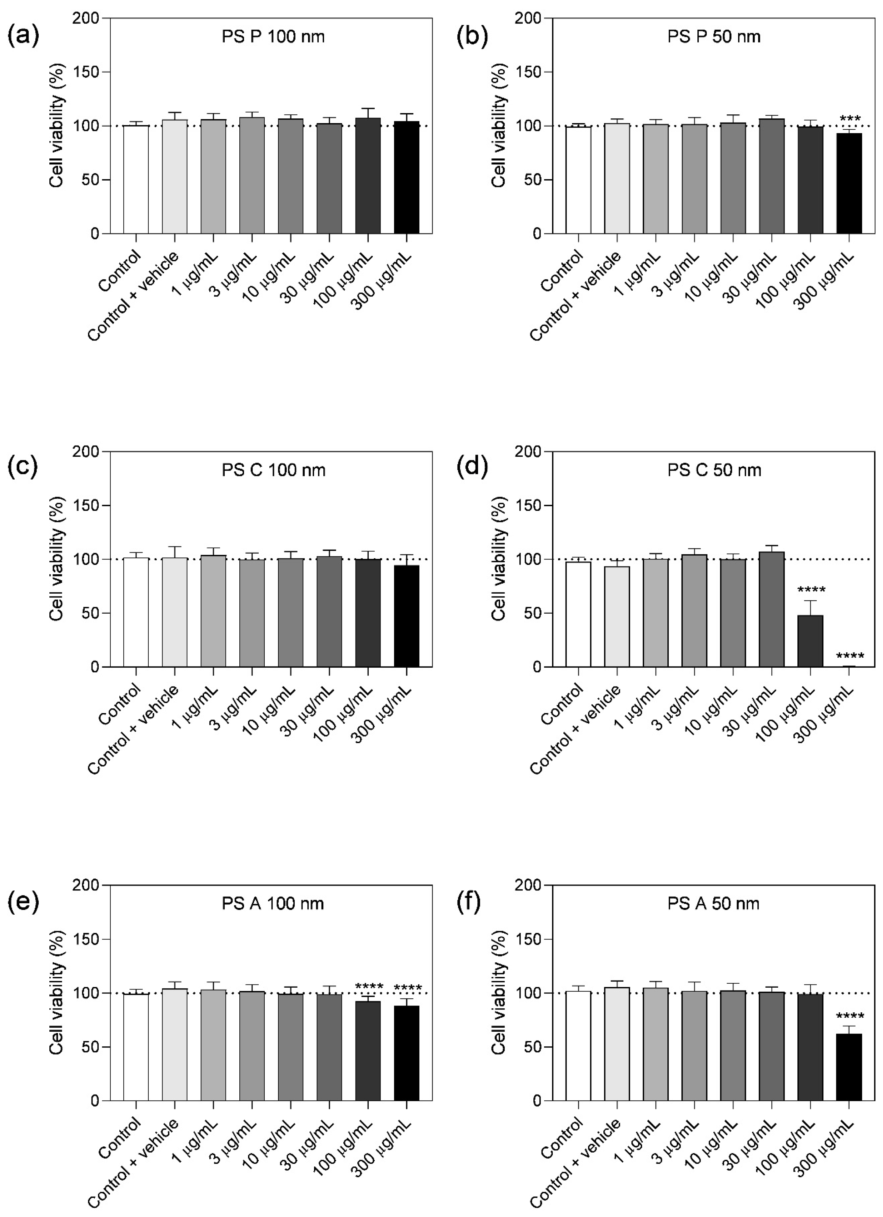
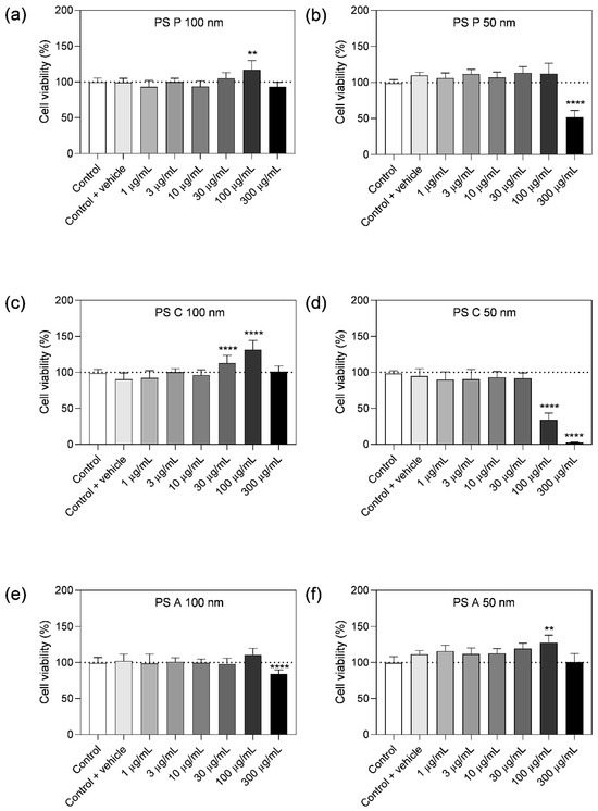
The impact of PS-NPs on cell viability in BECs (hCMEC/D3), human primary astrocytes, and pericytes was evaluated after exposure to pristine, carboxylated, or aminated particles with different sizes (50 nm, 100 nm). The particles were applied at concentrations from 1 to 300 µg/mL for 48 h. 100 nm pristine and carboxylated PS-NPs had no measurable effect on cell viability, only 100 nm aminated NPs led to a 12% decrease in viability at high concentrations (Figure 1a,c,e), whereas 50 nm PS-NPs significantly reduced hCMEC/D3 cell viability by 7% (pristine), 100% (carboxylated), and 38% (aminated) at the applied concentration of 300 µg/mL (Figure 1b,d,f).
Figure 1.
Effects of polystyrene (PS) nanoparticles on hCMEC/D3 cell viability after 48 h of incubation. (a,c,e) Viability after treatment with 100 nm PS nanoparticles; (b,d,f) viability after treatment with 50 nm PS nanoparticles, determined by MTT assay. Outliers were removed using Grubb’s test. Results are presented as percentage relative to the vehicle control; values represent mean ± SD, N = 10–18 (3–6 replicates per condition per experiment), n = 3. Data were analysed via one-way ANOVA on ranks (Kruskal–Wallis) with 95% confidence interval, followed by Dunn’s multiple comparisons test (* p < 0.05, ** p < 0.01, *** p < 0.001, **** p < 0.0001). P, pristine; C, carboxylated; A, aminated.
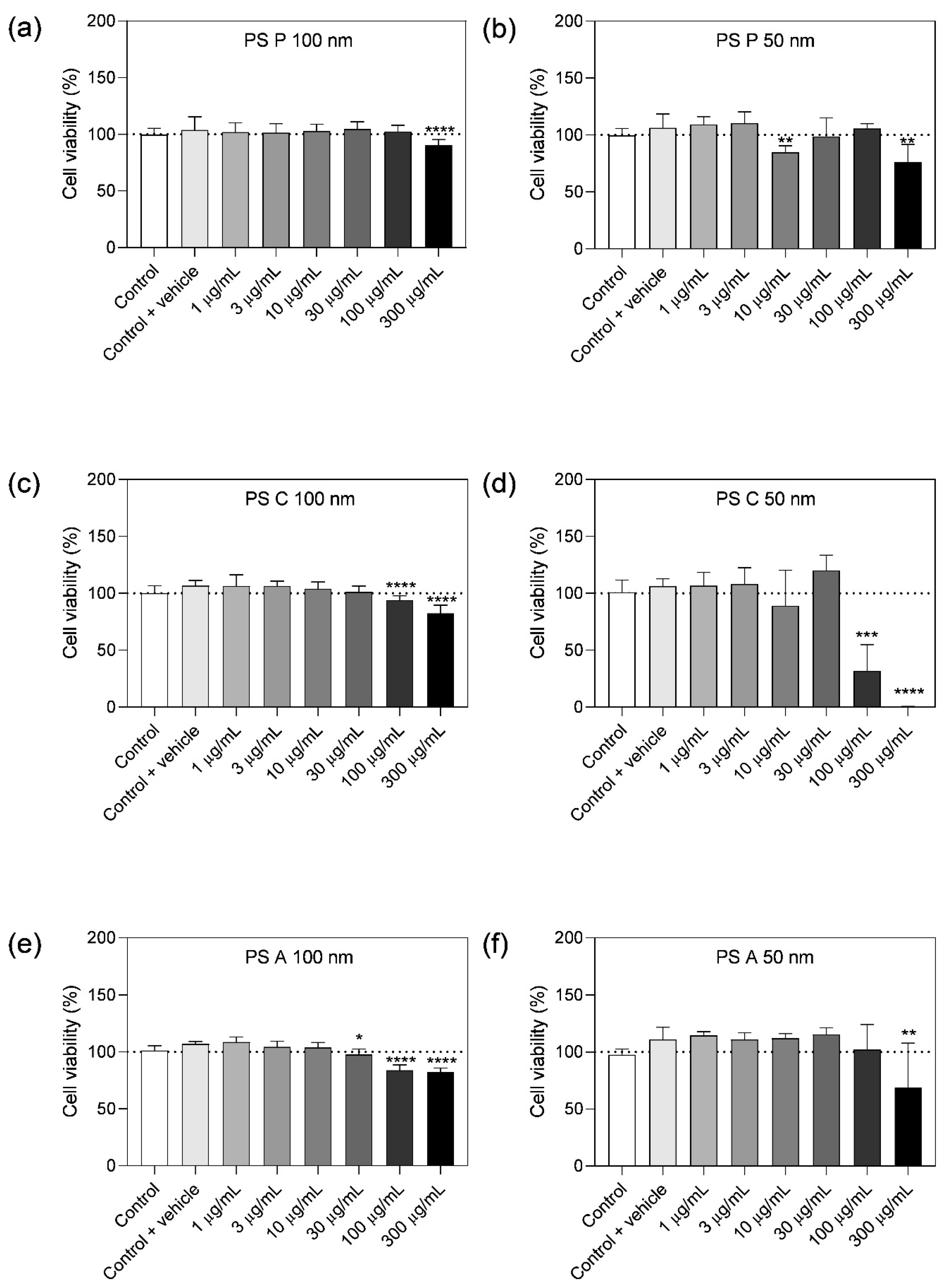
In human primary astrocytes, cell viability (or metabolic activity of cells) increased by up to 32% after exposure to 100 nm PS at 100 µg/mL for 48 h (Figure 2a). A similar effect was observed for 50 nm particles: both pristine and aminated PS significantly increased cell viability at 100 µg/mL (Figure 2d–f), whereas 300 μg/mL pristine 50 nm PS led to a reduction by 48%, and 50 nm carboxylated particles decreased viability to 34% at 100 μg/mL (Figure 2b–d). The highest applied concentration of 100 nm aminated PS led to a 16% viability decrease, while 50 nm aminated particles had no effect (Figure 2e,f).
Figure 2.
Effects of polystyrene (PS) nanoparticles on human primary astrocyte cell viability after 48 h of incubation. (a,c,e) Viability after treatment with 100 nm PS nanoparticles; (b,d,f) viability after treatment with 50 nm PS nanoparticles, determined by MTT assay. Outliers were removed using Grubb’s test. Results are presented as percentage relative to the vehicle control; values represent mean ± SD, N = 10–18 (3–6 replicates per condition per experiment), n = 3. Data were analysed via one-way ANOVA on ranks (Kruskal–Wallis) with 95% confidence interval, followed by Dunn’s multiple comparisons test (* p < 0.05, ** p < 0.01, *** p < 0.001, **** p < 0.0001). P, pristine; C, carboxylated; A, aminated.
Cell viability of human primary pericytes was reduced by 100 nm pristine and carboxylated PS at 300 μg/mL by 9% and 18%, respectively; aminated particles caused a similar decrease by 16–18% (Figure 3a–c). Consistent with previous results, 50 nm particles demonstrated higher toxicity: pristine PS reduced viability by up to 24%, carboxylated caused near-total cell death at 300 μg/mL, and aminated PS led to a 32% reduction at the same concentration (Figure 3d–f).
Figure 3.
Effects of polystyrene (PS) nanoparticles on human primary pericyte cell viability after 48 h of incubation. (a,c,e) Viability after treatment with 100 nm PS nanoparticles; (b,d,f) viability after treatment with 50 nm PS nanoparticles, determined by MTT assay. Outliers were removed using Grubb’s test. Results are presented as percentage relative to the vehicle control; values represent mean ± SD, N = 9–18 (3–6 replicates per condition per experiment), n = 3. Data were analysed via one-way ANOVA on ranks (Kruskal–Wallis) with 95% confidence interval, followed by Dunn’s multiple comparisons test (* p < 0.05, ** p < 0.01, *** p < 0.001, **** p < 0.0001). P, pristine; C, carboxylated; A, aminated.
3.3. Uptake of Stained Nanopolystyrene Particles Is Cell Model-Dependent
In order to visualise and compare the uptake of particles by hCMEC/D3, astrocytes and pericytes, 100 nm pristine, 50 nm carboxylated and 100 nm aminated NPs were selected. For fluorescent detection, the particles were stained with Atto665 and characterised by NTA. In comparison to the unstained particles, stained NPs exhibited larger hydrodynamic diametres and increased agglomeration in cell culture media (Tables S2–S4).
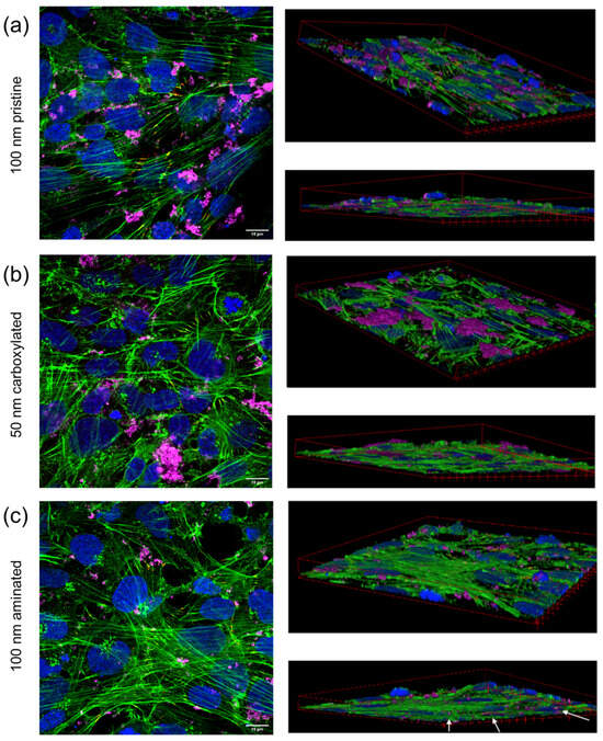
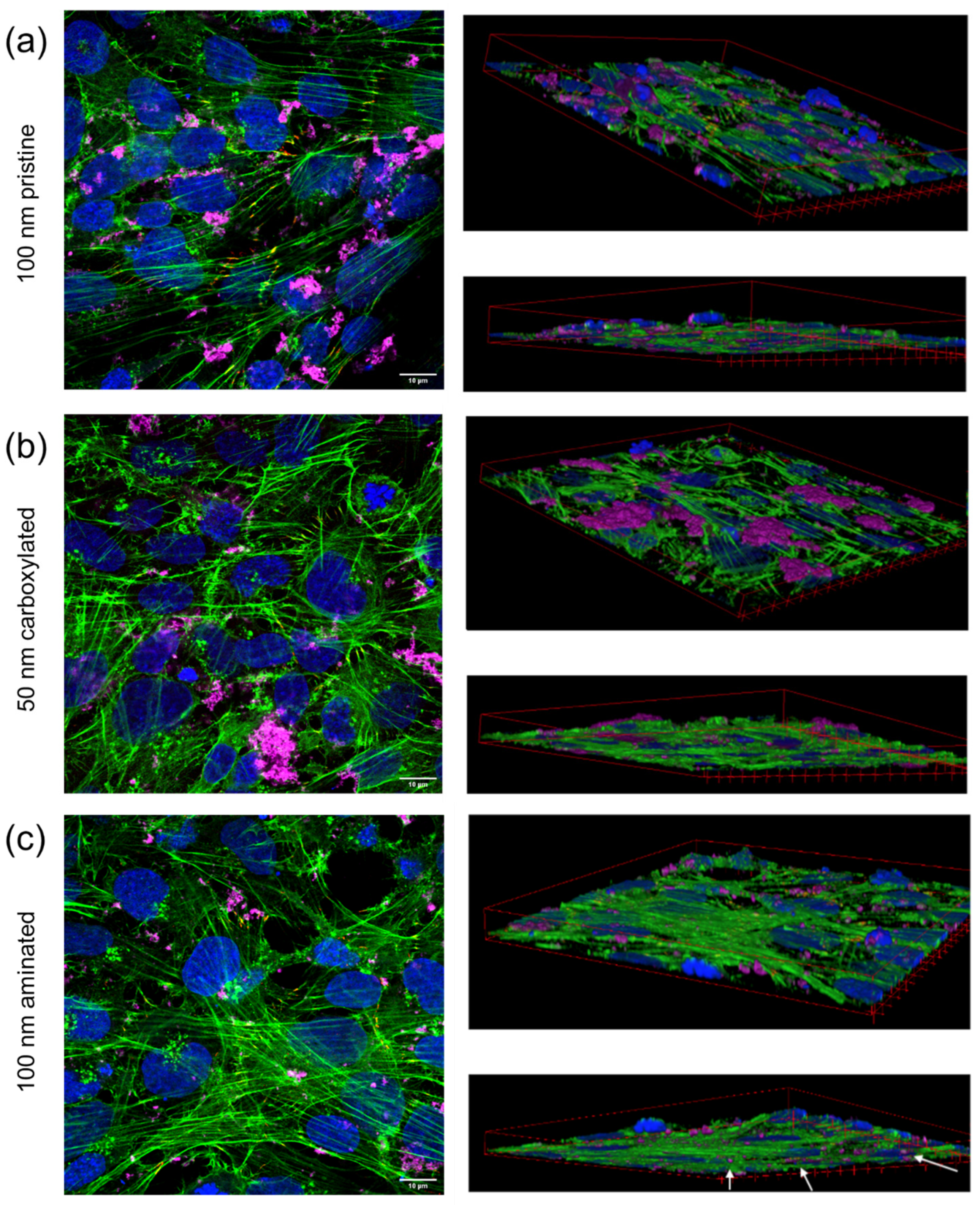
For microscopic analysis, hCMEC/D3 cells, human primary astrocytes and pericytes were cultured on glass cover slips and exposed to the labelled NPs for 48 h before fixation and counterstaining of DNA and actin cytoskeleton. Confocal images revealed that next to small uptaken particles, especially the 50 nm carboxylated and 100 nm pristine particles seemed to have settled as large agglomerates mostly on the cells’ surface, while 100 nm aminated particles showed less localisation in clusters and, hence, might have been more easily internalised (Figure 4c, white arrows).
Figure 4.
Confocal fluorescence images showing uptake of selected PS nanoparticles (PS-NPs) by hCMEC/D3 cells after 48 h: (a) 100 nm pristine PS-NPs; (b) 50 nm carboxylated PS-NPs; (c) 100 nm aminated PS-NPs. Each panel (a–c) contains three images: a top view showing all channels merged (left), and top and bottom z-stacks representing apical and basal views (right). Green: phalloidin staining for actin filaments (cytoskeleton); pink: nanopolystyrene; blue: DAPI staining for cell nuclei. Scale bar: 10 μm.
Similar observations were made in astrocytes and pericytes (Figures S3a–c and S4a–c). Formation of larger aggregates of particles may have been influenced by the respective cell media and the chemical modification introduced by the fluorescent label. Due to the pronounced agglomeration observed in several media conditions, it was not methodologically feasible to distinguish single from agglomerated particles and, consequently, to quantify the proportion between them. Interestingly, only in pericytes treated with 100 nm aminated NPs, cells containing micronuclei were detected, which could be indicative of direct or indirect DNA damage caused by the particles (Figure S4c, white arrows).
3.4. Nanoplastic Size and NP Staining Affect the Blood–Brain Barrier Integrity In Vitro
Two different in vitro ThinCert® BBB models were exposed to the three selected PS-NPs. The first model was based on human cell line hCMEC/D3 exhibiting moderate barrier tightness, whereas the second was based on hiPSC-BCELCs with significantly higher TEER values reflecting more in vivo-similar conditions with regard to paracellular barrier tightness.
Unstained PS particles had minimal impact on TEER of hCMEC/D3 cells, except for carboxylated 50 nm, which caused a 34% TEER decrease after 48 h (Figure 5a), corresponding to the previously observed effects on cell viability. Among stained particles, animated PS-NPs significantly reduced TEER by 22% from initial values (Figure 5c), while stained pristine and carboxylated PS initially raised TEER, which stabilised by 24 h. Altogether, unstained 50 nm carboxylated and stained 100 nm aminated PS exerted substantial (positive and negative) effects on hCMEC/D3 cell barrier integrity.
In hiPSC-BCELC layers, unstained PS particles showed similar effects. PS carboxylated 50 nm significantly reduced TEER by 38% relative to the control and by 46% compared to initial TEER values (Figure 5b). Stained PS-aminated particles decreased TEER by 34% (control) and 43% (initial values) after 48 h (Figure 5d). Consistent with hCMEC/D3 results, unstained 50 nm carboxylated and stained 100 nm aminated PS had the strongest impact on hiPSC-BCELC barrier integrity.
3.5. Endothelial Cells Responded to Carboxylated NP Treatment on mRNA Expression Level
To determine if mRNA expression of genes relevant for BBB functionality was affected by 48 h PS-NP exposure, hCMEC/D3 cells and hiPSC-BCELCs were lysed directly after TEER measurements.
In hCMEC/D3 cells, unstained and stained 50 nm carboxylated PS treatment led to downregulations of gene transcripts important for barrier function and integrity (Figure 6a), such as claudins: claudin-1 (unstained: 0.29-fold, stained: 0.32-fold), claudin-5 (0.29-fold, 0.42-fold), claudin-7 (0.37-fold, 0.34-fold), and claudin-9 (0.35-fold, 0.3-fold). Furthermore, transcripts essential for cell–cell junction formation, occludin (0.35-fold, 0.55-fold), ZO-1 (0.5-fold, 0.51-fold), and VE-Cadherin (unstained: 0.39-fold) were affected, aligning with the observed TEER reductions (Figure 5a). Only a few changes were found in samples treated with unstained 100 nm pristine and aminated PS NPs, including claudin-5 (pristine: 0.67-fold) and VE-Cadherin (pristine: 0.77-fold) (Table S5).
Figure 5.
Effects of 48 h treatment with 100 μg/mL of selected PS nanoparticles (PS-NPs) on blood–brain barrier integrity. (a,b) Unstained PS-NPs; (c,d) stained PS-NPs. (a,c) hCMEC/D3 cells; (b,d) hiPSC-derived brain capillary endothelial-like cells (hiPSC-BCELC), assessed by TEER measurements. Results are presented as percentage relative to the control with vehicle. The outliers were eliminated by performing Grubb’s tests. Value of each column is shown as mean ± SD, N = 6–13, n = 3. Absolute starting (‘Before’) TEER values (mean ± SD) for ‘Control + vehicle’ groups were: for hCMEC/D3 (stained and unstained) = 14.71 ± 4.96 Ω × m2, for hiPSC-BCELC (unstained) = 6336.56 ± 949.95 Ω × cm2, and for hiPSC-BCELC (stained) = 1975.70 ± 686.18 Ω × cm2. Data were established via mixed-effects model (REML—residual maximum likelihood) with confidence interval of 95% due to the unbalanced data set. The model fitting was followed by Tukey’s multiple comparisons tests (* p < 0.05, ** p < 0.01, *** p < 0.001 and # p < 0.05, ## p < 0.01, ### p < 0.001). P, pristine; C, carboxylated; A, aminated.
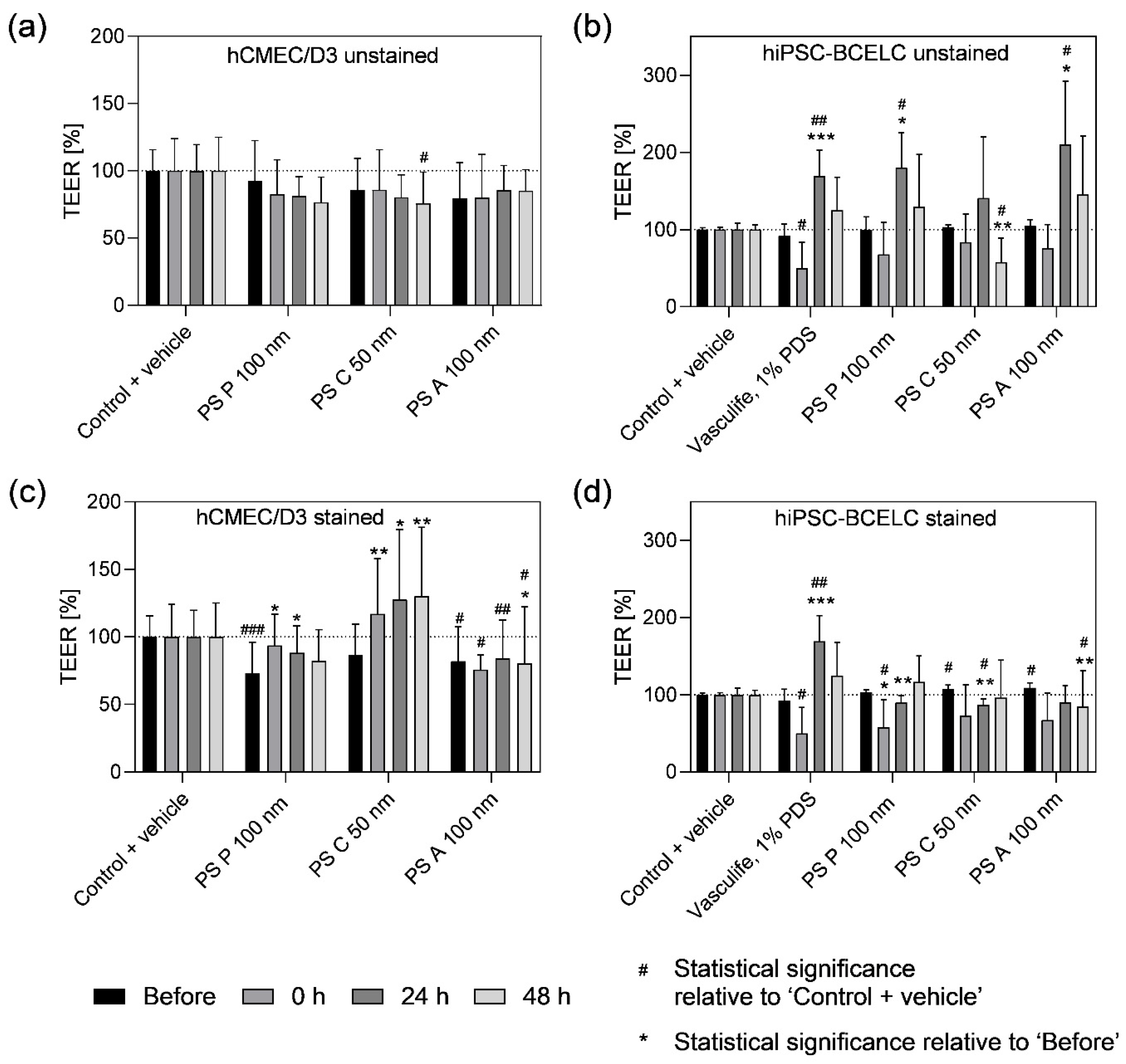
Figure 6.
Heatmap and hierarchical clustering of mRNA expression in endothelial cells treated with selected PS nanoparticles (PS-NPs). (a) hCMEC/D3 cells; (b) hiPSC-derived brain capillary endothelial-like cells (hiPSC-BCELCs) treated with unstained and stained (ST) versions of three PS-NPs. Threshold cycle (Ct) values were normalised to the endogenous housekeeping genes (PPIA) relative to the vehicle control in VascuLife® (VL + H2O). The colour code legend is formatted in log scale. Using the colour scale “viridis,” the X-fold expression of the mean values of three independent experiments is shown, with 2 (yellow) denoting a high expression and −2 (purple) denoting a low expression value. Data are presented as mean ± SD, N = 2–3, n = 2–3. P, pristine; C, carboxylated; A, aminated.
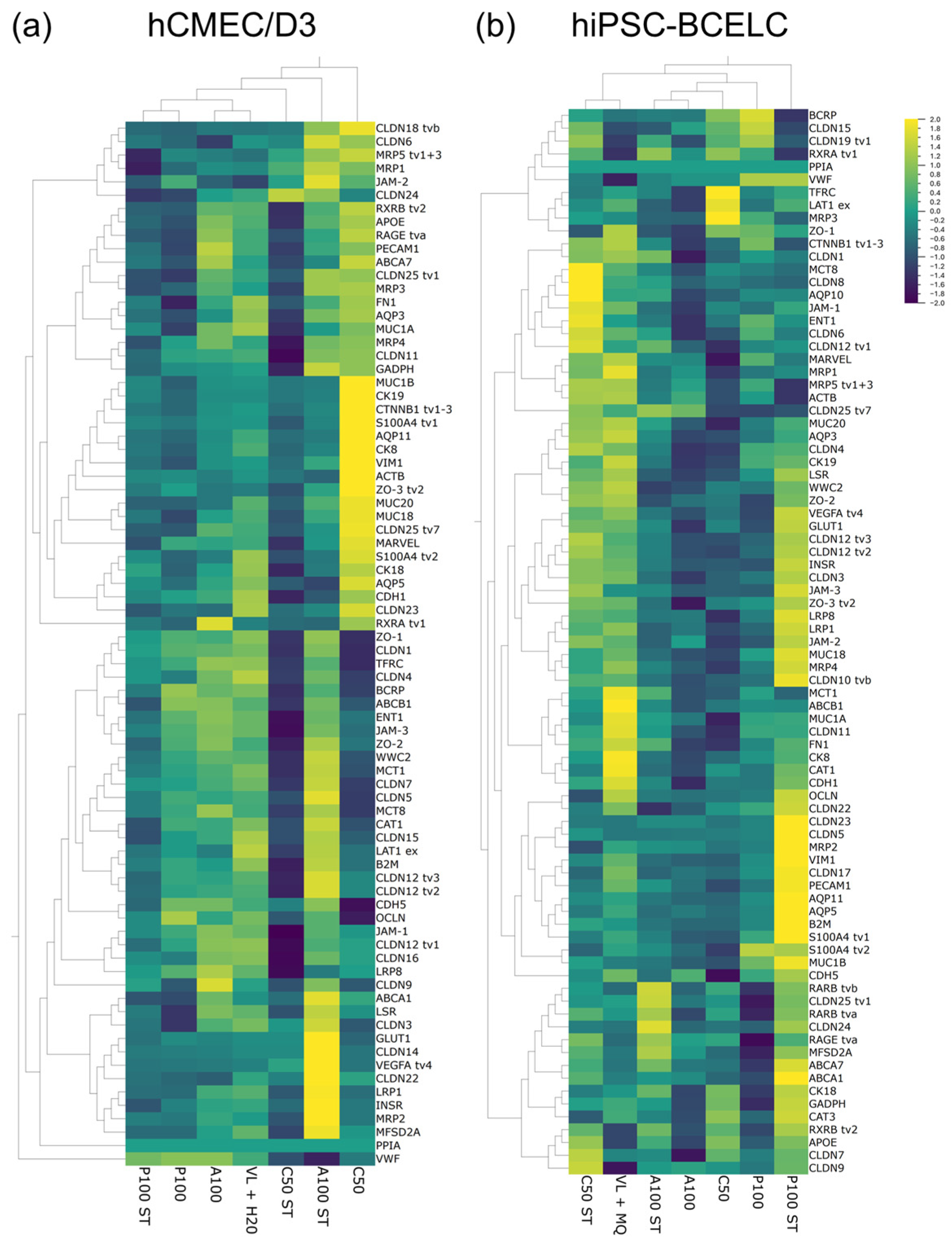
Concordantly, also in hiPSC-BCELCs, only 50 nm carboxylated PS particles caused downregulation in genes linked to BBB integrity, including tight and adherens junction proteins claudin-11 (unstained: 0.48-fold) and VE-Cadherin (unstained: 0.39-fold) (Table S6). Other PS-NPs did not cause significant changes in the expression of the assessed genes. Across both BBB models, hierarchical clustering confirmed that unstained 50 nm carboxylated PS had the most substantial impact on mRNA expression, reflecting its toxicity and effect on barrier integrity (Figure 6a,b).
4. Discussion
Micro- and nanoplastics are increasingly abundant in the environment [23]. They may enter the human body, penetrate biological membranes, and persist over time [24]. Due to their smaller size, nanoplastics pose a greater toxicity risk than microplastics [8,25]. The evidence of plastics, primarily PET and PS, in human blood has raised concerns about their ability to cross the BBB and potential adverse effects on NVU cells, and, consequently, cause further damage to neuronal cells [4]. This study investigated the impact of 50 and 100 nm sized PS-NPs with three chemical surface properties (pristine, carboxylation, amination) on human NVU cells: BECs, astrocytes and pericytes. Cell viability, fluorescently labelled PS-NP uptake, BBB integrity, and gene expression were analysed following PS-NP exposure.
NP characterisation involved measuring zeta potential and hydrodynamic diameter. While zeta potential served as a surface charge indicator, variations in functional group ratios across batches could make it challenging to standardise surface charge, and dispersing cell culture media further affected particle charge. Although aminated particles are expected to carry a positive charge, factors such as residual negatively charged sulfate ester groups from manufacturing (Polysciences Inc., Warrington, PA, USA; Technical Data Sheet 1002), adsorption of anionic biomolecules in cell culture media, and pH-dependent deprotonation can lead to observed negative or less positive zeta potentials under experimental conditions. Significant deviations in zeta potential from values from the literature were observed, likely due to differences in media composition, NP concentration, labelling, production, and measurement techniques [26,27].
Hydrodynamic diameter estimation is vital, as particle size correlates with nanotoxicity [8,25]. Importantly, EBM-2 medium promoted NP agglomeration; hence, an alternative endothelial medium, inducing less agglomeration, VascuLife®, was used throughout this study. Banerjee et al. reported significant size increases for PS 50 nm pristine (non-functionalised), 50 nm carboxylated and 100 nm aminated particles by 74-fold, 33-fold, and 37-fold after 24 h at 37 °C [27], confirming their agglomeration tendencies in the commonly used cell culture medium RPMI. One possible explanation could be the tendency of nanoparticles to agglomerate [9], further influenced by medium composition and the formation of protein coronas that can alter colloidal stability, surface charge, and ultimately NP-cell interactions. The adsorption of biomolecules onto NP surfaces can stabilise or destabilise dispersions depending on the corona composition, thereby influencing agglomeration behaviour and potentially modulating cellular responses and binding of protein-NP complexes through changes in particle structural integrity, identity and recognition [28,29].
PS-NPs were reported to impact cell viability by binding strongly to lipids, disrupting membrane integrity, and increasing paracellular permeability [30]. To comprehensively assess the effects of size and surface charge, PS pristine, carboxylated, and aminated NPs (100 nm and 50 nm) on cell viability of hCMEC/D3 cells, human primary astrocytes, and pericytes were tested via MTT assay. In hCMEC/D3, all three types of 50 nm PS-NPs, as well as PS 100 nm aminated and carboxylated NPs, reduced viability at the highest concentrations (100–300 μg/mL) applied in VascuLife®. In a related study using EBM-2 medium, PS 42 nm decreased hCMEC/D3 viability by 70% at 200 µg/mL after 24 h [31]. As expected, smaller particles revealed stronger effects on cell viability; however, contrary to the existing literature, carboxylated PS showed higher toxicity than aminated PS [26,32].
PS-NPs have the potential to interact with other biological barriers through direct contact with epithelial or endothelial cells or via distribution in the bloodstream. Consistent with our BBB cell viability findings, Wei et al. observed significant viability loss in human umbilical vein endothelial cells (HUVECs) exposed to 30 nm carboxylated and aminated PS-NPs at 50 μg/mL after 6 h, and 500 μg/mL after 3 h [33]. Similarly, size-dependent cytotoxicity in A549 alveolar epithelial cells was reported, where smaller PS-NPs rapidly accumulated and affected viability at 25–30 μg/mL, while larger particles decreased viability only at concentrations above 160–300 μg/mL after 24 h [7]. Dose-dependent viability reductions were found in lung epithelial cells (BEAS-2B, HPAEpiC) exposed to 40 nm pristine PS at 10–40 µg/cm2 (32–128 µg/mL) for 24 h [34].
In the current study, NVU cells exhibited similar sensitivity corresponding to the size and surface chemistry of the applied NPs. In astrocytes, carboxylated PS had the strongest impact on viability, followed by pristine PS 50 nm and aminated PS 100 nm at 100 and 300 μg/mL. All PS types reduced pericyte viability, with minor reductions for 100 nm particles (9–18%), and more substantial impacts for 50 nm particles (24–67%). Notably, carboxylated 50 nm PS proved to be lethal at 300 μg/mL. These results aligned with a study on NVU cells, which tested amine-modified 200 nm polystyrene beads on primary human brain endothelial cells, astrocytes and pericytes up to a concentration of 200 µg/mL [35]. Data revealed no significant effects on brain endothelial cells after 24 or 72 h, but showed a decrease in cell viability by about 20 to 25% for the highest concentration treating astrocytes and pericytes. In another study, SH-SY5Y neuroblastoma cells were treated with pristine 50 nm PS, resulting in a significant viability reduction at 50 and 500 μg/mL after 48 h [36]. The vulnerability of SH-SY5Y cells to carboxylated 50 nm PS was confirmed in this work as well (Figure S5). While the most pronounced effects on NVU cells were observed at higher concentrations (≥100 µg/mL), the applied concentration range also included environmentally relevant doses (1–3 µg/mL), which did not induce detectable toxicity. Nevertheless, as real-world exposures are likely to happen chronically at low levels, potential cumulative and adaptive cellular responses warrant further investigation to better contextualise these findings.
In contrast, some models did not reveal cytotoxic effects of NPs. Minimal effects (<3%) were reported in a lung epithelial cell model (MucilAirTM) exposed to pristine PS 50 and 100 nm particles at 1 mg/mL after 24 and 48 h [37]. Similarly, carboxylated 50 nm PS-NPs had no impact on cell viability in intestinal (Caco-2/HT29) and placental (BeWo b30 cell line) barrier models up to 100 μg/mL after 24 h [38]. These divergent results highlight model-specific and methodological variability in cytotoxic responses to NPs.
Microscopic evaluation of fluorescently labelled PS particles supported previous findings, showing that surface chemistry, particle size, and medium composition may also influence, amongst other properties, nanoparticle agglomeration. While fluorescent labelling was used to track NP uptake, it may alter NP surface properties and colloidal stability, potentially influencing cellular interactions. The particles for the labelling were chosen based on relevance: pristine PS was used as a standard due to its extensive prior testing, the 50 nm carboxylated PS was included due to its notably harmful effects observed on all tested cell lines, and the 100 nm aminated PS was selected to cover the missing surface chemistry type. Comparing, in this study, the effects of the selected labelled particles on cell viability of hCMEC/D3 cells (Figure S6) with the corresponding non-labelled particles (Figure 1) revealed altered impacts of the stained particles. For example, the harmful effect of PSC 50 nm particles was reduced after the labelling. Similarly, a decrease in barrier integrity determined by TEER was not detected with the labelled PSC 50 nm particles anymore (Figure 5).
In hCMEC/D3 cells (in VascuLife® medium), pristine 100 nm and carboxylated 50 nm particles appeared to settle as large clusters on top of the cell layer, while aminated 100 nm particles might have stayed dispersed. In pericytes, aminated 100 nm PS particles seemed to form smaller agglomerates, which might as well correspond to higher uptake levels. Interestingly, in these samples, more micronuclei were observed, indicating that DNA damage had occurred. Whether this was a direct or indirect effect of the applied NPs remained to be determined.
In recent years, the potential penetration of PS-NPs across the BBB has been a topic of ongoing discussion. Few studies have suggested that fluorescently dyed PS-NPs can breach the BBB under certain conditions. Evidence of BBB penetration in mice in vivo, detecting orally administered fluorescent PS micro- and nanoparticles (MNPs) in brain samples within two hours using fluorescence microscopy was reported [15]. As a counterstain for cell-specific (endothelial) markers on the brain sections was not included in this report, the permeation of the applied NPs across the BBB remained unproven. Another recent study reported the effects of long-term treatment (3 or 6 weeks) of rats with orally administered LDPE-MPs, which led to compromised BBB integrity, oxidative stress and declined BDNF levels in the brain [39]. However, a bona fide confirmation of particle permeation across the BBB was not provided. In an in vitro approach, hCMEC/D3 cells were apically exposed to 42 nm fluorescently labelled PS-NPs (25, 50 and 100 µg/mL) for 24 and 72 h and fluorescence was detected on the basolateral side of the insert model [31], representing an indirect confirmation of permeation, but leaving the possibility that the observed fluorescence originated from fluorescent dye leached from the particles. However, in our experimental setup, the fluorescence of the free dye control was negligible.
In order to address this limitation, in the presented study, particle concentrations in the donor and receiver compartments were measured immediately after the experiment using NTA. Overall, single, fluorescent permeated PS-NPs were undetectable immediately after the experiments (Figure S7a). However, after storage at −20 °C for several months, fluorescent particles were detected, but with significantly increased size (Figure S7, Table S7). This finding suggested not only the potential aggregation of NPs over time, but also underscored the importance of immediate particle analysis to accurately assess the permeability of fluorescently labelled NPs across biological barriers. Moreover, the development of newly fluorescent particles during storage could lead to overestimations and false positive interpretations of experimental results. However, due to the technical detection limits of NTA, we cannot exclude PS-NP permeation below 1% under our applied experimental setup. In this regard, optimised conditions (pore size bigger than 0.4 µm, addition of 1% BSA as a stabilising agent) to study the permeation of small extracellular vesicles across insert models could be considered for future studies on NP [40]. Altogether, the observations underscore that Atto665 labelling can induce particle aggregation and artifacts, highlighting the necessity to quantify dye loading, include free-dye controls, assess fluorescence loss of labelled particles, and perform immediate post-experiment analysis to avoid misinterpretation of NP permeability and uptake.
The BBB’s integrity was evaluated by TEER measurements in cell monolayers treated with NPs for 48 h. At a concentration of 100 μg/mL unstained NPs, PS pristine and aminated 100 nm had no substantial effect on TEER, while 50 nm carboxylated PS NPs decreased the barrier integrity over 48 h significantly, aligning with previous cell viability results. Concordantly, dose-dependent TEER reductions were reported upon exposure to 42 nm crimson-dyed PS-NPs at 50 μg/mL and 100 μg/mL for 24 and 48 h [31]. Similar results were obtained in a hiPSCs-BCELC model, where unstained 50 nm carboxylated PS and, to a lesser extent, stained aminated NPs, significantly reduced TEER after 48 h incubation. The permeation of paracellular tracer molecules such as lucifer yellow, fluorescein or FITC-labelled dextrans of different molecular weights is commonly used to complement TEER data to describe the tightness of the BBB models. Usage of both methods provides information on different levels. TEER describes the permeability of smaller ions, whereas the permeation of paracellular tracers is dependent on the occurrence of pores between the cells big enough to allow the permeation of the marker molecules. In the current study, fluorescent paracellular tracers were not applied because of their possible interference with the fluorescent NPs and consequent complicated data interpretation.
The study incorporated a Vasculife® + 1% PDS control for hiPSC-BCELC to adhere to standard operating procedures requiring serum during growth and treatment [17,18]. Interestingly, this serum-containing control increased TEER up to 110% after 24 h, followed by a decline after 48 h (Figure S8). In order to avoid interfering serum effects in this study, media were formulated serum-free.
Reported effects of NPs on biological barrier integrity are controversial. No change in transepithelial electrical resistance (TER) in epithelial cell layers after 24 and 48 h of PS-NP exposure at 1 mg/mL was found as compared to vehicle control [37]. Barrier integrity of in vitro gastrointestinal and placental barrier models remained unaffected by exposure to 10 and 100 μg/mL PS carboxylated 50 nm within 24 h [38] and, similarly, no TER reduction was observed in co-cultured intestinal cells (Caco-2, HT29 and Raji-B) exposed to PS pristine 50 nm at 1–100 μg/mL for 24 h [41]. On the other hand, a significant concentration-dependent TER reduction (by about 10%) was reported in lung models with PS pristine 40 nm at 7.5, 15, and 30 μg/cm2 [34].
Molecular analysis of BBB cells exposed to carboxylated NPs revealed biologically relevant effects, including reduced cell viability and barrier integrity, along with downregulation of genes encoding junctional proteins like claudins, occludin, and VE-cadherin in hCMEC/D3 cells, some of which were also affected in the hiPSC-BCELC model. Although the observed differences did not reach statistical significance, minor changes in transcription levels may still be sufficient to exert biological effects. Prior studies have largely focused on transcriptional changes in inflammatory markers following PS-NP exposure. For example, upregulation of pro-inflammatory genes (e.g., IL-6, IL-8, NF-κB, and TNFα) was observed after exposure to 25 and 70 nm PS-NPs for 24 h [7]. Similarly, a decreased barrier integrity was found in lung epithelial cells, evidenced by reduced ZO-1 protein expression and pathway activation in ECM-receptor interaction, focal adhesion, and cytokine pathways using 40 nm PS-NPs [34]. Protein expression studies also supported these findings. After treatment with 30 nm carboxylated and aminated PS-NPs for 6 h, reduced VE-cadherin in endothelial HUVEC cells was observed [33]. Additionally, protein levels of ZO-2, occludin, and claudin-11 were reduced in a mouse blood–testis barrier model exposed to 20 nm PS-NPs, though no mRNA changes were observed in the same timeframe [42].
The concentration of plastic in human blood was reported to be up to 1.6 µg/mL [4], and, presumably, this would represent the current, maximal exposure of the NVU towards NPs. In this study, plastic concentrations between 1 and 300 µg/mL were tested. Detrimental effects became only evident at the highest applied concentrations, which have (at the current level of environmental exposure) no physiological relevance. Nevertheless, even low concentrations may pose risks upon chronic or repeated exposure, as cumulative interactions with NVU cells over time could lead to subtle but biologically meaningful effects that are not captured in short-term experiments. PS was selected as a model nanoplastic because it is widely used in systematic in vitro studies and is available as a well-characterised reference material, allowing reproducible assessment of size, surface chemistry, and potential dose-dependent effects [43]. The most abundant plastic types reported in blood were polyethylene terephthalate (PET), polystyrene (PS), polyethylene (PE) and polymethyl methacrylate (PMMA) [4], suggesting that future studies should include also further polymers for a more comprehensive hazard assessment, since the data obtained in this study cannot fully capture the potential variability in biological responses among different polymer types, limiting the generalizability of conclusions regarding nanoplastic risks to the BBB. Additionally, even very low concentrations of NPs in the blood may have adverse effects on (human) health due to permanent exposure, and—if NPs permeate across biological barriers, even at very low levels—may also accumulate in tissues with yet unknown consequences. Taken together, these findings highlight the need for chronic low-dose studies, incorporation of additional polymer types, and the development of more physiologically relevant in vitro and in vivo BBB models to better assess potential long-term risks of environmentally relevant NP exposure.
5. Conclusions
This study compared the influence of different chemical surface modifications (pristine, aminated, carboxylated), particle sizes (50, 100 nm), and fluorescent labelling of polystyrene NPs systematically in several cell types of the NVU (brain endothelial cells, astrocytes, pericytes) on the molecular and functional level (cell viability, cellular uptake, barrier integrity).
It was demonstrated that smaller PS particles elicit more pronounced effects on cell viability across cell types compared to larger particles. To fully characterise particle attributes and their biological impact, it is essential to assess cellular responses alongside particle physicochemical properties. Our findings identified 50 nm carboxylated PS as the most toxic across cell lines, followed by Atto665-stained aminated 100 nm PS particles. The significant alterations caused by fluorescent labelling highlight that labelled particles should be considered a distinct particle type.
Media composition influenced particle agglomeration and internalisation. Indications of pericyte cell death with aminated 100 nm particles warrant further validation through viability assays. Molecular activity data for unstained 50 nm carboxylated PS mirrored changes in barrier integrity and tight junction gene expression.
Currently, assessing nanoplastic risks to humans remains challenging due to limited exposure data, a lack of clear definitions, standardised analytical methods, including SOPs to prevent particle contaminations, reference materials, and the complexity of nanoplastics’ diverse sizes and chemistries. Classical risk assessments, designed for dissolved chemicals, may need re-evaluation to account for particulate nanomaterials [3,44,45] complex.
Supplementary Materials
The following supporting information can be downloaded at: https://www.mdpi.com/article/10.3390/microplastics5010035/s1, Supplementary Figures (S1–S8) and Tables (S1–S7). Figure S1: sterilisation area for nanoparticles in a laminar flow cabinet with UV lamps. Figure S2: staining optimisation for 1 mg/mL nanoplastic solutions. Figure S3: confocal fluorescence images showing uptake of selected PS nanoparticles (PS-NPs) by human primary astrocytes after 48 h. Figure S4: confocal fluorescence images showing uptake of selected PS nanoparticles (PS-NPs) by human primary pericytes after 48 h. Figure S5: effects of polystyrene (PS) nanoparticles C (carboxylated) 50 nm on viability of differentiated SH-SY5Y cells determined by MTT test after 24 h. Figure S6: effects of stained selected polystyrene (PS) nanoparticles on viability of hCMEC/D3 cells determined by MTT test after 48 h. Figure S7: analysis of fluorescence particle development during storage of receiver media samples from hCMEC/D3 cell permeability experiments. Figure S8: effects of medium component—serum equivalent, 1% PDS in Vasculife on the blood–brain barrier integrity of hiPSC-BCELCs determined by TEER measurements. Table S1: list of high-throughput qPCR barrier chip and inflammatory markers. Table S2: characterisation of hydrodynamic particle size of PS pristine particles (100 μg/mL). Table S3: characterisation of hydrodynamic particle size of PS carboxylated particles (100 μg/mL). Table S4: characterisation of hydrodynamic particle size of PS aminated particles (100 μg/mL). Table S5: gene expression of housekeeping and target markers from hCMEC/D3 cells treated with the unstained and stained (ST) versions of the selected three different nanopolystyrenes using high-throughput qPCR barrier chip. Table S6: gene expression of housekeeping and target markers from hiPSC-BCELCs treated with the unstained and stained (ST) versions of the selected three different nanopolystyrenes using high-throughput qPCR barrier chip. Table S7: summary of fluorescent PS-NP size distribution in transport study samples after extended storage.
Author Contributions
Conceptualisation, A.J.C., A.K. and W.N.; methodology, A.J.C., A.K., A.Š., M.-T.L.-W., M.M.S., H.-P.F., S.B., D.F. and A.B.; validation, A.J.C., A.K. and W.N.; formal analysis, A.J.C. and A.K.; investigation, A.J.C., A.K., A.Š., M.-T.L.-W., H.-P.F., S.B. and A.B.; resources, D.F. and W.N.; writing—original draft preparation, A.J.C. and A.K.; writing—review and editing, A.B. and W.N.; visualisation, A.J.C. and A.K.; supervision, W.N.; project administration, W.N.; funding acquisition, W.N. All authors have read and agreed to the published version of the manuscript.
Funding
This research was funded by the project InChildHealth, receiving funding from the European Union’s Horizon Europe Research and Innovation Programme: Grant Agreement No. 101056883. Views and opinions expressed are, however, those of the author(s) only and do not necessarily reflect those of the European Health and Digital Executive Agency (HaDEA). Neither the European Union nor the granting authority can be held responsible for them. This research was funded in whole, or in part, by the Austrian Science Fund (FWF) [Grant project P 34137-B]. For the purpose of open access, the author has applied a CC BY public copyright licence to any Author Accepted Manuscript version arising from this submission.
Institutional Review Board Statement
Not applicable.
Informed Consent Statement
Not applicable.
Data Availability Statement
The data presented in this study are available on request from the corresponding author.
Conflicts of Interest
The authors declare no conflicts of interest.
Abbreviations
The following abbreviations are used in this manuscript:
| A | Aminated |
| AM | Astrocyte medium |
| BBB | Blood–brain barrier |
| BCELCs | Brain capillary endothelial-like cells |
| BDNF | Brain-derived neurotrophic factor |
| BECs | Brain endothelial cells |
| BSA | Bovine serum albumin |
| C | Carboxylated |
| FBS | Fetal bovine serum |
| hBFGF | Human basic fibroblast growth factor |
| hiPSC | Human-induced pluripotent stem cell |
| HPLC | High-performance liquid chromatography |
| IF | Immunofluorescence |
| MQ | Mili-Q |
| MWCO | Molecular weight cut-off |
| NP | Nanoplastic |
| NTA | Nanoparticle tracking analyser |
| NVU | Neurovascular unit |
| P | Pristine |
| PDS | Platelet-poor plasma-derived serum |
| PE | Polyethylene |
| PET | Polyethylene terephthalate |
| PM | Pericyte medium |
| PP | Polypropylene |
| PS | Polystyrene |
| P/S | Penicillin/streptomycin |
| RA | Retinoic acid |
| ST | Stained |
| TEER | Transendothelial electrical resistance |
| TER | Transepithelial electrical resistance |
| UV | Ultraviolet |
References
- Zhang, Y.; Pedersen, J.N.; Eser, B.E.; Guo, Z. Biodegradation of Polyethylene and Polystyrene: From Microbial Deterioration to Enzyme Discovery. Biotechnol. Adv. 2022, 60, 107991. [Google Scholar] [CrossRef]
- Gigault, J.; Halle, A.T.; Baudrimont, M.; Pascal, P.-Y.; Gauffre, F.; Phi, T.-L.; El Hadri, H.; Grassl, B.; Reynaud, S. Current Opinion: What Is a Nanoplastic? Environ. Pollut. 2018, 235, 1030–1034. [Google Scholar] [CrossRef]
- Brachner, A.; Fragouli, D.; Duarte, I.F.; Farias, P.M.A.; Dembski, S.; Ghosh, M.; Barisic, I.; Zdzieblo, D.; Vanoirbeek, J.; Schwabl, P.; et al. Assessment of Human Health Risks Posed by Nano-and Microplastics Is Currently not Feasible. Int. J. Environ. Res. Public Health 2020, 17, 8832. [Google Scholar] [CrossRef]
- Leslie, H.A.; van Velzen, M.J.M.; Brandsma, S.H.; Vethaak, A.D.; Garcia-Vallejo, J.J.; Lamoree, M.H. Discovery and Quantification of Plastic Particle Pollution in Human Blood. Environ. Int. 2022, 163, 107199. [Google Scholar] [CrossRef]
- Domenech, J.; Cortés, C.; Vela, L.; Marcos, R.; Hernández, A. Polystyrene Nanoplastics as Carriers of Metals. Interactions of Polystyrene Nanoparticles with Silver Nanoparticles and Silver Nitrate, and Their Effects on Human Intestinal Caco-2 Cells. Biomolecules 2021, 11, 859. [Google Scholar] [CrossRef]
- He, Y.; Li, J.; Chen, J.; Miao, X.; Li, G.; He, Q.; Xu, H.; Li, H.; Wei, Y. Cytotoxic Effects of Polystyrene Nanoplastics with Different Surface Functionalization on Human HepG2 Cells. Sci. Total Environ. 2020, 723, 138180. [Google Scholar] [CrossRef]
- Xu, M.; Halimu, G.; Zhang, Q.; Song, Y.; Fu, X.; Li, Y.; Li, Y.; Zhang, H. Internalization and Toxicity: A Preliminary Study of Effects of Nanoplastic Particles on Human Lung Epithelial Cell. Sci. Total Environ. 2019, 694, 133794. [Google Scholar] [CrossRef]
- Mattsson, K.; Johnson, E.V.; Malmendal, A.; Linse, S.; Hansson, L.-A.; Cedervall, T. Brain Damage and Behavioural Disorders in Fish Induced by Plastic Nanoparticles Delivered through the Food Chain. Sci. Rep. 2017, 7, 11452. [Google Scholar] [CrossRef]
- Zare, Y. Study of Nanoparticles Aggregation/Agglomeration in Polymer Particulate Nanocomposites by Mechanical Properties. Compos. Part A Appl. Sci. Manuf. 2016, 84, 158–164. [Google Scholar] [CrossRef]
- Patil, S.; Sandberg, A.; Heckert, E.; Self, W.; Seal, S. Protein Adsorption and Cellular Uptake of Cerium Oxide Nanoparticles as a Function of Zeta Potential. Biomaterials 2007, 28, 4600–4607. [Google Scholar] [CrossRef]
- Kadry, H.; Noorani, B.; Cucullo, L. A Blood–Brain Barrier Overview on Structure, Function, Impairment, and Biomarkers of Integrity. Fluids Barriers CNS 2020, 17, 69. [Google Scholar] [CrossRef]
- Claesson-Welsh, L.; Dejana, E.; McDonald, D.M. Permeability of the Endothelial Barrier: Identifying and Reconciling Controversies. Trends Mol. Med. 2021, 27, 314–331. [Google Scholar] [CrossRef]
- Abbott, N.J.; Revest, P.A.; Romero, I.A. Astrocyte-endothelial Interaction: Physiology and Pathology. Neuropathol. Appl. Neurobiol. 1992, 18, 424–433. [Google Scholar] [CrossRef]
- Weksler, B.B.; Subileau, E.A.; Perrière, N.; Charneau, P.; Holloway, K.; Leveque, M.; Tricoire-Leignel, H.; Nicotra, A.; Bourdoulous, S.; Turowski, P.; et al. Blood-brain Barrier-specific Properties of a Human Adult Brain Endothelial Cell Line. FASEB J. 2005, 19, 1872–1874. [Google Scholar] [CrossRef]
- Kopatz, V.; Wen, K.; Kovács, T.; Keimowitz, A.S.; Pichler, V.; Widder, J.; Vethaak, A.D.; Hollóczki, O.; Kenner, L. Micro- and Nanoplastics Breach the Blood–Brain Barrier (BBB): Biomolecular Corona’s Role Revealed. Nanomaterials 2023, 13, 1404. [Google Scholar] [CrossRef]
- Balistreri, C.R.; Magro, D.; Jadavji, N.M. Insights into the toxic effects of micro-nano-plastics on the human brain and their relationship with the onset of neurological diseases: A narrative review. Ageing Res. Rev. 2025, 111, 102836. [Google Scholar] [CrossRef]
- Appelt-Menzel, A.; Cubukova, A.; Günther, K.; Edenhofer, F.; Piontek, J.; Krause, G.; Stüber, T.; Walles, H.; Neuhaus, W.; Metzger, M. Establishment of a Human Blood-Brain Barrier Co-Culture Model Mimicking the Neurovascular Unit Using Induced Pluri- and Multipotent Stem Cells. Stem Cell Rep. 2017, 8, 894–906. [Google Scholar] [CrossRef]
- Lippmann, E.S.; Azarin, S.M.; Kay, J.E.; Nessler, R.A.; Wilson, H.K.; Al-Ahmad, A.; Palecek, S.P.; Shusta, E.V. Human Blood-Brain Barrier Endothelial Cells Derived from Pluripotent Stem Cells. Nat. Biotechnol. 2012, 30, 783–791. [Google Scholar] [CrossRef]
- Lin, G.C.; Leitgeb, T.; Vladetic, A.; Friedl, H.-P.; Rhodes, N.; Rossi, A.; Roblegg, E.; Neuhaus, W. Optimization of an Oral Mucosa in Vitro Model Based on Cell Line TR146. Tissue Barriers 2020, 8, 1748459. [Google Scholar] [CrossRef]
- Ramme, A.P.; Koenig, L.; Hasenberg, T.; Schwenk, C.; Magauer, C.; Faust, D.; Lorenz, A.K.; Krebs, A.; Drewell, C.; Schirrmann, K.; et al. Towards an Autologous iPSC-Derived Patient-on-a-Chip. bioRxiv 2018. [Google Scholar] [CrossRef]
- Gerhartl, A.; Pracser, N.; Vladetic, A.; Hendrikx, S.; Friedl, H.-P.; Neuhaus, W. The Pivotal Role of Micro-Environmental Cells in a Human Blood–Brain Barrier in Vitro Model of Cerebral Ischemia: Functional and Transcriptomic Analysis. Fluids Barriers CNS 2020, 17, 19. [Google Scholar] [CrossRef]
- Špilak, A.; Brachner, A.; Friedl, H.-P.; Klepe, A.; Nöhammer, C.; Neuhaus, W. Effects of Small Extracellular Vesicles Derived from Normoxia- and Hypoxia-Treated Prostate Cancer Cells on the Submandibular Salivary Gland Epithelium in Vitro. Tissue Barriers 2025, 13, 2347062. [Google Scholar] [CrossRef]
- Zhang, K.; Hamidian, A.H.; Tubić, A.; Zhang, Y.; Fang, J.K.H.; Wu, C.; Lam, P.K.S. Understanding Plastic Degradation and Microplastic Formation in the Environment: A Review. Environ. Pollut. 2021, 274, 116554. [Google Scholar] [CrossRef]
- Shen, M.; Zhang, Y.; Zhu, Y.; Song, B.; Zeng, G.; Hu, D.; Wen, X.; Ren, X. Recent Advances in Toxicological Research of Nanoplastics in the Environment: A Review. Environ. Pollut. 2019, 252, 511–521. [Google Scholar] [CrossRef]
- Lei, L.; Liu, M.; Song, Y.; Lu, S.; Hu, J.; Cao, C.; Xie, B.; Shi, H.; He, D. Polystyrene (Nano)Microplastics Cause Size-Dependent Neurotoxicity, Oxidative Damage and Other Adverse Effects in Caenorhabditis Elegans. Environ. Sci. Nano 2018, 5, 2009–2020. [Google Scholar] [CrossRef]
- Paget, V.; Dekali, S.; Kortulewski, T.; Grall, R.; Gamez, C.; Blazy, K.; Aguerre-Chariol, O.; Chevillard, S.; Braun, A.; Rat, P.; et al. Specific Uptake and Genotoxicity Induced by Polystyrene Nanobeads with Distinct Surface Chemistry on Human Lung Epithelial Cells and Macrophages. PLoS ONE 2015, 10, e0123297. [Google Scholar] [CrossRef]
- Banerjee, A.; Billey, L.O.; Shelver, W.L. Uptake and Toxicity of Polystyrene Micro/Nanoplastics in Gastric Cells: Effects of Particle Size and Surface Functionalization. PLoS ONE 2021, 16, e0260803. [Google Scholar] [CrossRef]
- Fleischer, C.C.; Payne, C.K. Nanoparticle–Cell Interactions: Molecular Structure of the Protein Corona and Cellular Outcomes. Acc. Chem. Res. 2014, 47, 2651–2659. [Google Scholar] [CrossRef]
- Gebauer, J.S.; Malissek, M.; Simon, S.; Knauer, S.K.; Maskos, M.; Stauber, R.H.; Peukert, W.; Treuel, L. Impact of the Nanoparticle–Protein Corona on Colloidal Stability and Protein Structure. Langmuir 2012, 28, 9673–9679. [Google Scholar] [CrossRef]
- Celebi Sözener, Z.; Cevhertas, L.; Nadeau, K.; Akdis, M.; Akdis, C.A. Environmental Factors in Epithelial Barrier Dysfunction. J. Allergy Clin. Immunol. 2020, 145, 1517–1528. [Google Scholar] [CrossRef]
- Shan, S.; Zhang, Y.; Zhao, H.; Zeng, T.; Zhao, X. Polystyrene Nanoplastics Penetrate across the Blood-Brain Barrier and Induce Activation of Microglia in the Brain of Mice. Chemosphere 2022, 298, 134261. [Google Scholar] [CrossRef]
- Schröter, L.; Ventura, N. Nanoplastic Toxicity: Insights and Challenges from Experimental Model Systems. Small 2022, 18, 2201680. [Google Scholar] [CrossRef]
- Wei, W.; Li, Y.; Lee, M.; Andrikopoulos, N.; Lin, S.; Chen, C.; Leong, D.T.; Ding, F.; Song, Y.; Ke, P.C. Anionic Nanoplastic Exposure Induces Endothelial Leakiness. Nat. Commun. 2022, 13, 4757. [Google Scholar] [CrossRef]
- Yang, S.; Cheng, Y.; Chen, Z.; Liu, T.; Yin, L.; Pu, Y.; Liang, G. In Vitro Evaluation of Nanoplastics Using Human Lung Epithelial Cells, Microarray Analysis and Co-Culture Model. Ecotoxicol. Environ. Saf. 2021, 226, 112837. [Google Scholar] [CrossRef]
- Cho, Y.; Seo, E.U.; Hwang, K.S.; Kim, H.; Choi, J.; Kim, H.N. Evaluation of Size-Dependent Uptake, Transport and Cytotoxicity of Polystyrene Microplastic in a Blood-Brain Barrier (BBB) Model. Nano Converg. 2024, 11, 40. [Google Scholar] [CrossRef]
- Huang, Y.; Liang, B.; Li, Z.; Zhong, Y.; Wang, B.; Zhang, B.; Du, J.; Ye, R.; Xian, H.; Min, W.; et al. Polystyrene Nanoplastic Exposure Induces Excessive Mitophagy by Activating AMPK/ULK1 Pathway in Differentiated SH-SY5Y Cells and Dopaminergic Neurons in Vivo. Part. Fibre Toxicol. 2023, 20, 44. [Google Scholar] [CrossRef]
- Donkers, J.M.; Höppener, E.M.; Grigoriev, I.; Will, L.; Melgert, B.N.; van der Zaan, B.; van de Steeg, E.; Kooter, I.M. Advanced Epithelial Lung and Gut Barrier Models Demonstrate Passage of Microplastic Particles. Microplast. Nanoplast. 2022, 2, 6. [Google Scholar] [CrossRef]
- Hesler, M.; Aengenheister, L.; Ellinger, B.; Drexel, R.; Straskraba, S.; Jost, C.; Wagner, S.; Meier, F.; von Briesen, H.; Büchel, C.; et al. Multi-Endpoint Toxicological Assessment of Polystyrene Nano- and Microparticles in Different Biological Models in Vitro. Toxicol. Vitr. 2019, 61, 104610. [Google Scholar] [CrossRef]
- Forutan, G.; Sarkaki, A.; Dehbandi, R.; Ghafouri, S.; Hajipour, S.; Farbood, Y. Chronic Exposure to Microplastics Induces Blood–Brain Barrier Impairment, Oxidative Stress, and Neuronal Damage in Rats. Mol. Neurobiol. 2025, 62, 13777–13785. [Google Scholar] [CrossRef]
- Špilak, A.; Klepe, A.; Kriwanek, S.T.; Friedl, H.P.; Brachner, A.; Nöhammer, C.; Neuhaus, W. Uptake of DU145 and LNCaP Prostate Cancer Cell Line Derived Extracellular Vesicles Is Inversely Correlated with Blood–Brain Barrier Integrity in Vitro. Fluids Barriers CNS 2025, 22, 70. [Google Scholar] [CrossRef]
- Domenech, J.; Hernández, A.; Rubio, L.; Marcos, R.; Cortés, C. Interactions of Polystyrene Nanoplastics with in Vitro Models of the Human Intestinal Barrier. Arch. Toxicol. 2020, 94, 2997–3012. [Google Scholar] [CrossRef]
- Hu, R.; Yao, C.; Li, Y.; Qu, J.; Yu, S.; Han, Y.; Chen, G.; Tang, J.; Wei, H. Polystyrene Nanoplastics Promote CHIP-Mediated Degradation of Tight Junction Proteins by Activating IRE1α/XBP1s Pathway in Mouse Sertoli Cells. Ecotoxicol. Environ. Saf. 2022, 248, 114332. [Google Scholar] [CrossRef]
- Sørensen, L.; Gerace, M.H.; Booth, A.M. Small Micro- and Nanoplastic Test and Reference Materials for Research: Current Status and Future Needs. Camb. Prism. Plast. 2024, 2, e13. [Google Scholar] [CrossRef]
- Alexy, P.; Anklam, E.; Emans, T.; Furfari, A.; Galgani, F.; Hanke, G.; Koelmans, A.; Pant, R.; Saveyn, H.; Sokull Kluettgen, B. Managing the Analytical Challenges Related to Micro- and Nanoplastics in the Environment and Food: Filling the Knowledge Gaps. Food Addit. Contam.-Part A Chem. Anal. Control Expo. Risk Assess. 2020, 37, 1–10. [Google Scholar] [CrossRef] [PubMed]
- Kuhlmann, R. Letter to the editor, discovery and quantification of plastic particle pollution in human blood. Environ. Int. 2022, 167, 107400. [Google Scholar] [CrossRef] [PubMed]

Disclaimer/Publisher’s Note: The statements, opinions and data contained in all publications are solely those of the individual author(s) and contributor(s) and not of MDPI and/or the editor(s). MDPI and/or the editor(s) disclaim responsibility for any injury to people or property resulting from any ideas, methods, instructions or products referred to in the content. |
© 2026 by the authors. Licensee MDPI, Basel, Switzerland. This article is an open access article distributed under the terms and conditions of the Creative Commons Attribution (CC BY) license.