


Abstract
The scientific literature lacks sufficient data on the transport of various toxic pollutants by polymer particles. Investigating how the structure of microplastic particles formed during the degradation of polymeric materials affects pollutant sorption processes will improve our ability to predict environmental behavior. General-purpose polystyrene, expanded polystyrene, ABS plastic (acrylonitrile–butadiene–styrene) and crosslinked polystyrene are produced on an industrial scale. Copolymers of styrene with divinylbenzene are used on a large scale as sorbents for gel permeation chromatography (Styragel brand sorbents), in the production of catalysts on a polymer substrate or ion-exchange resins. In this study, non-spherical, crosslinked polystyrene microparticles with varying polystyrene chain packing densities were used as model microplastic particles representative of crosslinked polystyrene. It was shown that the adsorption of a hazardous chemical rhodamine B was influenced by both the packing density of the polystyrene chains and the presence of ionic functional groups, i.e., the “degree of aging” of the microplastic particles. The sorption capacities of these model microparticles were compared with those of natural origin (silicon dioxide, quartz powder, and microcrystalline cellulose). A viability assay using HEK293 and HeLa cell lines exposed to leachates from both pristine and rhodamine B-loaded microparticles revealed that all unmodified microparticles, regardless of their nature, exhibited no cytotoxicity at concentrations up to 1000 μg/mL. In contrast, microparticles with adsorbed rhodamine B significantly reduced cell viability to 20–40% at concentrations of 100 μg/mL.
1. Introduction
Currently, research on the sorption of pollutants of various types by microplastic particles is highly relevant [1,2,3]. A deeper understanding of adsorption and desorption processes will help elucidate how microplastics transport pollutants in the environment, facilitate studies on the cellular uptake of polymer complexes, and enable tracking of their impacts on living organisms (plants, animals, and aquatic species such as fish). It is well established that polymer nanoparticles complexed with different toxic metal ions can either enhance or mitigate the toxic effects of these metals on living systems [2]. The effect of lead ion sorption on polystyrene microparticles was studied depending on the ionic strength of the solution [3]. Pollutants of concern include antibiotics, heavy metal ions, organic dyes and solvents, and non-steroidal anti-inflammatory drugs, among others [2,3,4,5,6]. Notably, not all organic pollutants exhibit the same environmental behavior: some are readily degraded by UV radiation, temperature, or microbial activity [7,8], while others persist in the environment for extended periods, exerting chronic toxic effects on biota [9]. These latter compounds are classified as persistent organic pollutants. In addition, a number of organic pollutants can accumulate in the organs of living organisms, but they do not exhibit high persistence in the environment. Rhodamine B, a polar and fluorescent dye, is a hazardous chemical and banned food coloring [10]. It is widely used in industrial and consumer applications, with annual global production exceeding several hundred tons. Rhodamine B has been shown to have toxic effects on animals, including gross liver damage [11], erythrocyte hemolysis [12], mutagenicity and potential carcinogenicity [13,14]. Due to its slow metabolic degradation, rhodamine B can interact with the DNA backbone, forming intermolecular hydrogen bonds via its carboxyl group. In 1987, the International Agency for Research on Cancer classified rhodamine B as a Group 3 carcinogen. The studies have shown that concentrations of 14–24 mg/L inhibit the growth of the green alga Raphidocelis subcapitata and cause embryonic lethality in the crustacean Daphnia magna and zebrafish (Danio rerio) [15]. In rats, the LD50 (lethal dose for 50% of the test population) is 89.5 mg/kg for intravenous administration and 500 mg/kg for oral administration of rhodamine B [10]. At a concentration of 5 mg/L, rhodamine B inhibits the photosynthetic activity of the aquatic macrophyte Hydrilla verticillata [16] and leads to the death of green algae. The lethal concentration for mosquitofish is reported as 171 ppm. Despite these findings, data on the effects of rhodamine B on human health remain limited. It is known, however, that at a concentration of 25 μg/mL, rhodamine B significantly suppresses the proliferation of human lip fibroblasts [17], and use of this fluorescent dye in decorative cosmetics is prohibited in both the European Union and the United States [18].
Since a portion of the produced rhodamine B is used to color polymeric materials, these polymers, once released into the environment, gradually degrade, releasing the fluorescent dye back into the ecosystem. An additional pathway for rhodamine B to associate with microplastics exists through wastewater discharge. The dye is widely employed in the textile industry, leading to significant contamination of industrial effluents. Conventional wastewater treatment often relies on sorbents such as Zeolites CTR-Sorb [19], activated carbons like AG-3 [20], and kaolinite [21]. However, even under optimized conditions, complete removal is not achieved. For instance, study [20] reports a maximum removal efficiency of 95%, resulting in a colorless effluent but leaving a residual rhodamine B concentration of 0.46 mg/L. Given that rhodamine B is continuously introduced into aquatic environments via incompletely treated wastewater, it can readily adsorb onto microplastic particles upon contact. Being a hazardous chemical, rhodamine B tends to accumulate in the environment over time, leading to increasing environmental concentrations. This raises urgent concerns about its long-range transport, accumulation potential, and ecological impact. Investigating these processes presents a complex challenge; to date, studies have been limited to controlled laboratory experiments aimed at elucidating the adsorption mechanisms of rhodamine B onto polymer microparticles.
The study [22] demonstrated that rhodamine B was chemisorbed onto microplastic particles of polystyrene (PS), polyvinyl chloride (PVC), and polyethylene terephthalate (PET) in the size range of 74–125 μm. The concentration of the adsorbed dye depended on the polymer type, as well as on the salinity and pH of the surrounding medium. Maximum sorption capacities were reported as approximately 2.4 mg/g for PS and 4.6 mg/g for PVC microparticles. It should be noted that the study used “aged” polymer waste, which was crushed and sieved before the study. However, the term “aged waste” was not clearly defined, and the degree of polymer degradation could not be reliably assessed from the provided Fourier-transform infrared spectra. Nevertheless, it is well established that prolonged exposure of microplastics to natural environmental conditions can significantly alter their sorption and desorption behavior toward various contaminants.
In the present study, we investigated how the degree of aging of model microplastic particles based on crosslinked polystyrene affected their adsorption capacity for the hazardous chemical rhodamine B. We also compared the sorption capacity of these synthetic microparticles with that of naturally occurring microparticles, including silicon dioxide, quartz powder, and microcrystalline cellulose. Furthermore, we characterized the physicochemical properties of all microparticles: chemical structure was analyzed by Fourier-transform infrared (FTIR) spectroscopy; surface morphology was examined using scanning electron microscopy (SEM) and transmission electron microscopy (TEM); and the molecular weight distribution of non-crosslinked polymer chains was estimated via gel permeation chromatography (GPC). Finally, we assessed the cytotoxicity of these microparticles by evaluating the viability of HEK293 and HeLa cell lines. Our results demonstrate that cell viability is influenced by the particle nature, the degree of aging, and the presence of hazardous chemical rhodamine B adsorbed onto the particle surface.
2. Materials and Methods
2.1. Materials and Microparticles Used
The following chemical reagents were purchased from Vekton LLC (Saint-Petersburg, Russia): rhodamine B (reagent grade), sodium chloride (reagent grade), toluene, tetrahydrofuran (THF) and N,N-dimethylformamide (DMF). Ethanol was purchased from the Chemical and Pharmaceutical Plant PAO Bryntsalov-A (Elektrogorsk, Russia). All aqueous solutions were prepared using deionized water with an electrical conductivity of 1 μS/cm.
Crosslinked polystyrene microparticles synthesized by the method previously described [23] were used as the initial model polymer microparticles. These microparticles were further modified in either toluene or N,N-dimethylformamide (DMF) according to the procedure outlined in Section 2.2. Microparticles prepared using the method reported in [24] were used as a model of “fully aged” microplastics. Microparticles of microcrystalline cellulose were obtained from Vekton LLC (Russia). Microparticles of silicon dioxide and quartz powder were obtained from Acros Organics (Darmstadt, Germany). The key physicochemical characteristics of all microparticles are summarized in Table 1.
Table 1.
Characteristics of microparticles.
2.2. Procedure for Obtaining Aged Microparticles
The packing density of polymer chains in model non-spherical polystyrene microparticles was varied by immersing them in toluene or DMF. A known mass of predried microparticles was placed in a test tube, and the appropriate organic solvent (either DMF or toluene) was added to achieve a microparticle concentration of 0.05 g/mL. The microparticles were immersed in the solvent for three months. Then the microparticles were centrifuged (Eppendorf 5424 centrifuge, Hamburg, Germany), the supernatant was decanted, and the microparticles were washed with ethanol three to four times. Notably, microparticles immersed in toluene were easily freed of residual solvent, as confirmed by the absence of solvent peaks in the FTIR spectra. In contrast, to remove DMF which may reside in both the surface layer and the bulk of the microparticles, additional drying was performed under reduced pressure at 45 °C using a Heidolph Hei-VAP Advantage rotary evaporator (Hamburg, Germany) after ethanol washing. The particles were then dried at 25 °C and weighed using an OHAUS Pioneer PR224 analytical balance (Nanikon, Switzerland). Furthermore, the amount of leached polystyrene oligomeric chains in the supernatant, after solvent removal, was quantified by GPC (see Section 2.3).
2.3. Determination of the Molecular Weight of Oligomers by GPC and the ζ-Potential of Microparticles
GPC was performed on a Prominence LC-20AD (Shimadzu, Kyoto, Japan) chromatograph equipped with a refractometric detector and PLgel MIXED-C column (300, 7.5 mm, 5 µm particles, linear molecular weight ranges up to 2000 kg/mol based on polystyrene, Agilent Technologies, Amstelveen, The Netherlands). Runs were performed in tetrahydrofuran (THF) at 40 °C and 1.0 mL/min flow rate, P = 4.2–4.3 MPa. Polymer solutions (3 mg/mL) were filtered through 0.22 µm PTFE filters. Weight average molar masses (Mw) and dispersities (Ð = Mw/Mn) were calculated from GPC traces using the LCSolution software (Version 1.22; Shimadzu, Kyoto, Japan). A cubic calibration curve was built using a set of polystyrene standards (500–250,000 g/mol).
The ζ-potential of polystyrene microparticles was measured in a supporting electrolyte (10−2 M NaCl) after the microparticles were allowed to equilibrate for 2 h. Measurements were performed using a Brookhaven Zeta Plus instrument (Holtsville, NY, USA).
2.4. Study of Microparticles by SEM, TEM and FTIR Spectroscopy
SEM: A Zeiss Merlin scanning electron microscope (Zeiss, Oberkochen, Germany) with a field emission cathode was used to study the surface layer structure of both model polystyrene microparticles and naturally occurring microparticles. The powders of the samples were mounted on conductive adhesive tape. It should be noted that the SEM samples were not gold-coated.
TEM:Transmission electron microscopy (Jeol JEM-1400 microscope, Akishima, Japan) was used to study the structure of the surface layer. Before TEM measurements, an aqueous dispersion of particles was applied to molding substrates treated with a 1% sodium dodecyl sulfate solution. Photographing was performed after the substrate had completely dried.
FTIR: Fourier-transform infrared (FTIR) spectroscopy (IRPrestige-21, Shimadzu, Kyoto, Japan) was used to investigate the changes in surface structure before and after immersing in DMF and toluene in KBr pellets in the range of 400–4500 cm−1. All the spectra were recorded and represented an average of 60 scans at a resolution of 2 cm−1 taken.
2.5. Adsorption of a Hazardous Chemical Rhodamine B on Microparticles
Rhodamine B sorption was carried out using both the original microparticles of various nature (see Table 1) and microparticles that had been immersed in DMF or toluene. The sorption of the organic dye Rhodamine B was studied not only in the presence of salts but also in the presence of a controlled amount of a co-solvent (ethanol), which reduces the surface tension of water to 55–65 mN/m. The final ethanol concentration was 17% vol. Since crosslinked model polystyrene particles are poorly redispersible in an aqueous-salt dispersion medium after aging, ethanol was first added to all dried particles, followed by the aqueous-salt dispersion medium. Thus, all synthesized microparticles with different surface layer structures were redispersed in the dispersion medium, and the adsorption process occurred under identical and controlled conditions. First, 0.5 mL of ethanol was added to a weighed portion of particles (30 mg), and the mixture was dispersed in an ultrasonic bath (Sapphire 2.8, Moscow, Russia) for 10–15 min. Next, 2.5 mL of a 10−2 M NaCl solution (simulating the salinity of freshwater) was added, and the suspension was sonicated again for 10–15 min. A rhodamine B stock solution in ethanol (0.24 g/L) was prepared in advance. This solution was then added to the aqueous saline dispersion of particles to achieve final rhodamine B concentrations ranging from 0 to 5 mg/L. The adsorption process was allowed to proceed for 2 h. Afterward, the particles were centrifuged at 10,000 rpm for 10 min using a centrifuge (Eppendorf 5424, Hamburg, Germany). The concentration of rhodamine B remaining in the supernatant was determined spectrophotometrically at 530 nm using a pre-established calibration curve (UV-1700 Plus spectrophotometer, Shimadzu, Kyoto, Japan). Each adsorption point was measured three times. Statistical analysis and graph plotting were performed using Origin 9.0 software.
2.6. Study of Cell Viability in the Presence of Microparticles of Various Origins
The HeLa and HEK293 cell lines were kindly provided by the Cell Culture Collection of the Institute of Cytology RAS. Cell lines were cultured in complete DMEM growth medium supplemented with 2 mM L-glutamine and 10% fetal bovine serum (FBS) (all from Biolot, Saint-Petersburg, Russia). MTT assay was performed in complete growth medium additionally supplemented with antibiotic-antimycotic solution: 100 U/mL penicillin, 100 μg/mL streptomycin, and 0.25 µg/mL amphotericin B (all from Biolot, Russia). Powders of the studied microparticles were dispersed in complete growth medium to a final concentration of 50 mg/mL and incubated for 4 days at +4 °C with periodic vortex mixing. One day before the analysis, the studied cell lines were seeded in the inner wells of 96-well microplates (Wuxi NEST Biotechnology Co., Wuxi, Jiangsu, China) at a density of 1.5 × 10³ cells per well. Edge wells were filled with 200 µL of sterile phosphate-buffered saline (PBS, Biolot, Russia) to avoid edge effects. Cells were incubated overnight at 37 °C with 5% CO2 in a humidified incubator. To avoid non-specific results due to complete coverage of the plate bottom and other steric problems with microparticles larger than 1 µm, the microparticles were centrifuged at 4427 g for 10 min (OHAUS FC5706, Parsippany, NJ, USA). The supernatant was carefully collected and added to the plate wells to final concentrations of 1, 10, 100, 250, 500, and 1000 μg/mL. Each concentration point was performed in four technical replicates. After 4 days incubation at 37 °C with 5% CO2 in a humidified incubator, the medium in each well was replaced with 200 µL of fresh culture medium containing 0.5 mg/mL MTT (PanEko, Moscow, Russia), and the plates were incubated for 2 h to allow formazan crystal formation. The medium was then carefully removed, and the formazan crystals were dissolved in 150 µL of DMSO (Helicon, Moscow, Russia). The plates were mixed, and absorbance was measured at 570 nm using a SPECTROstar Nano microplate reader (BMG LABTECH, Ortenberg, Germany). Cell viability was calculated as the ratio of the optical density of the sample to the optical density of the untreated control sample. Data are presented as mean ± SD from three independent experiments. Statistical analysis and graph plotting were performed using GraphPad Prism 7 software (GraphPad Software, San Diego, CA, USA).
3. Results
3.1. Physicochemical Properties of Model Polymer Microparticles and Microparticles of Natural Origin
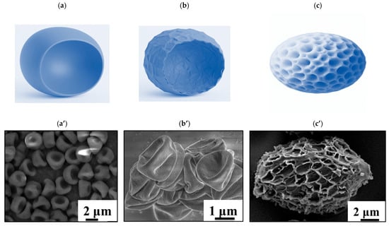
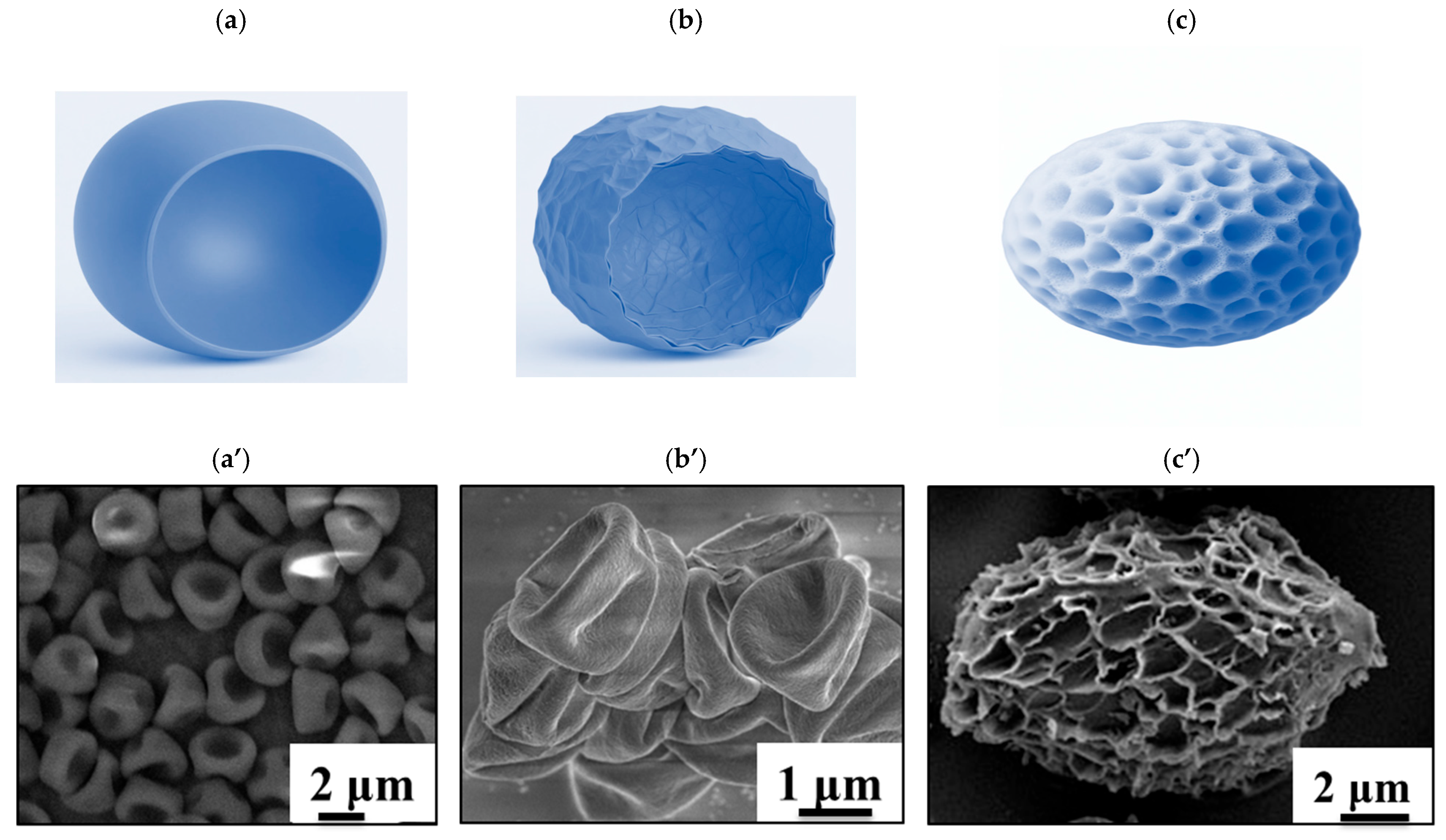
General-purpose polystyrene, expanded polystyrene, ABS plastic and crosslinked polystyrene are produced on an industrial scale. Copolymers of styrene with divinylbenzene are used on a large scale as sorbents for gel permeation chromatography (Styragel brand sorbents), in the production of catalysts on a polymer substrate or ion-exchange resins. In real-world environments, microplastic particles generated during the degradation of polymeric products are typically non-spherical in shape [25]. In this study, non-spherical, crosslinked polystyrene microparticles are used as a model for crosslinked polystyrene microparticles (Figure 1). There is insufficient data in the scientific literature to draw conclusions about the influence of the shape and structure of the surface layer of polystyrene particles on their adsorption behavior and toxicological properties [26]. Some studies report that polystyrene microparticles exhibit toxicity both to cell lines and cause various adverse reactions when they enter the bodies of experimental animals, while other experimental results show no significant toxicity for particles in the 1–40 μm size range across various cell lines. In addition, there are insufficient experimental data describing and explaining the process of sorption of small molecules (Rhodamine B—the theoretical area occupied by one molecule of rhodamine B is ≈1.4 nm2) in the surface layer of model polystyrene microparticles.
Figure 1.
Model of polystyrene microparticles: (a) original non-spherical polystyrene microparticles MPI, (b) “aged” non-spherical polystyrene microparticles with loosely packed polymer chains MPD, (c) fully “aged” polystyrene model microparticles consisting of hydrophilic polystyrene chains with ionic functional groups MPS. SEM data of model polystyrene particles MPI (a’), MPD (b’), MPS (c’).
It is undeniable that fibrous or fragmented microparticles interact differently with organic molecules and living organisms. However, it must be acknowledged that fibers and fragments often originate from different polymer types. Consequently, comparing the effects of particle shape (fibers or fragments) alone without accounting for inherent differences in polymer chemistry (e.g., hydrophobicity/hydrophilicity, elastic modulus, surface charge, and other physicochemical properties) can lead to misleading conclusions.
If one limits the scope of the study to fragment-shaped microparticles formed during the degradation of crosslinked polystyrene, the most commonly used model systems are spherical polystyrene microparticles. However, some researchers believe that such spherical models do not yield reliable results [25]. In a previous study [27], we demonstrated that the shape of model polystyrene microparticles does not significantly affect their sorption behavior toward hazardous chemical. Instead, the key factor governing interactions between polystyrene microparticles and environmentally relevant organic polar dye is the packing density of the polymer chains. In the present work, we investigate how both the packing density of polymer chains in model crosslinked polystyrene microparticles and the hydrophilicity/hydrophobicity of those chains as well as the presence of functional groups (Figure 1) influence the sorption of the hazardous chemical rhodamine B. To mimic “aged” polystyrene microparticles, we employed several preparation methods. As an intermediate aging approach, we generated polystyrene microparticles with loosely packed polymer chains by immersing the original non-spherical polystyrene microparticles (MPI) in N,N-dimethylformamide (DMF) for three months.
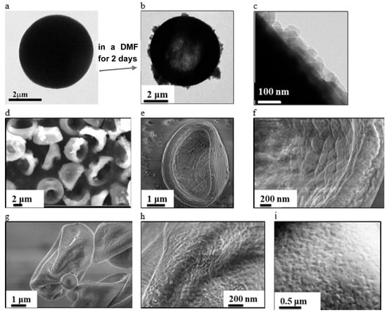
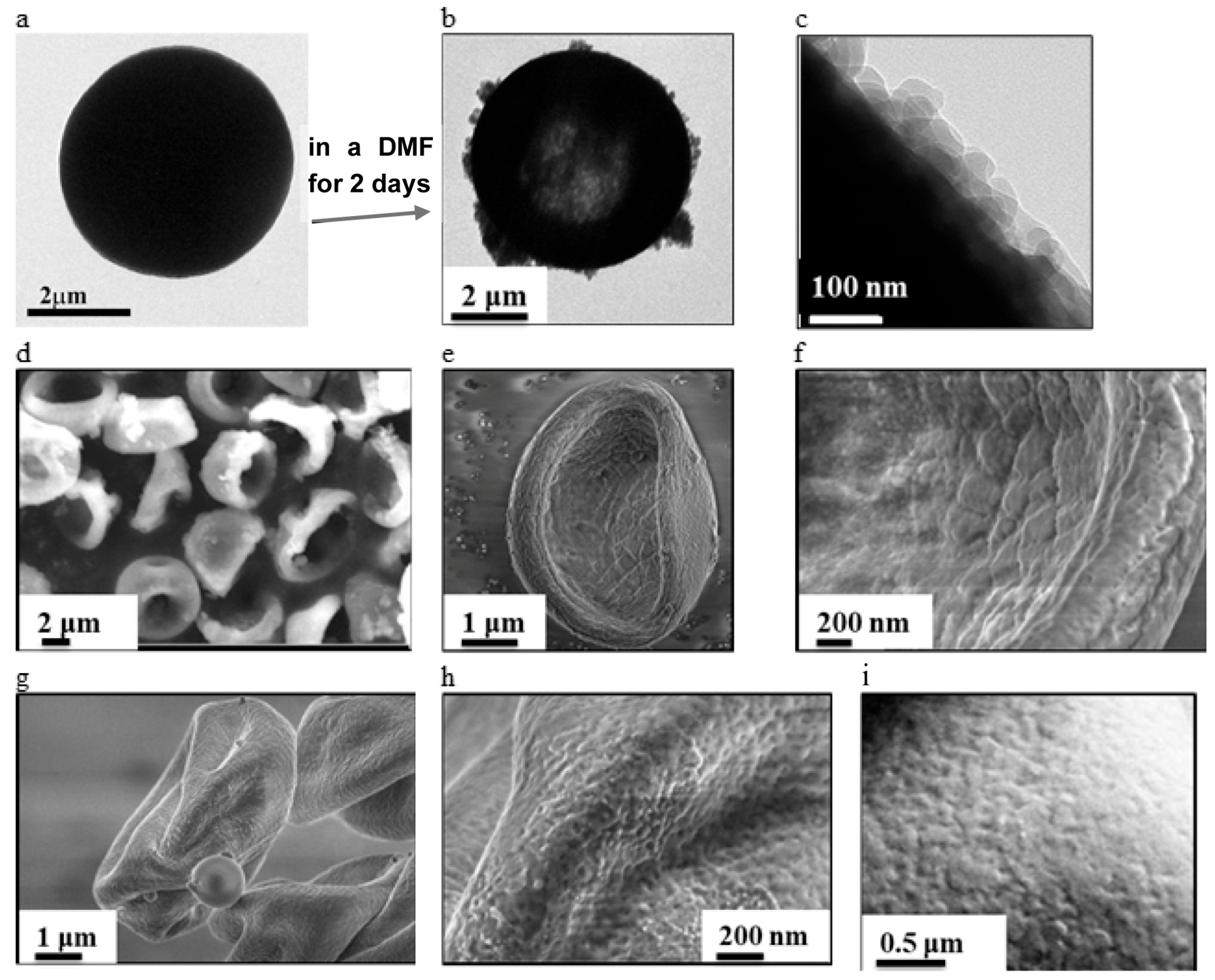
In our earlier work [27], we showed that after just two days of immersing in DMF, the surface layer of these model microparticles develops a “raspberry-like” morphology due to the diffusion of non-crosslinked polymer chains from the particle interior into the solvent. This process significantly reduces chain packing density, resulting in a looser particle structure, as confirmed by transmission electron microscopy (TEM) and scanning electron microscopy (SEM) (Figure 2a–c). Notably, up to 29 wt.% of polystyrene chains with a weight-average molecular weight of 13,000 and a polydispersity index of 1.7 diffuse out of the particles within this 2-day period. In the current study, we further examined how the nature of the solvent and the duration of exposure affect microparticle structure. Prolonged exposure (3 months) of polystyrene microparticles to the aprotic polar solvent DMF leads to the complete extraction of all non-crosslinked polystyrene chains from the particle matrix. Consequently, the surface layer undergoes dramatic restructuring: cracks and micropores (20–50 nm) form throughout the surface, and no residual unbound polymer chains are detectable in the surface layer (Figure 2e,f).
Figure 2.
TEM (a–c) and SEM (d–i) images of non-spherical polystyrene microparticles: kept in a DMF for 2 days (b–d) and 3 months (e,f), in a toluene for 3 months (g,h), as well as the structure of the surface layer of the original polystyrene model microparticles (a,i).
In the surface layer of the initial crosslinked model polystyrene microparticles, no cracks or large micropores were observed (Figure 2i), consistent with literature data [28].
To further investigate the influence of solvent nature on the packing density of polystyrene chains, the initial MPI microparticles were also immersed in toluene for three months. After this treatment, the surface layer of polystyrene microparticles exhibited no extended cracks, but displayed macropores ranging from 20 to 60 nm (Figure 2g,h). Notably, the particle shape changed dramatically upon treatment in toluene for three months: the microparticles became compressed into disc-like shapes with surface dents (Figure 2g).
Gel permeation chromatography (GPC) analysis revealed that up to 34 wt.% of non-crosslinked polystyrene chains characterized by a specific average molecular weight 10,000 ÷ 14,000 diffused out of the microparticles over the three-month period. Importantly, the nature of the solvent (DMF vs. toluene) did not significantly affect the total mass of extracted polymer chains.
As a model for fully “aged” crosslinked polystyrene, we employed polystyrene-based microparticles synthesized using a previously described method [24,29]. These microparticles consist of crosslinked sodium polystyrene sulfonate (MPS) chains. During the aging process of polymeric materials, both oxygen-containing groups and sulfonate groups are formed in their surface layer as a result of UV irradiation of polymers in the presence of sulfur dioxide (SO2) and oxygen [30,31]. Since it is not possible to model all the processes that occur in the environment in a single study, we chose a model of aged polystyrene particles containing sulfonate groups (moreover, it is impossible to obtain model polymer particles of fully oxidized polystyrene, i.e., containing carboxyl groups in each polymer unit). It is well established that during environmental aging of polystyrene, its degradation leads to chain scission reducing packing density, and the formation of ionic functional groups along the polymer backbone occurs due to oxidation.
These changes result in microparticles with increased hydrophilicity of the polymer chains [32]. The model MPS microparticles are composed of crosslinked, hydrophilic sodium polystyrene sulfonate chains; they are permeable to aqueous media and swell in water, with ionic functional groups distributed throughout both the surface layer and the interior of the particles (Figure 1c’).
A study of the ζ-potential of model polystyrene microparticles reveals that the original MPI microparticles exhibit a ζ-potential of −5 mV. Aging these microparticles in DMF or toluene alters their surface electrokinetic properties: the resulting MPD and MPT microparticles display ζ-potentials of −31 mV and −30 mV, respectively. 4,4′-Azobis(4-cyanovaleric acid), which promotes the formation of a carboxyl group at the end of the polymer chain, was used as an initiator (see Supporting Information). When solvents (DMF or toluene) are used to remove oligomeric chains from polystyrene particles, the mobile polymer chains in the surface layer of the particles alter their arrangement. As a consequence, more carboxyl groups are concentrated at the interface, which leads to an increase in the zeta potential. Furthermore, the ζ-potential of the model MPS microparticles was also measured, reaching −22 mV.
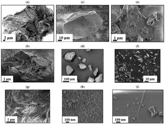
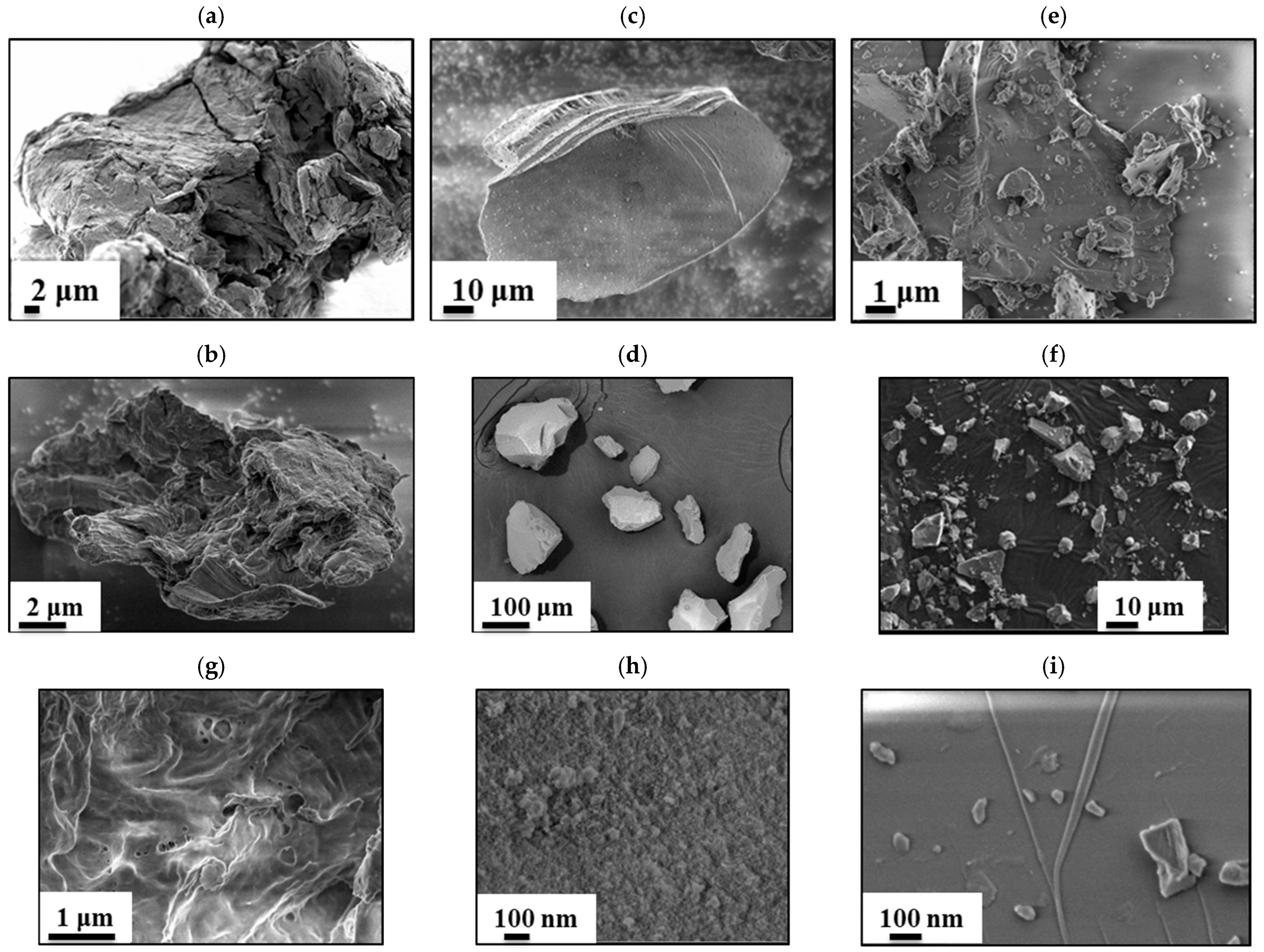
For comparison, the study included naturally occurring microparticles commonly found in the environment: silicon dioxide (SiO2) particles (MSi, 10–150 μm), quartz powder (MQ, 1–50 μm), and microcrystalline cellulose (MCC, 1–200 μm) (Figure 3). Quartz powder microparticles are produced by crushing sand followed by size fractionation and are widely used in construction materials, so it is quite common in the environment [33].
Figure 3.
SEM images of MCC (a,b), MSi (c,d) and MQ (e,f) microparticles. Images of the original microparticles before (a,c,e) and after exposure to DMF (b,d,f). The structure of the surface layer of microparticles after exposure to DMF for MCC, MSI and MQ respectively (g–i).
Microcrystalline cellulose (MCC) is widely used in the pharmaceutical industry as a tablet excipient, as well as in creams and food products [34]. In this study, all naturally derived microparticles were also subjected to aging by immersing in DMF or toluene for three months. After three months in DMF, MCC exhibited a negligible mass loss of 0.40 wt.%, while silicon dioxide (MSi) and quartz powder (MQ) microparticles lost 0.10 wt.% and 0.15 wt.%, respectively. Notably, the mass loss observed for MSi and MQ microparticles is unlikely to result from chemical change, but it is most likely attributable to the detachment and release of loosely bound nanoparticles from the particle surfaces during washing and centrifugation following the aging. The presence of such nanoparticles on the surface layer was confirmed by SEM (Figure 3h,i).
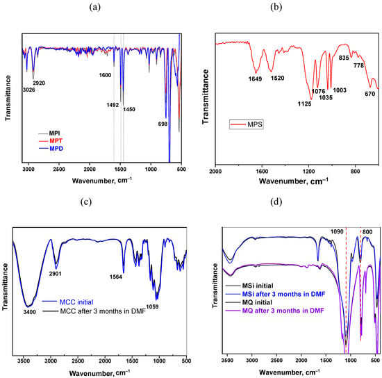
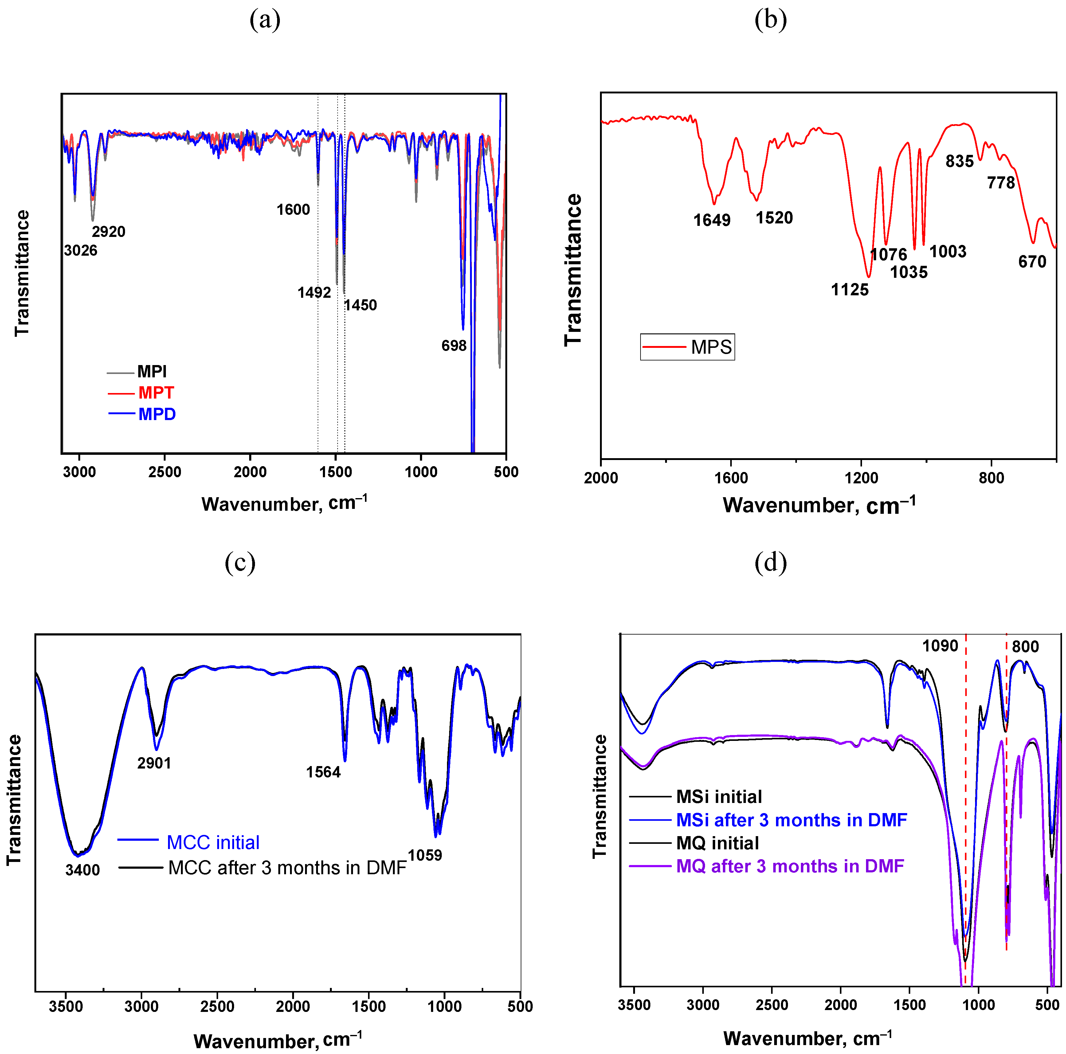
The structure of the model microparticles both before and after treatment in DMF or toluene was confirmed by Fourier-transform infrared (FTIR) spectroscopy. All spectra were recorded in the range of 400–4500 cm−1 and represent the average of 60 scans (Figure 4). The FTIR spectrum of the model polystyrene microparticles (Figure 4a) exhibits characteristic bands of polystyrene: the presence of an aromatic ring leads to the appearance of transmission bands at 3026, 3054 and 3077 cm−1, which correspond to the stretching vibrations of aromatic C–H bond, and bands at 1600 and 1492 cm−1, caused by the stretching vibrations of the C–C bonds in the ring; bending vibrations of the C–H bond in the spectrum appear as a transmission band at 698 cm−1.
Figure 4.
FTIR spectra of model polystyrene particles (a,b), MCC (c), MSi and MQ (d) microparticles.
The FTIR-spectrum contains transmission bands at 2920 and 2850 cm−1, related to asymmetric and symmetric C–H stretching vibrations in the methylene group. Importantly, the FTIR spectra of polystyrene microparticles remained unchanged across samples with varying degrees of “aging” (packing density) of polymer chains. This confirms that prolonged immersion for three months in either toluene or DMF affects only the packing of polystyrene chains [27]. The chemical structure of the fully “aged” crosslinked sodium polystyrene sulfonate (MPS) microparticles was also verified by FTIR (Figure 4b).
The band at 670 cm−1 is assigned to C–S and C–H vibrations associated with the sulfonate group attached to aromatic ring. Bands at 835 and 778 cm−1 correspond to aromatic C–H bending modes; while bands at 1125 cm−1 and 1076 cm−1 are characteristic of –SO3H and –SO3− of sulfonate group. S=O stretching vibrations are observed at 1003 and 1035 cm−1. A broad band at 1649 cm−1 arises from overlapping contributions: C=C stretching of the aromatic ring and C=O stretching from the cross-linking agent (methylenebisacrylamide). Additionally, N–H bending vibrations appear at 1520 cm−1. In the FTIR spectrum of microcrystalline cellulose (MCC, Figure 4c), the following features are observed: a broad, intense band centered at ~3400 cm−1, attributed to O–H stretching vibrations; a band at 1564 cm−1, assigned to O–H bending modes; asymmetric C–H stretching of the pyranose ring appears at 2901 cm−1; and a strong, broad band at ~1059 cm−1 corresponds to C–O stretching vibrations in the cellulose. The obtained FTIR spectra of MCC are in good agreement with those reported in the literature [35,36].
Figure 4d shows the FTIR spectra of silicon dioxide (MSi) and quartz powder (MQ) microparticles. The spectra display characteristic bands at 800 cm−1 and 1090 cm−1, assigned to Si–O–Si bending and stretching vibrations, respectively. An intense broad band at ~3500 cm−1 is attributed to hydroxyl groups (Si–OH) and adsorbed water molecules [37]. These results indicate that the chemical structure of naturally occurring microparticles is only minimally affected by exposure to toluene or DMF.
3.2. Sorption of Rhodamine B on Micron-Sized Solids of Different Origins
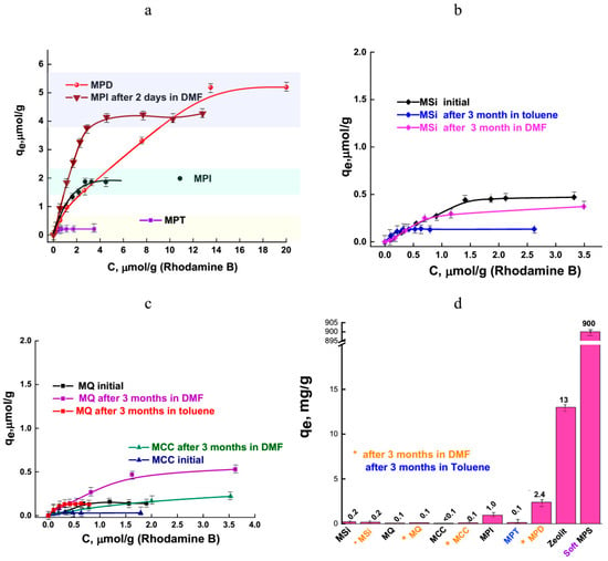
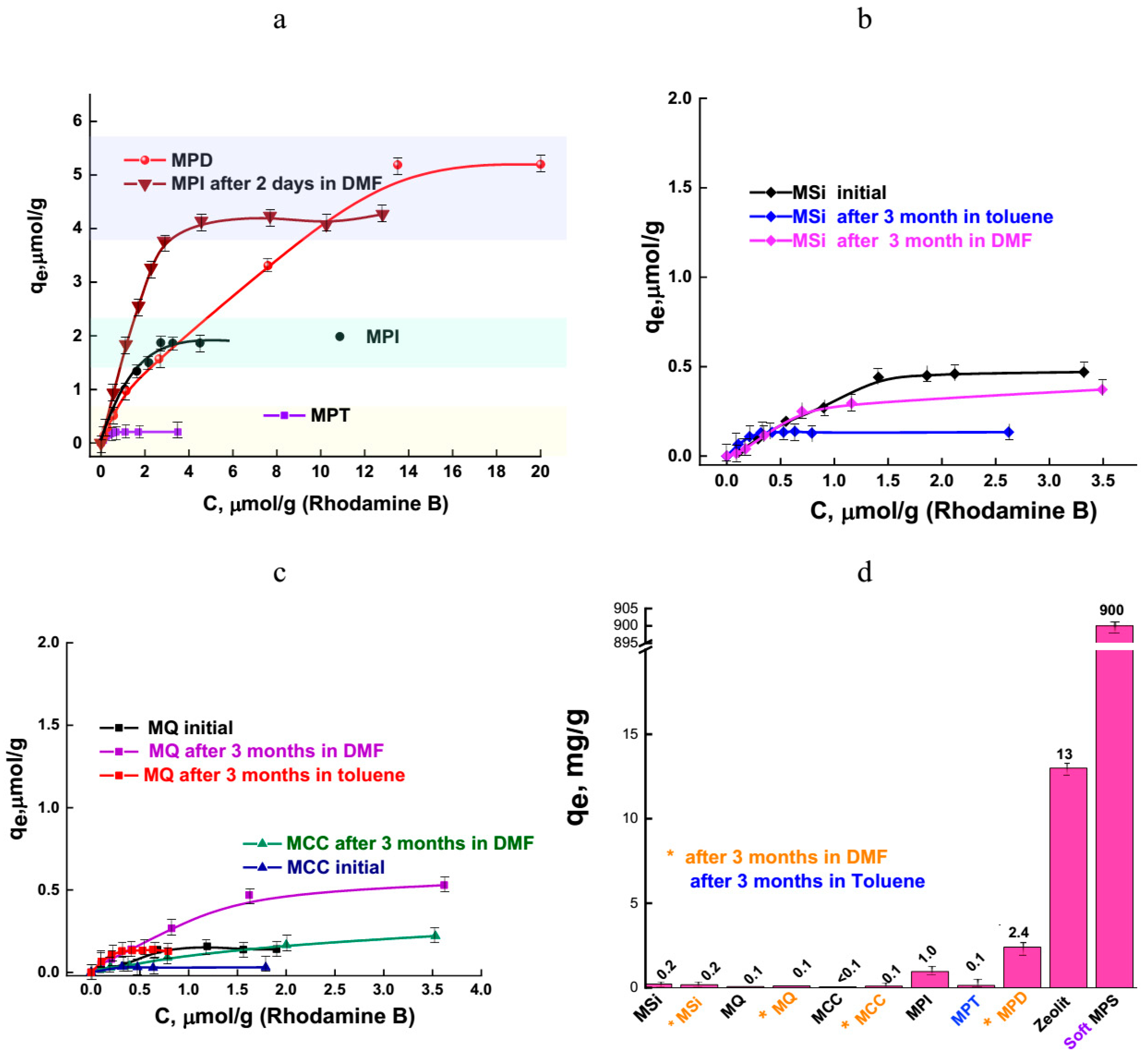
The sorption capacity of microparticles of various origins was evaluated using the hazardous chemical rhodamine B. It was found that the packing density of polymer chains in model non-spherical polystyrene microparticles significantly influenced their sorption capacity (Figure 5a). Microparticles aged in toluene for three months (MPT) exhibited the highest polymer chain packing density. Although approximately 30 wt.% of non-crosslinked polymer chains diffused out of the particles into toluene, subsequent solvent exchange (first to ethanol and then to an aqueous dispersion medium) resulted in a pronounced shape change: the particles collapsed into irregularly shaped disks (see Supplementary Figure S1). This behavior reflects the solvent quality difference: toluene is a good solvent for polystyrene, causing the microparticles to swell. Upon addition of ethanol (a poor solvent) the polystyrene chains collapse, leading to maximal compaction of the MPT microparticles. In contrast, crosslinked polystyrene microparticles aged in DMF for three months (MPD) displayed a looser chain packing. Since DMF is a polar solvent that is miscible with ethanol, its removal occurs under milder conditions, without inducing polystyrene chain collapse. Consequently, the MPD microparticles retain their original shape and exhibit a loose packing throughout the particle volume (Figure 2b). Sorption experiments revealed that the initial MPI microparticles used as a model for primary crosslinked polystyrene microplastics adsorbed up to 2 μmol/g (0.96 mg/g) of rhodamine B (Figure 5a).
Figure 5.
Adsorption of rhodamine B by model polystyrene particles (a), MSi (b), MQ and MCC microparticles (c). The comparison of RB adsorption on the initial particles and particles after modification (d).
As the degree of “aging” (i.e., chain loosening) increased, the sorption capacity rose to 5 μmol/g (2.4 mg/g) for the MPD sample (Figure 5a,d). Conversely, the increased chain packing density in MPT microparticles reduced sorption capacity to only 0.3 μmol/g (0.14 mg/g). As can be seen from Figure 5, the sorption capacity of MPD and MPT particles differs significantly, while their ζ-potential is comparable, which indicates a significant influence of the packing density of polymer chains on the sorption process, rather than the electro-surface properties of the particles.
Microparticles modeling fully “aged” polystyrene composed of hydrophilic sodium polystyrene sulfonate chains bearing ionic functional groups (MPSs) exhibit the highest sorption capacity: 1880 μmol/g (900 mg/g) of rhodamine B (Figure 5d). In contrast, microparticles of natural origin (MCC, MSi, MQ) showed low sorption capacities, not exceeding 0.5 μmol/g (0.2 mg/g) of rhodamine B (Figure 5b,c). These values are comparable to those of model polystyrene microparticles with densely packed polymer chains (MPT), suggesting that sorption in these systems is limited to surface only. These conclusions regarding the influence of polymer chain packing density on sorption behavior are further supported by additional experimental data.
As shown, hyper-crosslinked polystyrene microspheres (20–300 μm in diameter) exhibited a sorption capacity of only 0.02 μmol/g (0.01 mg/g) for rhodamine B (see Supporting Information and Figure S2 for details about hyper-crosslinked polystyrene microspheres). Moreover, the sorption capacity of naturally occurring microparticles was found to be slightly affected by prior exposure to toluene or DMF (Figure 5).
To gain deeper insight into the sorption mechanism, the adsorption capacity of rhodamine B on various microparticles was analyzed using four isotherm models: Langmuir, Freundlich, Temkin, and Dubinin–Radushkevich (see Table 2 and Supplementary Table S1).
Table 2.
Comparison of maximum sorption capacities for rhodamine B on micron-sized particles of different origins.
The Freundlich model assumes heterogeneous surface energetics and can describe both monolayer and multilayer adsorption. In contrast, the Langmuir model assumes homogeneous surface sites with identical energy and is applicable to monolayer coverage. Fitting of the experimental data yielded high correlation coefficients (R2 ≥ 0.94) for the Langmuir model across all tested microparticles, indicating that rhodamine B adsorption occured predominantly via monolayer coverage. The obtained values of qmax of model crosslinked polystyrene particles are statistically different (Table 2). The Temkin model accounts for adsorbate–adsorbent interactions and provides an estimate of the adsorption potential. However, this model provided a good fit (R2 > 0.90) only for MPI microparticles, MSi (before and after modification), as well as MQ and MCC aged in DMF (Supplementary Table S1). The Temkin constant Bt reflects changes in adsorption energy and can indicate whether the process is endothermic (Bt < 1) or exothermic (Bt > 1). According to the fitted parameters, rhodamine B adsorption on both polymeric and natural microparticles is endothermic. The Dubinin–Radushkevich model provides insight into the physicochemical nature of the adsorption process by estimating the apparent free energy of adsorption (E). Values of E > 40 kJ/mol typically suggest chemisorption, whereas E < 40 kJ/mol indicates physisorption. In this study, the calculated E values ranged around 4 kJ/mol (i.e., <40 kJ/mol) confirming that rhodamine B adsorption on both polymer and natural microparticles proceeds via physical interactions.
The calculated p values for the Langmuir model were below 0.008, while those for the Freundlich model ranged from 0.045 to 0.067.
3.3. Evaluation of the Toxicity of Microparticles of Different Origins Depending on the Presence of Sorbed Rhodamine B
In the scientific literature, findings on the cytotoxicity of polystyrene microparticles (>1 μm) are contradictory, with some studies reporting cytotoxic effects [38,39], and others showing no impairment of cell viability [40,41,42]. To resolve this discrepancy for our model polystyrene particles, we first evaluated their own effect on cell viability.
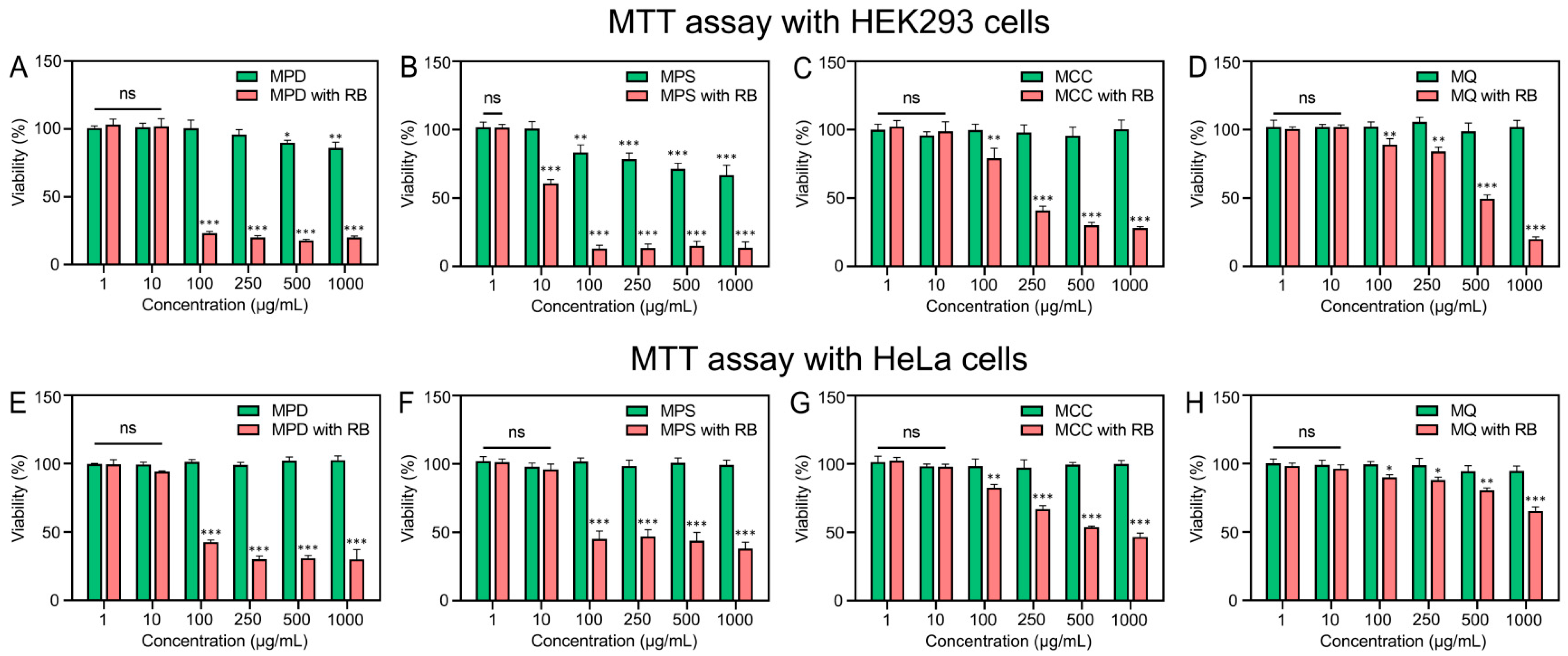
For nanoscale samples, direct addition of polymeric particles to the cultured cells is appropriate, as they can penetrate cell membranes and affect the intracellular organization. However, for microparticles larger than 1 μm, we used an indirect exposure method to avoid artifacts: particles were preincubated in growth medium for 4 days, removed, and the resulting particle-free conditioned medium (containing potential leachates—organic additives, oligomeric chains, or environmental contaminants sorbed onto the particles) was applied to cells. This prevents false-positive results arising from the steric hindrance of cell proliferation by particles of comparable size [43] (see Supplementary Figure S3). Viability assays using HeLa and HEK293 cell lines revealed that neither the pristine polystyrene microparticles (MPI) nor the “aged” ones (MPD, Figure 6) nor the non-crosslinked oligomeric polystyrene chains released during aging (see Supplementary Figure S4) exhibited cytotoxicity or inhibited cell proliferation. The decreased viability of HEK293 cells in the presence of MPS microparticle extract (a model of fully aged crosslinked polystyrene) is likely due to the presence of nanoparticles in the 200 nm ÷ 1 µm size range (see Supplementary Figure S5). Such nanoparticles are difficult to remove by centrifugation because they are permeable to water, and thus may also have a toxic effect on cell proliferation upon physical contact. Given that naturally occurring microparticles are orders of magnitude more abundant in the environment than microplastics, we also evaluated the viability of HeLa and HEK293 cell lines using microcrystalline cellulose and quartz powder extracts. These natural microparticles showed no toxicity and demonstrated no adverse effects on cell viability. Studies of the toxic properties of microparticles were carried out at concentrations from 1 to 1000 μg/mL. Notably, the maximum concentration (1000 μg/mL) far exceeds current environmental levels of microplastics, even in aquatic organism gastrointestinal tracts [44]. Therefore, fragments ≥1 µm generated during crosslinked polystyrene degradation do not appear to release substances exhibiting cytotoxic properties into the environment.
Figure 6.
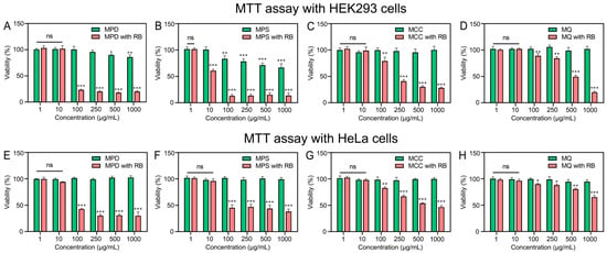
Viability of HeLa and HEK293 cells measured by MTT assay after 4 days of cultivation with extracts from microparticles of different origins: (A) MPD, (B) MPS, (C) MCC, (D) MQ, (E) MPD, (F) MPS, (G) MCC, (H) MQ (see Table 1). The data are shown as mean ± SD of three independent experiments (ns, not significant; *, p ≤ 0.05; **, p ≤ 0.01; ***, p ≤ 0.001).
In the second stage, we have performed the sorption of the hazardous chemical—rhodamine B—onto both aged crosslinked polystyrene models and natural particles and assessed the resulting cytotoxicity as described above. The presence of rhodamine B molecules on the surface layer (for partially aged model MPD microparticles) or in the bulk of polymer microparticles (for fully aged crosslinked polystyrene model—MPS microparticles) was shown to reduce the viability of HeLa and HEK293 cells by 60–90% (Figure 6) (the concentration of polymer microparticles in the nutrient medium was equal to or higher than 100 µg/mL). Thus, by sorbing rhodamine B, polymer microparticles become potent factors that significantly reduce cell viability. This effect is attributed to the release of rhodamine B into the growth medium during desorption, not to the particles themselves. The adsorbed rhodamine B concentrations were 2.4 mg/g (MPD) and 2.8 mg/g (MPS). Assuming complete dye desorption at a microparticle dose of 100 μg/mL, the corresponding rhodamine B concentrations in the medium would be approximately 0.24 μg/mL and 0.28 μg/mL, respectively (Theoretically calculated, taking into account complete desorption of rhodamine B molecules. Glucose, protein, and other molecules in the growth medium determine the desorption rate. More research needs to be done on desorption processes in biological environments). Thus, these in vitro experiments demonstrate that rhodamine B can significantly suppress kidney cell proliferation at hazardous chemical concentrations as low as 0.24 μg/mL. This toxic threshold for kidney cells is comparable to the reported toxic concentration for lip fibroblasts [17].
Given the far greater environmental abundance of natural microparticles compared to microplastics, they represent a major potential vector for pollutants like rhodamine B. However, as demonstrated, their maximum sorption capacity for rhodamine B is low (<0.1 mg/g).
Cell viability assays revealed that extracts from rhodamine B-loaded microcrystalline cellulose reduced HeLa cell viability to ~50% at particle concentrations ≥ 500 μg/mL (equivalent to ≥0.1 μg/mL rhodamine B (theoretically calculated)). HEK293 cells were more sensitive, with viability dropping to ~40% at 250 μg/mL MCC (~0.05 μg/mL rhodamine B (theoretically calculated)).
Therefore, despite their lower sorption capacity, rhodamine B-loaded natural microparticles can induce cytotoxicity comparable to that of plastic microparticles. This finding is mechanistically supported by desorption experiments in aqueous media of varying salinity, which showed that rhodamine B leaches more readily from microcrystalline cellulose than from aged polystyrene microparticles (see Supplementary Table S2).
Extracts from rhodamine B-loaded quartz powder, used as a model inorganic natural microparticle, affected HeLa and HEK293 cell proliferation only at concentrations ≥ 500 μg/mL (Figure 6), corresponding to ~0.05 μg/mL rhodamine B upon full desorption. Therefore, rhodamine B desorbing from inorganic microparticles induces toxicity at concentrations as low as 0.05 μg/mL. The MTT assay data (Figure 6) were consistent with optical microscopy data (See Supplementary Figure S6).
Collectively, our data establish a clear toxicity threshold: rhodamine B-loaded microparticles of all types reduced cell viability by 20–40% at concentrations ≥ 100 μg/mL, while no effect was observed at 10 μg/mL (Figure 6). For context, a 10 μg/mL dose of ~5 μm particles corresponds to a particle density on the order of 12 × 109 particles per liter. This experimental concentration is several orders of magnitude higher than current environmental microplastic levels found in ocean waters.
4. Discussion
Model polystyrene microparticles of 1–50 μm in diameter are widely used to study the adsorption of environmental pollutants (heavy metal ions, pharmaceuticals, and persistent organic pollutants) as well as to assess their biological impacts (cytotoxicity, induction of oxidative stress, etc.). However, the literature contains diverse reports on the toxicity of such model polystyrene particles. Some studies report that polystyrene microparticles exhibit toxicity both to cell lines and cause various adverse reactions when they enter the bodies of experimental animals [38,39], while other experimental results show no significant toxicity for particles in the 1–40 μm size range across various cell lines [40,41,42]. A key limitation in many of these studies is the lack of detailed characterization of particle surface properties. Commercially available spherical polystyrene microparticles are often used without reporting surface charge, functional group density and nature, or the presence of surfactants and stabilizers, etc. This omission makes it difficult to make generalizations and trace both the individual contributions of polystyrene particle size, and surface properties to observed toxicological influence on cells.
This study addresses two central questions. First, we synthesized non-spherical, crosslinked polystyrene microparticles and comprehensively characterized their shape, chemical structure, and surface electrokinetic properties (ζ-potential) both before and after exposure to organic solvents (DMF or toluene). We demonstrated that model particles based on polystyrene with controlled polymer chain packing density as well as model microparticles with ionic functional groups localized both at the surface and throughout the particle bulk may be prepared. Using rhodamine B as an example of cationic hazardous chemical, we showed that sorption capacity of microparticles with the same chemical structure (crosslinked polystyrene) depends primarily on chain packing density: in the series hyper-crosslinked polystyrene > MPT > MPI > MPD decreasing chain packing density correlates with increasing rhodamine B uptake. The highest sorption capacity was observed for microparticles modeling “fully aged” crosslinked polystyrene, which feature a permeable, hydrophilic network with sulfonate groups enabling both surface and bulk sorption of rhodamine B. These findings align with prior reports on the sorption of such particles regarding other organic molecules [45]. When comparing with naturally occurring microparticles (silicon dioxide, quartz powder, and microcrystalline cellulose), polymeric microparticles exhibited markedly higher sorption capacities. This allows us to assume that, as a result of aging, polystyrene microparticles can act as sorbents in relation to the hazardous chemical rhodamine B. It is worth noting that environmental abundance of naturally occurring microparticles vastly exceeds that of microplastics, which does not allow us to assess the actual negative potential of microplastics in the transport of rhodamine B in the environment. The coexistence of natural and plastic microparticles in the environment introduces complexity: competitive or synergistic sorption effects could alter the particle sorption process itself, and can also lead to competitive sorption by microparticles of a certain type. It remains unclear whether rhodamine B (or similar pollutants) will preferentially accumulate on microplastics or natural particles under environmentally relevant conditions and this issue requires further investigation.
Second, the influence of the nature of microparticles and the presence of rhodamine B in their surface layer on their toxic properties in relation to two human cell lines was investigated. Notably, all initial microparticles both polymeric and natural origin devoid of adsorbed hazardous chemical rhodamine B even at concentrations as high as 1000 μg/mL showed no reduction in cell viability. This indicates that toxic substances that would reduce cell viability were not desorbed from the surface of the studied microparticles. Given the particle size is around 5 μm, the 1000 μg/mL microparticle concentration is equivalent to ~1.2 × 10⁷ particles/mL (or ~1.2 × 10¹³ particles/m3). These concentrations appear to be excessively high, as the average microplastic concentration in all five oceans is known to be ~2.76 pieces/m3 [46]. However, a recent analysis of microplastic contamination in China reported that soil concentrations can reach up to 4.4 × 10¹¹ particles/km2 [47]. Furthermore, another study on microplastic content in China has shown that there are high concentrations of microplastics (3.5 × 10⁹ particles/m3) [48]. It should be noted that so far, only a few publications have reported such high values.
When rhodamine B was adsorbed onto surface of polymer microparticles (or sorbed by their volume) or MCC particles, significant reduced cell viability by 60–90% is observed at concentrations ≥ 100 μg/mL (≥1.2 × 10¹² particles/m3). At the same time no effects on HeLa и HEK293 cell viability were observed at 10 μg/mL (0.12 × 10¹² particles/m3). Thus, the toxic effect on cells is exerted by rhodamine B molecules, which are desorbed into the cell growth medium. Importantly, the rhodamine B loading was 5–10 times higher on MPS and MPD microparticles respectively than that on MCC. It can be assumed that when comparing polystyrene microparticles with MCC the toxic effect on cells of the particles under consideration is comparable.
Our study of rhodamine B sorption was conducted under conditions mimicking freshwater (salinity < 0.5‰). Future work should examine sorption behavior of rhodamine B on differently originated particles in higher-salinity environments. Moreover, it is necessary to directly compare competitive sorption among coexisting microparticle types involving polymer particles, silicon dioxide, and microcrystalline cellulose.
The key finding of this study is that toxic substances are not desorbed into the growth medium from the surface layer or volume of 5 μm polystyrene microparticles, nor from the oligomeric chains, and thus no toxic effects on the proliferation of HEK293 cells in vitro were demonstrated. However, when rhodamine B is adsorbed onto these microparticles, a toxic effect on human cells is observed at concentrations exceeding 10 μg/mL (equivalent to ~1.2 × 10¹² particles/m3), and this toxicity is attributable to rhodamine B itself.
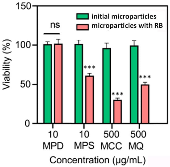
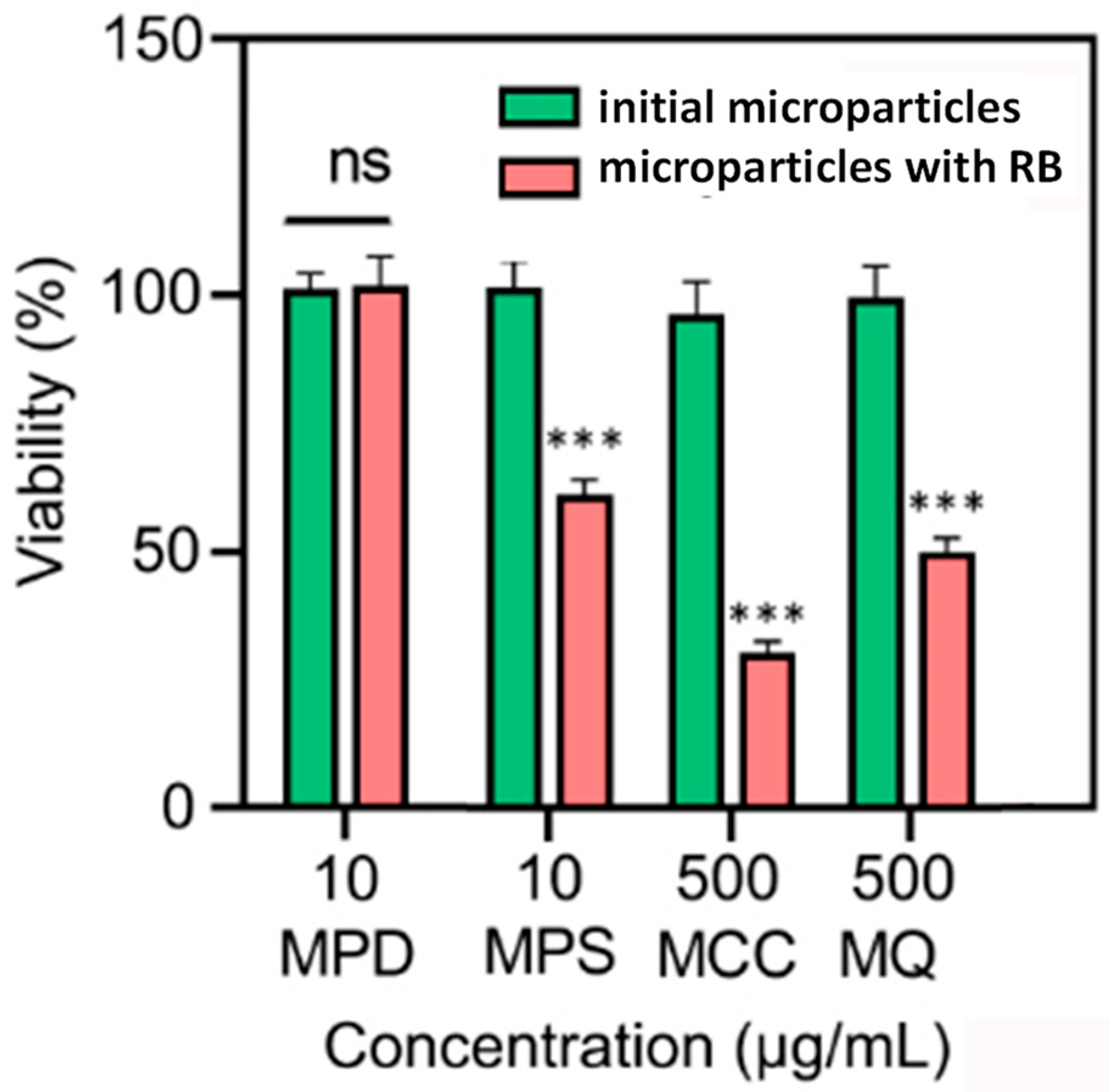
The experimental data revealed that polymer microparticles have significantly better sorption capacity for rhodamine B than naturally occurring microparticles such as microcrystalline cellulose and quartz powder. To cause a toxic effect while desorbing rhodamine B, the amount of naturally occurring microparticles need to increase by an order of magnitude (Figure 7).
Figure 7.
Viability of HEK293 cells measured by MTT assay after 4 days of cultivation with extracts from microparticles of different origins: MPD, MPS, MCC, and MQ (the concentration of RB is the same for all microparticles). The data are shown as mean ± SD of three independent experiments (ns, not significant; ***, p ≤ 0.001).
It should be noted that the studied processes of rhodamine B sorption on model polystyrene particles and particles of natural origin took place under laboratory conditions. Therefore, to assess the risks of sorption of a hazardous chemical on particles of various natures, it is necessary to study more complex dispersion media compositions. Furthermore, the experimental data obtained pertain to microparticles larger than 1 µm. The adsorption properties of polymer particles smaller than 500 nm should also be investigated, as well as their adsorption properties compared to those of naturally occurring nanoparticles and the cytotoxicity of these nanoparticles.
5. Conclusions
This study demonstrates that non-spherical, crosslinked polystyrene microparticles can serve as model systems for crosslinked microparticles. The packing density of polystyrene chains was shown to significantly influence the sorption of the hazardous chemical rhodamine B, enabling predictions of environmental pollutant uptake by both virgin and environmentally aged plastic particles. The sorption capacity of rhodamine B on model microparticles bearing ionic functional groups representing fully aged crosslinked microplastics was found to be substantially higher than that on both pristine polymer microparticles and naturally occurring microparticles (silicon dioxide, quartz powder, and microcrystalline cellulose). Toxicity assessments of various microparticle types both before and after rhodamine B adsorption revealed that, at concentrations up to 10 μg/mL (~12 × 1012 particles/m3), none of the tested microparticles reduced the viability of HeLa or HEK293 cells. The observed cytotoxicity was attributable primarily to the adsorbed rhodamine B molecules themselves, rather than the carrier particles. Nevertheless, based on the current data, it is not yet possible to fully assess the potential risks to human health posed by environmental microplastics loaded with adsorbed rhodamine B. Future studies should go beyond cell viability assays and evaluate particle effects on cell-specific functions, such as the production of surfactants, mucins, and cytokines.
Supplementary Materials
The following supporting information can be downloaded at https://www.mdpi.com/article/10.3390/microplastics5020110/s1, Figure S1. SEM images of non-spherical polystyrene microparticles: kept in a DMF for 3 months (a,b), in a toluene for 3 months (c,d). Figure S2. SEM images of spherical crosslinked polystyrene microspheres: kept in a DMF for 3 months (a), in a toluene for 3 months (b), as well as the structure of the surface layer of the microspheres (c). Figure S3. Viability of HeLa and HEK293 cells measured by MTT assay after 4 days of cultivation with extracts from microparticles of MQ. The data are shown as mean ± SD of three independent experiments. (PC is positive control). Figure S4. Viability of HeLa and HEK293 cells measured by MTT assay after 4 days of cultivation with extracts from non-crosslinked oligomeric polystyrene chains released during microparticle aging. The data are shown as mean ± SD of three independent experiments. (PC is positive control; ns, not significant; * p ≤ 0.05; ** p ≤ 0.01; *** p ≤ 0.001). Figure S5. SEM images of crosslinked microparticles based on polystyrene sulfonate (a,b)–model of fully aged microplastic. Figure S6. Optical microscopy images of cells in the presence of medium after 4 days of cultivation after incubation with extract from microparticles. Table S1. Comparison of maximum sorption capacities for rhodamine B on micron-sized particles of different origins. Table S2. Desorption of rhodamine B after 1 h, percentage of maximum sorption.
Author Contributions
Conceptualization, formal analysis, investigation N.S. and O.I.; GPC investigation, P.C.; cytotoxicity investigation, V.K.-V. and S.S.; FTIR investigation, O.I. and E.S.; provision of reagents, V.I.; visualization N.S., O.I. and V.I.; writing—original draft preparation N.S., O.I. and V.K.-V.; synthesis, supervision N.S. All authors have read and agreed to the published version of the manuscript.
Funding
This study was supported by the Ministry of Science and Higher Education of the Russian Federation (state contract no. 075-15-2025-016 from 28 February 2025 MegaGrant).
Data Availability Statement
The original contributions presented in the study are included in the article/Supplementary Materials; further inquiries can be directed to the corresponding authors.
Acknowledgments
Part of the experimental studies was carried out using equipment of the Research Park of St. Petersburg State University (Centers for Chemical Analysis and Materials Research, and Nanotechnologies).
Conflicts of Interest
The authors declare no conflicts of interest.
References
- Zadereev, E.S.; Lopatina, T.S.; Yaskelyainen, D.D.; Lyubimtseva, S.A. The effect of microplastics and co-occurring toxicants on survival and life-history traits of the cladoceran Moina macrocopa. Limnol. Freshw. Biol. 2025, 4, 1083–1101. [Google Scholar] [CrossRef]
- Gavlová, A.; Jachimowicz, P.; Praus, P.; Bednár, P. Environment changes everything. How relevant are laboratory studies of sorption of pollutants on microplastics? A critical review. J. Environ. Chem. Eng. 2025, 13, 115655. [Google Scholar] [CrossRef]
- Zhao, W.; Zhao, Y.; Geng, T.; Tian, Y.; Zhao, P. Co-transport behavior and Trojan-horse effect of colloidal microplastics with different functional groups and heavy metals in porous media. J. Hazard. Mater. 2023, 459, 131892. [Google Scholar] [CrossRef]
- Yuzhanin, K.I.; Panova, I.G.; Kozhunova, E.Y.; Shevchenko, N.N.; Yaroslavov, A.A. Micro-Sized Polymer Hydrogels as Model Microplastics: Interaction with Polycationic Toxins in Solution and Precipitate. Langmuir 2025, 41, 12645–12654. [Google Scholar] [CrossRef] [PubMed]
- Brennecke, D.; Duarte, B.; Paiva, F.; Caçador, I.; Canning-Clode, J. Microplastics as vector for heavy metal contamination from the marine environment. Estuar. Coast. Shelf Sci. 2016, 178, 189–195. [Google Scholar] [CrossRef]
- Yeo, B.G.; Takada, H.; Hosoda, J.; Kondo, A.; Yamashita, R.; Saha, M.; Maes, T. Polycyclic Aromatic Hydrocarbons (PAHs) and Hopanes in Plastic Resin Pellets as Markers of Oil Pollution via International Pellet Watch Monitoring. Arch. Environ. Contam. Toxicol. 2017, 73, 196–206. [Google Scholar] [CrossRef]
- Sodhi, K.K.; Kumar, M.; Singh, D.K. Potential application in amoxicillin removal of Alcaligenes sp. MMA and enzymatic studies through molecular docking. Arch. Microbiol. 2020, 202, 1489–1495. [Google Scholar] [CrossRef]
- Chia, X.K.; Hadibarata, T.; Kristanti, R.A.; Jusoh, M.N.H.; Tan, I.S.; Foo, H.C.Y. The function of microbial enzymes in breaking down soil contaminated with pesticides: A review. Bioprocess. Biosyst. Eng. 2024, 47, 597–620. [Google Scholar] [CrossRef]
- Ashraf, M.A. Persistent organic pollutants (POPs): A global issue, a global challenge. Environ. Sci. Pollut. Res. 2017, 24, 4223–4227. [Google Scholar] [CrossRef]
- Fisher, P. Review of Using Rhodamine B as a Marker for Wildlife Studies. Wildl. Soc. Bull. 1999, 27, 318–329. [Google Scholar]
- Combes, R.D.; Haveland-Smith, R.B. A review of the genotoxicity of food, drug and cosmetic colours and other azo, triphenylmethane and xanthene dyes. Mutat. Res. 1982, 98, 101–248. [Google Scholar] [CrossRef]
- Gangolli, S.D.; Grasso, P.; Goldberg, L. Physical factors determining the early local tissue reactions produced by food colorings and other compounds injected subcutaneously. Food Cosmet. Toxicol. 1967, 5, 601–621. [Google Scholar] [CrossRef]
- Levis, I.L.; Patterson, R.M.; McBay, H.C. The effect of rhodamine B on the chromosomes of Muntacs muntiac. Mutat. Res. 1981, 8821, 211–216. [Google Scholar] [CrossRef]
- Webb, J.M.; Hansen, W.H.; Desmond, A.; Fitzhugh, O.G. Biochemical and toxicologic studies of rhodamine B and 3,6-diaminofluroan. Toxicol. Appl. Pharmacol. 1961, 3, 696–706. [Google Scholar] [CrossRef]
- Skjolding, L.M.; Jørgensen, L.V.; Dyhr, K.S.; Köppl, C.J.; McKnight, U.S.; Bauer-Gottwein, P.; Mayer, P.; Bjerg, P.L.; Baun, A. Assessing the aquatic toxicity and environmental safety of tracer compounds Rhodamine B and Rhodamine WT. Water Res. 2021, 197, 117109. [Google Scholar] [CrossRef] [PubMed]
- Sharma, J.; Sharma, S.; Bhatt, U.; Soni, V. Toxic effects of Rhodamine B on antioxidant system and photosynthesis of Hydrilla verticillata. J. Hazard. Mater. Lett. 2022, 3, 100069. [Google Scholar] [CrossRef]
- Toshiyuki, K.; Tomotsugu, K.; Chika, Y.; Michiko, S.; Youichi, K.; Fumitomo, K. Reversibility of the inhibitory effect of rhodamine B on the proliferation of cultured human lip fibroblasts. Toxicol. Lett. 1992, 60, 69–74. [Google Scholar] [CrossRef] [PubMed]
- Regulation (EC) No 1223/2009 and Federal Food, Drug, and Cosmetic Act, Chapter II (USA); Food and Drug Administration (FDA): Silver Spring, MD, USA, 2009.
- You-ji, L.; Wei, C. Photocatalytic degradation of Rhodamine B using nanocrystalline TiO2–zeolite surface composite catalysts: Effects of photocatalytic condition on degradation efficiency. Catal. Sci. Technol. 2011, 1, 802–809. [Google Scholar] [CrossRef]
- Nakagawa, Y.; Mitamura, S.; Fujiwara, Y.; Nishitani, T. Decolorization of Rhodamine B in Water by Pulsed High-Voltage Gas Discharge. Jpn. J. Appl. Phys. 2003, 42, 1422. [Google Scholar] [CrossRef]
- He, H.; Chai, K.; Wu, T.; Qiu, Z.; Wang, S.; Hong, J. Adsorption of Rhodamine B from Simulated Waste Water onto Kaolin-Bentonite Composites. Materials 2022, 7, 4058. [Google Scholar] [CrossRef]
- Du, H.; Zhang, Y.; Jiang, H.; Wang, H. Adsorption of rhodamine B on polyvinyl chloride, polystyrene, and polyethylene terephthalate microplastics in aqueous environments. Environ. Technol. Innov. 2022, 27, 102495. [Google Scholar] [CrossRef]
- Shevchenko, N.N.; Evseeva, T.G.; Shevaldysheva, D.I.; Shabsel’s, B.M.; Skurkis, Y.O.; Men’shikova, A.Y. The seed heterophase polymerisation as a method of forming crosslinked monodisperse microspheres. Russ. J. Appl. Chem. 2013, 86, 242–252. [Google Scholar] [CrossRef]
- Shevchenko, N.; Tomšík, E.; Laishevkina, S.; Iakobson, O.; Pankova, G. Cross-linked polyelectrolyte microspheres: Preparation and new insights into electro-surface properties. Soft Matter 2021, 4, 2290–2301. [Google Scholar] [CrossRef]
- Rozman, U.; Kalčíková, G. Seeking for a perfect (non-spherical) microplastic particle—The most comprehensive review on microplastic laboratory research. J. Hazard. Mater. 2022, 15, 127529. [Google Scholar] [CrossRef] [PubMed]
- Yu, Y.; Ma, R.; Qu, H.; Zuo, Y.; Yu, Z.; Hu, G.; Li, Z.; Chen, H.; Lin, B.; Wang, B.; et al. Enhanced adsorption of tetrabromobisphenol a (TBBPA) on cosmetic-derived plastic microbeads and combined effects on zebrafish. Chemosphere 2020, 248, 126067. [Google Scholar] [CrossRef]
- Shevchenko, N.; Iakobson, O.; Isakov, V.; Zorin, I. Effects of Particle Shape and Surface Structure on the Adsorption Properties of Polystyrene Microplastics. Polymers 2024, 16, 3159. [Google Scholar] [CrossRef] [PubMed]
- Mao, R.; Lang, M.; Yu, X.; Wu, R.; Yang, X.; Guo, X. Aging mechanism of microplastics with UV irradiation and its effects on the adsorption of heavy metals. J. Hazard. Mater. 2020, 393, 122515. [Google Scholar] [CrossRef]
- Laishevkina, S.G.; Iakobson, O.D.; Ivankova, E.M.; Shabsels, B.M.; Shevchenko, N.N. Influence of the structure of sulfo-containing polyelectrolyte matrices on the adsorption of Cu2+ ions. Colloid J. 2024, 86, 94–105. [Google Scholar] [CrossRef]
- Kaneko, M.; Sato, H. Photosulfonation of Low-Density Polyethylene Films. Macromol. Chem. Phys. 2004, 205, 173–178. [Google Scholar] [CrossRef]
- Jellinek, H.H.G.; Flajsman, F.; Kryman, F.J. Reaction of SO2 and NO2 with polymers. J. Appl. Polym. Sci. 1969, 13, 107–116. [Google Scholar] [CrossRef]
- Sen, A.K.; Roy, S.; Juvekar, V.A. Effect of structure on solution and interfacial properties of sodium polystyrene sulfonate (NaPSS). Polym. Int. 2007, 56, 145–289. [Google Scholar] [CrossRef]
- Kumar, B.N.; Sunil, B.S.; Bhat, P.K. Exploring the synergistic effects of quartz powder and GGBS in sustainable standard strength concrete: Insights from carbonation and electrochemical impedance spectroscopy (EIS). Discov. Mater. 2025, 5, 147. [Google Scholar] [CrossRef]
- Thoorens, G.; Krier, F.; Leclercq, B.; Carlin, B.; Evrard, B. Microcrystalline cellulose, a direct compression binder in a quality by design environment—A review. Int. J. Pharm. 2014, 473, 64–72. [Google Scholar] [CrossRef] [PubMed]
- Sun, C.C. Mechanism of moisture induced variations in true density and compaction properties of microcrystalline cellulose. Int. J. Pharm. 2008, 346, 93–101. [Google Scholar] [CrossRef]
- Kian, L.K.; Jawaid, M.; Ariffin, H.; Karim, Z. Isolation and characterization of nanocrystalline cellulose from roselle-derived microcrystalline cellulose. Int. J. Biol. Macromol. 2018, 114, 54–63. [Google Scholar] [CrossRef]
- Thiébaud, F.; Kneubühl, F.K. Infrared properties of quartz fibres. Int. J. Infrared Milli Waves 1981, 2, 1015–1028. [Google Scholar] [CrossRef]
- Brown, D.M.; Wilson, M.R.; MacNee, W.; Stone, V.; Donaldson, K. Size-Dependent Proinflammatory Effects of Ultrafine Polystyrene Particles: A Role for Surface Area and Oxidative Stress in the Enhanced Activity of Ultrafines. Toxicol. Appl. Pharmacol. 2001, 175, 191–199. [Google Scholar] [CrossRef]
- Deng, Y.; Zhang, Y.; Lemos, B.; Ren, H. Tissue accumulation of microplastics in mice and biomarker responses suggest widespread health risks of exposure. Sci. Rep. 2017, 7, 46687. [Google Scholar] [CrossRef]
- Hwang, J.; Choi, D.; Han, S.; Jung, S.Y.; Choi, J.; Hong, J. Potential toxicity of polystyrene microplastic particles. Sci. Rep. 2020, 10, 7391. [Google Scholar] [CrossRef]
- Rudolph, J.; Völkl, M.; Jérôme, V.; Scheibel, T.; Freitag, R. Noxic effects of polystyrene microparticles on murine macrophages and epithelial cells. Sci. Rep. 2021, 11, 15702. [Google Scholar] [CrossRef]
- Zhao, B.; Liu, R.; Guo, S.; Li, S.; Huang, Z.; Wang, Y.; Yu, C.; Hou, Z.; Zhang, Y.; Zhang, Y.; et al. Large-sized polystyrene microplastics induce oxidative stress in AML12 cells. Sci. Rep. 2025, 15, 26616. [Google Scholar] [CrossRef] [PubMed]
- Petersen, E.J.; Barrios, A.C.; Henry, T.B.; Johnson, M.E.; Koelmans, A.A.; Montoro Bustos, A.R.; Matheson, J.; Roesslein, M.; Zhao, J.; Xing, B. Potential Artifacts and Control Experiments in Toxicity Tests of Nanoplastic and Microplastic Particles. Environ. Sci. Technol. 2022, 15, 15192–15206. [Google Scholar] [CrossRef]
- Roch, S.; Friedrich, C.; Brinker, A. Uptake routes of microplastics in fishes: Practical and theoretical approaches to test existing theories. Sci. Rep. 2020, 10, 3896. [Google Scholar] [CrossRef]
- Laishevkina, S.G.; Druian, L.M.; Iakobson, O.D.; Ivan’kova, E.M.; Shabsel’s, B.M.; Shevchenko, N.N. Spherical Polymer Gels Containing Sulfonate Groups: Synthesis and Adsorption Properties. Colloid J. 2025, 87, 22–31. [Google Scholar] [CrossRef]
- Mutuku, J.; Yanotti, M.; Tocock, M.; Hatton MacDonald, D. The Abundance of Microplastics in the World’s Oceans: A Systematic Review. Oceans 2024, 5, 398–428. [Google Scholar] [CrossRef]
- Wei, H.; Wu, L.; Liu, Z.; Saleem, M.; Chen, X.; Xie, J.; Zhang, J. Meta-analysis reveals differential impacts of microplastics on soil biota. Ecotoxicol. Environ. Saf. 2022, 15, 113150. [Google Scholar] [CrossRef] [PubMed]
- En-Nejmy, K.; EL Hayany, B.; Al-Alawi, M.; Jemo, M.; Hafidi, M.; El Fels, L. Microplastics in soil: A comprehensive review of occurrence, sources, fate, analytical techniques and potential impacts. Ecotoxicol. Environ. Saf. 2024, 288, 117332. [Google Scholar] [CrossRef]

Disclaimer/Publisher’s Note: The statements, opinions and data contained in all publications are solely those of the individual author(s) and contributor(s) and not of MDPI and/or the editor(s). MDPI and/or the editor(s) disclaim responsibility for any injury to people or property resulting from any ideas, methods, instructions or products referred to in the content. |
© 2026 by the authors. Licensee MDPI, Basel, Switzerland. This article is an open access article distributed under the terms and conditions of the Creative Commons Attribution (CC BY) license.