


Abstract
Plastic pollution is an increasing concern in freshwater ecosystems, yet the roles of polymer chemistry, environmental context, and microbial community composition in governing degradation remain poorly resolved. This study examined plastic–microbe interactions across river, creek, and pond environments using gravimetric mass loss, scanning electron microscopy (SEM), and 16S rRNA gene sequencing. Four polymers were evaluated: biodegradable polyhydroxyalkanoate (PHA) and polylactic acid (PLA), and conventional low-density polyethylene (LDPE) and polyethylene terephthalate (PET). Rapid biofilm formation occurred on all plastic surfaces, indicating widespread microbial colonization; however, measurable degradation was strongly polymer-dependent. PHA exhibited rapid and extensive mass loss across environments, approaching complete degradation after four months in river and pond settings, whereas PLA, LDPE, and PET showed limited mass loss despite substantial colonization. Environmental context influenced degradation intensity, but these effects amplified degradation only when polymer chemistry permitted breakdown. Microbial community analyses showed that substrate presence influenced beta diversity more than alpha diversity, and differential abundance patterns revealed overlapping enriched taxa across polymers. Overall, degradation was governed primarily by polymer chemistry and environmental conditions, while microbial composition played a secondary, indirect role.
1. Introduction
Plastic pollution is widespread in freshwater environments, where plastics represent one of the most abundant forms of anthropogenic waste. Microplastics, defined as plastic particles smaller than 5 mm, pose particularly severe ecological risks [1]. Plastic pollution in freshwater systems originates from urban waste, industrial discharge, agricultural runoff, wastewater effluents, stormwater runoff, landfills, atmospheric deposition, and natural disasters before entering rivers and lakes [2,3]. With ongoing urbanization and industrialization, inputs of plastic into freshwater systems are expected to increase [3,4].
The most prevalent polymers detected in freshwater include polyethylene, polypropylene, polystyrene, and polyethylene terephthalate, which are highly persistent and resistant to biodegradation [5]. As plastics fragment, they release chemical additives such as plasticizers, stabilizers, and flame retardants that become more bioavailable, increasing the likelihood of environmental and biological uptake [6,7]. Microplastics have been shown to induce oxidative stress in aquatic organisms, leading to cellular damage and long-term health effects [8]. These risks extend beyond aquatic ecosystems because freshwater systems serve as drinking water sources and can contribute to human exposure to plastic-associated chemicals and particles [6].
A critical aspect of the microplastic problem is microbial degradation, in which microorganisms break down plastics in natural environments. Recent studies have identified microbial communities associated with synthetic polymer alteration, suggesting potential bioremediation applications [9]. However, the efficiency of microbial degradation depends on polymer structure. Commercial plastics are typically semi-crystalline, containing both ordered and amorphous domains; higher crystallinity can limit microbial and enzymatic accessibility, slowing degradation rates [10,11,12].
1.1. Microbial Communities in Rivers, Ponds, and Creeks
Freshwater ecosystems host diverse microbial communities, with molecular surveys identifying tens to hundreds of thousands of distinct microbial taxa [13]. The dominant bacterial phyla commonly include Proteobacteria, Bacteroidetes, Cyanobacteria, Firmicutes, and Actinobacteria. However, community composition varies among habitats. Ponds typically harbor higher proportions of Cyanobacteria and Actinobacteria, whereas rivers and creeks are often dominated by Proteobacteria and Bacteroidetes [14]. Within freshwater systems, β-proteobacteria frequently prevail and are positively correlated with dissolved organic carbon (DOC) and related dissolved organic matter (DOM) fractions, indicating their association with terrestrially derived organic matter inputs [15]. Their abundance in lotic environments reflects continual watershed inputs of organic carbon from soils and vegetation, highlighting the role of hydrological connectivity in structuring microbial communities [14,16].
Because freshwater microbial communities regulate organic matter turnover, some taxa may also participate in polymer surface alteration or degradation. Although polyolefins such as polyethylene (PE) and polypropylene (PP) dominate global plastic production, enzymatic degradation activities have been reported across multiple polymer types, including PE, PVC, and polystyrene (PS) [17]. These organisms may offer potential long-term bioremediation applications, but degradation potential likely depends on both polymer chemistry and the environmental conditions that shape microbial activity [18].
Marine environments have historically been the primary focus of plastic biodegradation research, where microbial communities have been shown to colonize plastic surfaces and contribute to polymer alteration and degradation processes [19,20]. In contrast, fewer studies have examined the fate and degradation of plastics in freshwater systems despite their direct importance to human populations and drinking water resources [21]. Freshwater and marine environments differ in several key characteristics, including salinity, nutrient availability, temperature regimes, and organic matter composition [22]. These environmental factors strongly influence microbial community composition and metabolic activity, highlighting the importance of investigating plastic degradation within specific aquatic contexts [23].
Freshwater habitats often support highly diverse microbial communities and may exhibit greater taxonomic richness and community heterogeneity than marine environments [24]. Freshwater microbial assemblages often display greater species richness, habitat heterogeneity, and niche overlap, which may intensify competition among microorganisms colonizing plastic debris [1,25]. Such competition within the plastisphere—the microbial ecosystem associated with plastic surfaces—may influence microbial activity and degradation potential in ways that differ from patterns observed in marine plastisphere studies [25].
Freshwater systems, particularly rivers and lakes, are increasingly recognized as key reservoirs and processing environments for plastic debris, where physical, chemical, and biological processes—including microbial colonization—contribute to the transformation and environmental fate of plastics [26]. The microbial communities that develop on plastic surfaces, often referred to as the plastisphere, can differ from surrounding planktonic communities and are shaped by substrate properties, seasonality, geography, and local environmental conditions [23,27]. Field-based freshwater investigations have demonstrated that microbial communities colonizing plastics in lake environments can contribute to measurable biodegradation and polymer surface alteration under natural conditions [28]. However, many studies investigating microbial plastic degradation rely on laboratory experiments, and relatively few integrate field exposure experiments with controlled laboratory analyses of microbial community composition and degradation processes. Integrating field and laboratory approaches is therefore important for understanding how natural environmental conditions influence plastisphere formation, microbial community dynamics, and plastic degradation in freshwater ecosystems [29].
1.2. Research Questions
The overarching goal of this study is to investigate the potential for the microbial degradation of conventional and biodegradable plastics in freshwater environments and to evaluate how polymer type and environmental conditions influence degradation processes. The primary objective was to determine how polymer chemistry and freshwater environmental context influence plastic degradation, while also assessing whether microbial community composition corresponds with measured mass loss. We hypothesized that polymer chemistry would exert a stronger influence on degradation dynamics than microbial taxonomic composition, while environmental conditions would modulate the intensity of degradation across freshwater sites. To address this objective, the study was designed around three central research questions:
- Which microbial communities are associated with LDPE, PET, PLA, and PHA plastics in freshwater environments?
- How do degradation patterns differ between non-biodegradable (LDPE and PET) and biodegradable (PLA and PHA) plastics?
- Do degradation rates differ among freshwater environments, including pond, river, and creek systems?
To address these research questions, this study combined field-based enrichment experiments with laboratory analyses to examine microbial colonization and plastic degradation across freshwater environments. Plastic substrates representing both biodegradable (PHA and PLA) and non-biodegradable polymers (LDPE and PET) were deployed in three freshwater systems (river, creek, and pond) for four months to allow natural microbial colonization. These polymers were selected to compare conventional plastics with biodegradable polyesters that differ in chemical structure and expected environmental degradation behavior. Plastic degradation was evaluated through measurements of mass loss and scanning electron microscopy to assess physical surface changes. Microbial communities associated with plastics and surrounding water were characterized using 16S rRNA gene sequencing, and statistical analyses were conducted to evaluate differences in degradation rates, microbial community composition, and environmental influences among sites and polymer types. Together, these approaches allowed assessment of both the biological communities colonizing plastics and the environmental conditions influencing degradation in freshwater systems.
2. Materials and Methods
2.1. Environmental Measurements
2.1.1. Water Quality Parameters
Environmental conditions were measured at each sampling site to characterize the physicochemical context of the freshwater environments. Measurements were collected in situ using a YSI ProDSS multiparameter water quality meter (YSI Inc., Yellow Springs, OH, USA). Parameters measured included water temperature, nitrate concentration, turbidity, electrical conductivity, total dissolved solids (TDS), total suspended solids (TSS), pH, chlorophyll-a, and phycocyanin. Chlorophyll-a and phycocyanin concentrations were determined using fluorescence-based optical sensors integrated into the sonde, with phycocyanin fluorescence serving as an indicator of cyanobacterial biomass. Conductivity measurements were used to estimate TDS, while turbidity and TSS were measured using optical sensors designed to quantify suspended particulate matter. Atmospheric pressure was recorded using the integrated barometric sensor within the YSI system. Incident solar radiation was measured separately using a Daystar DS-05 solar radiation meter (Daystar Inc., Phoenix, AZ, USA), which records irradiance in watts per square meter (W m−2). Environmental measurements were collected during each sampling event at approximately 30-day intervals throughout the four-month deployment period at the pond, river, and creek study sites.
2.1.2. Water Sampling and Filtration
Surface water samples were collected from the pond, river, and creek sampling sites at approximately 30-day intervals throughout the study period. At each site, 180 mL of surface water was filtered directly on site through 0.22 µm pore-size Sterivex filter cartridges (MilliporeSigma, Burlington, MA, USA) using sterile 60 mL Luer-lock syringes (Fisher Scientific, Hanover Park, IL, USA), consistent with established methods for aquatic microbial community sampling [30]. Following filtration, cartridges were removed from the syringe, placed in sterile tubes, and transported to the laboratory, where they were stored at −80 °C until nucleic acid extraction [31,32]. Negative controls consisting of sterile ultrapure water produced using a Milli-Q IQ Element water purification unit (MilliporeSigma, Burlington, MA, USA).
2.2. Plastic Enrichment Trap Installation
2.2.1. Location of Film Enrichment
Plastic enrichment traps were deployed at three freshwater sites in eastern Tennessee representing contrasting environmental conditions: a river, an urban stream, and a pond (Figure 1). The river sampling location was at Seven Islands State Birding Park in Kodak, Tennessee along the French Broad River (Figure 1A), representing a relatively low-disturbance lotic environment surrounded primarily by agricultural and protected conservation land. The second site was located along Third Creek in Knoxville, Tennessee, an urban stream influenced by municipal runoff and wastewater inputs (Figure 1B). This stream is listed under a bacteriological advisory due to persistently elevated bacterial contamination levels [33]. The site was selected to represent a freshwater system with elevated microbial inputs and anthropogenic disturbance. The third sampling site was a small pond located adjacent to the University of Tennessee compost facility in Knoxville, Tennessee (Figure 1C). Unlike the flowing river and creek sites, this pond represents a lentic freshwater environment with limited water movement. The pond also receives runoff from the nearby compost facility and is located within an active construction zone where a highway off-ramp is currently being built.
Figure 1.
Locations of freshwater sampling sites in eastern Tennessee. The regional map shows the positions of the French Broad River site at Seven Islands State Birding Park (A), the Third Creek urban stream site in Knoxville (B), and the pond adjacent to the University of Tennessee compost facility (C). Panels (A–C) show aerial imagery of each sampling location where plastic enrichment traps were deployed. Black boxed letters identify the study sites, blue map pins indicate the approximate sampling locations, and white scale bars indicate distance. Imagery source: Google Earth.
2.2.2. Plastic Film Enrichment Trap
Plastic enrichment experiments were conducted to examine microbial colonization and degradation of different polymer types in freshwater environments. Biodegradable polymers included polylactide (PLA) and polyhydroxyalkanoate (PHA), while non-biodegradable polymers included polyethylene terephthalate (PET) and low-density polyethylene (LDPE). Plastic films were prepared at approximately 200 µm thickness and cut into 5 × 5 cm squares [28]. PLA and PHA films were not commercially available in this format and were therefore produced using 3D printing following previously described procedures [34]. All plastic substrates consisted of ≥99% pure polymer. Prior to field deployment, plastic films were sterilized using 70% ethanol to remove pre-existing microorganisms [19,35].
One independent deployment unit was used for each polymer type at each site and retrieval interval, with each mesh bag containing a single plastic sample. Each plastic sample was weighed prior to deployment. Control plastic samples were incubated in dark deionized water to provide a baseline comparison for non-environmental mass change while minimizing microbial activity, natural water chemistry effects, suspended particle interactions, and ultraviolet-driven photodegradation. Mass loss was monitored at approximately 30-day intervals by re-weighing retrieved samples [36]. Weight loss was evaluated relative to a ≥20% degradation threshold reported in previous polymer degradation studies [37,38]. Plastic film squares were enclosed within sewn polyethylene (PE) mesh bags to allow water flow and microbial colonization while preventing loss of the plastic films and limiting access by larger organisms (Figure 2). The mesh bags were composed of PE to minimize potential metal contamination [39]. Each bag contained a single plastic sample and was deployed for a four-month exposure period. Additional mesh bags without plastic were included as blank controls to monitor background microbial attachment. Plastic enrichment traps containing PET, PLA, PHA, and LDPE substrates were deployed at the three freshwater environments described above (Figure 1).
Figure 2.
Preparation of mesh, enclosed plastic substrates used for enrichment experiments.
Deployment methods differed between lentic and lotic environments. In the lentic pond environment (Figure 3), mesh bags were attached to rope and anchored to cement blocks to prevent movement or flotation of the samples. In the lotic river and creek environments (Figure 4), mesh bags were placed within protective cages secured to nearby trees to prevent displacement by water currents. Each cage contained one plastic sample along with a blank mesh bag control [40]. The locations of all deployed samples were recorded to ensure accurate retrieval after each exposure period. One sample of each plastic type and one blank mesh bag was retrieved approximately every 30 days over a four-month period. A total of 70 samples were collected, as two sites required redeployment following extreme rainfall events that washed away several traps. The compost pond site did not require redeployment and therefore had one fewer sampling interval. Additional samples were collected during the first and final retrieval periods for scanning electron microscopy (SEM) analysis.
Figure 3.
Enrichment units containing PHA, PLA, PET, and LDPE substrates were suspended in the water column to promote microbial colonization. The inset illustrates the trap configuration, including plastic containing bags (black rectangles) and blank control bags (white rectangles).
Figure 4.
Enrichment cages containing PET, PLA, PHA, and LDPE substrates were submerged in flowing water to promote microbial colonization. The inset illustrates the trap configuration, including six plastic containing bags and two blank control bags.
2.3. Scanning Electron Microscopy
After the initial 30-day period and after the final 30-day period, one sample of each plastic type and the corresponding blank control were retrieved from each sampling site to assess temporal changes in microbial colonization and surface degradation [28]. Retrieved plastic substrates were examined using TM3030 tabletop scanning electron microscopy (SEM) (Hitachi High-Tech America, Inc., Schaumburg, IL, USA) at the University of Tennessee Core Laboratory facilities to evaluate surface degradation and microbial attachment [41]. Plastic substrates retrieved from the enrichment experiments were chemically fixed and prepared for SEM following established protocols [42]. Samples were fixed in a solution containing 2.5% glutaraldehyde and 1.25% paraformaldehyde prepared from EM-grade stock solutions (Electron Microscopy Sciences, Hatfield, PA, USA) and diluted with deionized water. Each sample was immersed in 10 mL of fixation solution overnight under a fume hood [43]. Following fixation, samples were dehydrated through a graded series of 200-proof ethanol (Electron Microscopy Sciences, Hatfield, PA, USA) at concentrations of 30%, 50%, 70%, 90%, and 100%, with each step lasting approximately 15 min [44,45]. After the final rinse in 100% ethanol, samples were stored at −32 °C overnight prior to critical point drying [44]. Samples were then dried using a LADD CPD3 CO2 critical point dryer (LADD Research Industries, Williston, VT, USA) following manufacturer guidelines. After drying, specimens were mounted onto aluminum SEM stubs using conductive double-sided carbon tape to stabilize the samples and ensure electrical grounding [46,47]. Mounted samples were examined under high vacuum conditions using SEM to visualize microbial colonization and surface degradation of the plastic substrates [46,48].
2.4. Liquid Degradation Assay
To evaluate microbial degradation potential under controlled conditions, a liquid degradation assay was established using microorganisms recovered from enriched plastic film samples [49]. Glass Petri dishes were used to minimize contamination and prevent the introduction of additional organic carbon sources. Petri dishes were disinfected with 70% ethanol and dried overnight prior to use. Unenriched plastic samples were prepared by cutting the original 5 × 5 cm films in half to fit within the Petri dishes. Plastic substrates were similarly cleaned with 70% ethanol and air dried overnight prior to use [28]. To detach microbial biomass from plastic films, samples were placed in sterile test tubes containing 5 mL of 100% ethanol and vortexed to remove adherent cells from the polymer surface following previously described biofilm extraction methods [50]. The resulting suspension was serially diluted in sterile water by transferring 100 µL into 900 µL (10−1) and repeating the process to obtain 10−2 and 10−3 dilutions [51]. Optical density at 600 nm (OD600) was measured to ensure bacterial concentrations fell within the linear detection range of the spectrometer. A blank vial containing sterile water was used as a reference, and diluted samples were measured for 10 s. Samples with OD600 values between 0.05 and 0.50 absorbance units were used for subsequent assays [52,53,54]. Spectrometer measurements are provided in Table A1.
The degradation medium consisted of an M9-based salt solution prepared from a 5× stock (Table 1). Stock solutions were prepared by dissolving salts in Milli-Q water to a final volume of 1 L and sterilized by autoclaving at 121 °C for 15 min. The working medium (1×) was prepared by diluting the stock solution 1:5 with sterile deionized water. This formulation followed protocols for plastic polymer degradation assays [54] and standard M9 medium preparation [55]. Ammonium sulfate ((NH4)2SO4) was used as the nitrogen source rather than NH4Cl based on previous studies reporting improved performance in plastic biodegradation assays [54]. No additional carbon sources or surfactants were added so that the plastic substrates served as the sole carbon source. For degradation assays, bacterial suspensions were diluted with M9 salts at ratios of 1:10 (1 culture to 9 mL medium) or 1:100 (0.1 mL culture to 9.9 mL medium) following standard microbiological dilution procedures [51]. The resulting suspensions were added to Petri dishes containing the corresponding plastic substrates. Assays were incubated at 30 °C for two months to evaluate microbial degradation of the plastic polymers [54,56].
Table 1.
Adapted composition of M9 salts (5× stock solution, 1 L) [55,56].
2.5. Microbial Community
2.5.1. DNA Extraction
DNA was extracted from all plastic and Sterivex filter samples using the QIAamp PowerFecalPro DNA kit (QIAGEN, Hilden, Germany) following the manufacturer’s Quick-Start Protocol [57]. All DNA extraction and processing were conducted by the University of Tennessee’s Genomic Core Lab Facility.
2.5.2. 16S rRNA Gene Amplification and Library Preparation
Amplicon libraries targeting the V3–V4 region of the 16S rRNA gene were prepared following the Illumina 16S Metagenomic Sequencing Library Preparation Guide (Part #15044223 Rev. B) [58]. Targeted amplification was performed using locus-specific primers with Illumina overhang adapters. Each 25 µL PCR reaction contained microbial genomic DNA (5 ng µL−1), KAPA HiFi HotStart ReadyMix (2×), and 1 µM of each forward and reverse primer. Thermal cycling conditions consisted of an initial denaturation at 95 °C for 3 min; 30 cycles of 95 °C for 30 s, 55 °C for 30 s, and 72 °C for 30 s; and a final extension at 72 °C for 5 min. The expected amplicon size was approximately 550 bp.
PCR products were purified using AMPure XP magnetic beads (Beckman Coulter, Inc., Brea, CA, USA) according to the manufacturer’s recommended ratio. Beads were combined with PCR products and incubated for 5 min to allow DNA binding, followed by magnetic separation. The supernatant was removed, and beads were washed twice with 80% ethanol. After air drying for 10 min, DNA was eluted in 10 mM Tris (pH 8.5), and 50 µL of eluate was carried forward for indexing. Index PCR was performed to attach dual indices and Illumina sequencing adapters using the Nextera XT Index Kit (Illumina, Inc., San Diego, CA, USA) [59]. Each 50 µL reaction contained KAPA HiFi HotStart ReadyMix (2×) (Roche Sequencing Solutions, Inc., Pleasanton, CA, USA) and 10 µL PCR-grade water. Thermal cycling conditions included an initial denaturation at 95 °C for 3 min; 8 cycles of 95 °C for 30 s, 55 °C for 30 s, and 72 °C for 30 s; and a final extension at 72 °C for 5 min. Indexed libraries were purified using 56 µL AMPure XP beads (Beckman Coulter, Inc., Brea, CA, USA), followed by two washes with 80% ethanol, air drying for 10 min, and elution in 27.5 µL of 10 mM Tris (pH 8.5). A final volume of 25 µL per sample was used for downstream quantification.
2.5.3. Instrument Loading
Samples were individually quantified on a NanoDrop spectrophotometer (Thermo Fisher Scientific, Waltham, MA, USA), normalized, and pooled. The final pool was checked for quality and quantity on an Agilent TapeStation (Agilent Technologies, Santa Clara, CA, USA). The library was diluted to 300 pM, spiked with 1% PhiX, and sequenced using 2 × 250 bp paired end reads on an Illumina NovaSeq (Illumina, Inc., San Diego, CA, USA) at the University of Tennessee Genomics Core Facility. Computational analyses were performed in collaboration with High Performance and Scientific Computing (HPSC) staff at the University of Tennessee, Knoxville using the Infrastructure for Scientific Applications and Advanced Computing-Next Generation (ISAAC-NG) cluster.
2.6. Statistical Analyses
2.6.1. Environmental Conditions
Monthly environmental parameters were measured at each sampling site to characterize the conditions under which plastic degradation occurred. Data for each parameter were analyzed individually by site using R, while Excel was used to calculate mean values for cross-site comparisons. Because these measurements represent generalized environmental conditions rather than continuous temporal datasets, the results were summarized as averaged values. This data provides contextual insight into how local environmental factors may have influenced plastic degradation patterns.
2.6.2. Plastic Weight Loss
Plastic degradation was first assessed qualitatively through scanning electron microscopy (SEM) to identify visible surface alterations indicative of either microbial or environmental degradation. Quantitative analysis of plastic weight loss was then conducted by comparing initial and final sample weights following retrieval from each freshwater site along with a control site. Percent mass loss was calculated as: Mass loss (%) = [(initial dry mass − final dry mass)/initial dry mass] × 100. Statistical analyses were performed using Excel and R to determine whether the 20% degradation threshold was achieved, as commonly used by biodegradation assessments [37]. Weight loss trends were visualized for each site to evaluate temporal and spatial differences in degradation across the study environments.
2.6.3. Microbial Community Composition
Amplicon sequencing was performed at the University of Tennessee Genomics Core Facility. Binary Base Call (BCL) files generated by the Illumina sequencing platform were converted to FASTQ format using Illumina BCL Convert software prior to downstream analyses. Processed sequence data were imported into R for microbial community analyses. Microbial community composition was evaluated using Bray–Curtis dissimilarity and visualized using non-metric multidimensional scaling (NMDS). Permutational multivariate analysis of variance (PERMANOVA) was used to test for differences among plastic substrates, sampling environments, and sampling months. Differential abundance analyses were performed using DESeq2 version 1.46.0 on unrarefied count data with size factors estimated using the poscounts method. Models evaluated contrasts among plastic substrates, water controls, and environments across the four-month sampling period. Statistical significance was determined using Benjamini–Hochberg adjusted p-values (padj < 0.05). All statistical analyses were conducted in R version 4.4.1. Microbial community and differential abundance analyses were conducted in R using the phyloseq version 1.50.0, DESeq2 version 1.46.0, tidyverse version 2.0.0, tibble version 3.3.0, ggplot2 version 4.0.1, pheatmap version 1.0.13, and grid packages included with R version 4.4.1. Additionally, the microbial communities associated with plastic substrates were compared to those detected in corresponding water samples to assess potential overlap in taxa and community composition. Cross-site comparisons were conducted to evaluate whether microbial diversity and community composition influenced plastic degradation efficiency. Relationships between microbial community characteristics and plastic weight loss were further examined using cross-correlation analyses in R to explore potential microbial contributions to biodegradation dynamics.
3. Results
3.1. Environmental Test Analysis
Environmental parameters were measured during monthly site visits across the river, creek, and pond locations over the four-month sampling period to assess physicochemical differences among sites. Mean ± standard deviation values for each environmental parameter are summarized in Table 2, and site-specific trends showed temporal variation in temperature, UV exposure, nutrients, turbidity, and conductivity across the sampling period (Figure A1, Figure A2 and Figure A3). The river site had a pH slightly below neutral (pH = 6.08 ± 0.17) and mean water temperatures of 23.5 ± 4.2 °C (Figure A1). UV intensity averaged 441.1 ± 324.1 W m−2. Conductivity averaged 194.7 ± 137.8 µS cm−1 and total dissolved solids (TDS) averaged 116.0 ± 93.9 mg L−1. Nitrate concentrations averaged 1.82 ± 1.62 mg L−1. Turbidity averaged 412.1 ± 216.7 FNU. Chlorophyll concentrations averaged 1.38 ± 0.60 mg L−1 and phycocyanin averaged 5.09 ± 5.06 µg L−1.
Table 2.
Mean ± standard deviation of environmental conditions at each sampling site.
The creek site exhibited slightly lower pH (5.98 ± 0.52) and cooler temperatures (19.9 ± 1.4 °C) relative to the river (Figure A2). UV intensity averaged 36.0 ± 9.6 W m−2. Conductivity averaged 523.1 ± 12.9 µS cm−1 and TDS averaged 341.8 ± 10.1 mg L−1. Nitrate concentrations averaged 2.59 ± 0.94 mg L−1. Turbidity averaged 263.0 ± 25.1 FNU. Chlorophyll concentrations averaged 1.69 ± 0.26 mg L−1 and phycocyanin averaged 4.99 ± 4.82 µg L−1. The pond site exhibited slightly higher pH (6.23 ± 0.25) and the highest mean water temperature among the sampling locations (25.6 ± 4.1 °C) (Figure A3). UV intensity averaged 671.8 ± 200.0 W m−2. Conductivity averaged 303.8 ± 91.6 µS cm−1 and TDS averaged 191.2 ± 52.6 mg L−1. Nitrate concentrations averaged 0.83 ± 0.67 mg L−1. Turbidity averaged 248.9 ± 253.0 FNU. Although dissolved oxygen was not measured during the original summer deployment period, later field measurements were taken April 8th and 9th in 2026; they indicated that oxygen conditions differed among sites. These later observations are presented here as contextual information only and were not included in statistical analyses of the original deployment period. Chlorophyll concentrations averaged 37.5 ± 66.4 mg L−1 and phycocyanin averaged 14.71 ± 13.17 µg L−1. Comparisons among sites (Table 2; Figure A1, Figure A2 and Figure A3) showed that the pond had the highest chlorophyll and phycocyanin concentrations, while the creek exhibited the highest conductivity and nitrate concentrations. The river displayed intermediate values for most measured parameters.
3.2. Plastic Weight Loss Analyses
3.2.1. Measured Plastic Weight Loss
Plastic degradation differed among polymer types and freshwater environments over the four-month incubation period (Figure 5, Figure 6, Figure 7 and Figure 8). In contrast, plastic substrates incubated under laboratory control conditions showed no measurable mass loss over the same exposure period. Percent weight loss values reported below correspond to the mean ± SEM displayed in the figures. Across all environments, PHA exhibited substantially greater degradation than all other plastic types (Figure 5). After four months of exposure, PHA degradation approached complete mass loss in the river and exceeded 80% in the pond, while degradation in the creek remained lower but still substantial. In comparison, PET, LDPE, PLA, and the blank PE mesh bags generally exhibited lower degradation levels, with most values remaining near or below the 20% degradation benchmark indicated in Figure 5.
Figure 5.
Percent weight loss of plastic substrates after 4 months across freshwater sites. Bars represent mean ± SEM. Different letters above bars indicate significant differences among sites within each plastic type (Tukey HSD, p < 0.05); bars sharing the same letter are not significantly different. The dotted horizontal line represents the 20% degradation benchmark.
Figure 6.
Mean percent weight loss of five plastic types (PET, LDPE, PHA, PLA, and PE blank controls) incubated under pond conditions for 4 months. Shaded bands represent 95% confidence intervals. Different lowercase letters above data points indicate significant differences among months within each plastic type based on one-way ANOVA followed by Tukey’s HSD test (p < 0.05); data points sharing the same letter are not significantly different. The dotted horizontal line represents the 20% degradation benchmark. Linear regression coefficients of determination (R2) are shown for each plastic type to quantify temporal trends in weight loss.
Figure 7.
Mean percent weight loss of five plastic types (PET, LDPE, PHA, PLA, and PE blank controls) incubated under creek conditions for 4 months. Shaded bands represent 95% confidence intervals. Different lowercase letters above data points indicate significant differences among months within each plastic type based on one-way ANOVA followed by Tukey’s HSD test (p < 0.05); data points sharing the same letter are not significantly different. The dotted horizontal line represents the 20% degradation benchmark. Linear regression coefficients of determination (R2) are shown for each plastic type to quantify temporal trends in weight loss.
Figure 8.
Mean percent weight loss of five plastic types (PET, LDPE, PHA, PLA, and PE blank controls) incubated under river conditions for 4 months. Shaded bands represent 95% confidence intervals. Different lowercase letters above data points indicate significant differences among months within each plastic type based on one-way ANOVA followed by Tukey’s HSD test (p < 0.05); data points sharing the same letter are not significantly different. The dotted horizontal line represents the 20% degradation benchmark. Linear regression coefficients of determination (R2) are shown for each plastic type to quantify temporal trends in weight loss.
In the pond environment (Figure 6), PHA degradation increased rapidly after the second month, reaching the highest overall degradation among all plastics by the end of the sampling period. LDPE and PLA also showed measurable degradation by month four, while PET and the blank PE mesh bags remained closer to the 20% degradation threshold. Statistical comparisons indicated significant differences among sites within several plastic types (Tukey HSD, p < 0.05), as indicated by the letters shown above the bars in Figure 5. In the creek environment (Figure 7), PHA again showed the greatest degradation over time, increasing steadily throughout the four-month incubation period. LDPE displayed moderate weight loss by the final sampling month, while PET, PLA, and blank PE bags exhibited comparatively limited degradation. Regression analyses indicated strong temporal trends for several polymers, particularly PHA.
In the river environment (Figure 8), PHA degradation was greatest overall, approaching complete mass loss by the end of the study period. Other plastics showed comparatively limited degradation in this environment, with LDPE and the blank PE mesh bags showing moderate losses and PET and PLA remaining relatively resistant to degradation over the sampling period. Comparisons across all sites (Figure 5) demonstrate that PHA degraded significantly faster than the other polymer types (p < 0.05). LDPE showed moderate and site-dependent degradation, exceeding the 20% degradation threshold in some environments, whereas PLA, PET, and PE exhibited comparatively limited weight loss across sites.
To further evaluate the relative contributions of polymer type and environmental context to plastic degradation, a linear model (Figure 9) was used to partition variance in percent weight loss after four months. As shown, polymer type accounted for a large proportion of variation in degradation (≈92%), while the site explained a smaller component (≈2%). Visual patterns in Figure 9 further illustrate that degradation varied across both polymer types and sites. PHA exhibited the greatest weight loss overall, with higher degradation observed in the river compared to the creek and pond. In contrast, PLA and PET showed lower overall degradation but still exhibited variation among sites. These patterns indicate that while differences among polymers are prominent, environmental conditions and material properties both contribute to observed degradation outcomes. Because only one observation per site-polymer combination was available, interaction effects were interpreted visually rather than statistically; however, Figure 9 provides a clear synthesis of how variation in degradation is distributed across polymer types and environmental contexts.
Figure 9.
Percent weight loss of five plastic types (PET, LDPE, PHA, PLA, and PE blank controls) after 4 months of incubation across freshwater sites. Points represent percent weight loss for each polymer type at each site, with lines connecting values within each site. The dotted horizontal line represents the 20% degradation benchmark. Linear model results shown on the figure summarize the relative contributions of polymer type and site to explained variation in percent weight loss.
3.2.2. Visually Observed Plastic Changes with Time
Scanning electron microscopy (SEM) revealed clear differences in surface colonization and physical alterations of plastic substrates across environments, polymer types, and enrichment duration. Distinct patterns were evident after 30 days compared to 120 days of enrichment along with variation between natural aquatic sites and the DI water control. After 30 days of deployment, plastic substrates exposed to the three natural aquatic environments (river, creek, and pond) exhibited visible biofilm formation and surface alteration, including microbial attachment and early physical roughening, compared to DI water control samples (Figure A4, Figure A5, Figure A6, Figure A7, Figure A8, Figure A9, Figure A10 and Figure A11). By the final retrieval period, visible surface alteration was more pronounced on PHA than on the other polymers, including greater roughening, cracking, and structural deterioration. PET, LDPE, and PLA showed biofilm coverage and localized surface roughening, but less extensive physical breakdown than PHA. DI water controls showed comparatively limited surface alteration.
3.3. Alpha Diversity of Plastics Associated with Microbial Communities
Genus-level alpha diversity was evaluated using the Shannon Diversity Index and varied across substrate types and environments (Figure A12). Across all three freshwater environments, microbial communities associated with plastic substrates generally exhibited higher Shannon alpha diversity compared to the blank mesh bag controls. However, substantial overlap in Shannon diversity values were observed among substrate types. Differences in Shannon diversity among plastic polymer types (LDPE, PET, PHA, and PLA) were modest and inconsistent across environments, indicating that alpha diversity alone did not strongly distinguish microbial communities among polymer types [28,59].
In contrast, analyses of microbial community composition revealed clearer treatment related patterns. Non-metric multidimensional scaling (NMDS) ordination demonstrated separation between microbial communities associated with incubated plastic substrates and those present in the surrounding water samples (Figure 10). This compositional differentiation was statistically supported by PERMANOVA, which indicated that sample type explained a substantial proportion of variation in microbial community structure (PERMANOVA, R2 = 0.36, p = 0.001; Table 3 summarized in Table 4). Microbial communities associated with plastic substrates were also compared to those detected in the corresponding water samples to evaluate potential overlap in taxa and community composition. Cross-site comparisons further examined whether microbial diversity and community composition varied among freshwater environments and plastic polymer types. Together, these results indicate that substrate presence influenced microbial community composition more strongly than Shannon alpha diversity, highlighting the importance of beta diversity analyses for identifying plastisphere-associated microbial assemblages [25,28].
Figure 10.
Non-metric multidimensional scaling (NMDS) ordination of genus-level microbial communities from freshwater water samples and plastic substrates after incubation. Ordination was performed using Bray–Curtis dissimilarity on relative abundance data. Samples cluster by substrate type and environment, with points colored by substrate (BagBlank, LDPE, PET, PHA, PLA) and shaped by environment (Creek, Pond, River).
Table 3.
PERMANOVA results for genus-level microbial community composition between water column and enriched plastic samples based on Bray–Curtis dissimilarity. Analysis conducted using relative abundance data.
Table 4.
PERMANOVA results evaluating the effects of environment, polymer type, and enrichment duration on genus-level microbial community composition associated with deployed plastics, based on Bray–Curtis dissimilarity. R2 represents variance explained. Significance was assessed using 999 permutations.
3.4. Beta Diversity and Community Composition
3.4.1. Water Versus Enriched Plastic Communities
Beta diversity analyses were conducted to examine differences in microbial community composition between water samples and plastic-associated communities (Figure 10). Non-metric multidimensional scaling (NMDS) ordination revealed clear separation between planktonic microbial communities in water samples and those associated with enriched plastic substrates. PERMANOVA analysis confirmed that sample type explained a significant proportion of variation in genus-level community composition (R2 = 0.36, p = 0.001; Table 3, summarized in Table 4).
3.4.2. Incubated Plastics: Environment, Substrate, and Time Effects
To further evaluate factors structuring plastisphere microbial communities, beta diversity analyses were performed using only samples collected from incubated plastic substrates, excluding the water samples. NMDS ordination revealed partial clustering of microbial communities according to both sampling environment and plastic polymer type, although considerable overlap among polymer types remained (Figure 11). Communities associated with plastics from different freshwater environments showed greater separation than communities associated with different polymer types within the same environment. Temporal variation was also observed across the enrichment period, with later sampling months showing shifts in community composition relative to earlier samples. PERMANOVA analysis indicated that the combined effects of environment, polymer substrate, and enrichment time explained a moderate proportion of variance in genus-level microbial community composition (R2 = 0.28), although this effect was not statistically significant at α = 0.05 (p = 0.069; Table 5 summarized in Table 4). These results indicate that multiple environmental and experimental factors contributed to structuring microbial assemblages associated with plastic substrates.
Figure 11.
NMDS ordination of genus-level microbial communities associated with deployed plastic substrates and bag blanks across freshwater environments. Ordination is based on Bray–Curtis dissimilarity of relative abundance data. Samples exhibit clustering by substrate type with additional variation attributable to environmental context. Points are colored by substrate and shaped by environment.
Table 5.
PERMANOVA results for genus-level microbial community composition based on Bray–Curtis dissimilarity. Models tested sample type, environment, substrate, polymer type, and enrichment duration; R2 represents variance explained. Significance was assessed using 999 permutations.
3.4.3. Plastic Community Patterns
Analysis restricted to plastic substrates revealed weak separation among polymer types in NMDS ordination (Figure 11). Communities associated with different plastics overlapped substantially; this indicates that polymer identity alone did not drive strong compositional divergence. However, PERMANOVA testing among plastics only identified a moderately significant effect of polymer type, environment, and month (R2 = 0.41, p = 0.003; Table 6 summarized in Table 4). These findings suggest a subtly detectable influence from the plastic polymer type when it comes to the microbial community structures [23].
Table 6.
PERMANOVA results evaluating microbial community differences among specific plastic polymers based on Bray–Curtis dissimilarity of genus-level relative abundance data. R2 represents variance explained. Significance was assessed using 999 permutations.
3.5. Differential Abundance Analyses
3.5.1. Differential Abundance of Plastisphere Communities
Differential abundance analysis using DESeq2 on genus-level taxonomic counts identified pronounced compositional differences between microbial communities associated with plastic substrates and those present in surrounding water samples. Differential abundance results (Table A2) showed that multiple bacterial genera were significantly enriched on plastic substrates relative to surrounding water samples, indicating clear compositional differences between plastisphere and water column communities. Numerous taxa were significantly enriched on plastic surfaces relative to controls, indicating selective enrichment within plastisphere communities. A broader genus-level differential abundance analysis further demonstrated consistent enrichment patterns across polymer types (Table A3, Table A4, Table A5 and Table A6). Although the same general set of taxa was frequently observed across LDPE, PET, PLA, and PHA, the magnitude and statistical significance of enrichment varied among polymers. This pattern suggests that polymer type did not determine which taxa could colonize plastic surfaces but instead influenced the degree to which specific genera were favored following colonization. To provide environmental context for these plastics-versus-water comparisons, the relative abundance of enriched genera was compared between Sterivex-filtered environmental water samples and deployed plastic communities across the creek, pond, and river environments (Figure 12). This comparison was used to determine whether genera enriched on deployed plastics were also present in the surrounding water column and whether their abundance differed between environmental water samples and deployed plastic communities. Several genera identified in the plastisphere community analyses were also detected in environmental water samples, indicating overlap between ambient freshwater microbial communities and deployed plastic biofilms. However, these taxa generally occurred at lower relative abundance in the water samples than on deployed plastics. In contrast, deployed plastic communities showed consistently greater representation of several genera across environments, suggesting that plastic surfaces supported selective enrichment rather than simply reflecting the surrounding water community. This difference in genera may be influenced by the plastics being deployed near the benthic substrate, where local environmental conditions may have influenced the composition of microbial communities observed on the incubated plastics.
Figure 12.
Bubble size and color indicate mean relative abundance of genera previously identified as enriched on deployed plastic substrates. Values are shown for both Sterivex-filtered environmental water samples and deployed plastic communities across creek, pond, and river environments.
To further visualize patterns of enrichment across polymer types, the top 15 genera with the strongest differential abundance were compared using a heatmap (Figure 13). The heatmap displays log2 fold change values across polymers, with asterisks indicating taxa that were significantly enriched (padj < 0.05). This visualization shows that several enriched genera occurred across multiple substrates, but the strength and direction of enrichment varied by polymer type. Genera shown in Figure 13, including Bacillus, Meiothermus, Ochrobactrum, Acinetobacter, Nitrobacter, Streptomyces, Achromobacter, and Rhodococcus, illustrate this pattern. Although some taxa met significance thresholds, enrichment did not consistently correspond with measurable polymer degradation, particularly for LDPE, PET, and PLA. Overall, these results indicate that plastisphere communities shared several enriched taxa across polymer types, while polymer chemistry influenced the magnitude of enrichment rather than producing entirely distinct genus-level communities, reinforcing the distinction between microbial colonization and functional degradation.
Figure 13.
Genus-level differential abundance across plastic polymers relative to bag blank controls, shown as log2 fold change from DESeq2 analysis. The genera displayed represent the top 15 taxa with the strongest differential abundance across all polymer comparisons. Red indicates relative enrichment and blue indicates relative depletion. Values near zero (white) indicate minimal change in abundance. Asterisks denote significant differential abundance (adjusted p < 0.05).
This figure illustrates overlap between ambient freshwater microbial communities and plastic-associated taxa while also highlighting genera that were more strongly represented on plastics than in surrounding water.
To further evaluate the influence of environmental context on these patterns, site-specific differential abundance was visualized using a heatmap of the same top 15 genera (Figure 14). When stratified by environment (Creek, Pond, River), similar genera were observed across polymers; however, the magnitude and direction of enrichment varied substantially among sites. Several genera that were consistently enriched across polymers exhibited contrasting responses depending on environment, with stronger enrichment observed in pond communities and more variable or reduced responses in river and creek samples. These patterns indicate that while polymer type does not strongly determine which taxa colonize plastic surfaces, environmental conditions play a significant role in shaping the extent and direction of enrichment within plastisphere communities.
Figure 14.
Heatmap showing log2 fold change (log2FC) in genus-level abundance for plastic substrates (PHA, PLA, LDPE, PET) relative to bag blank controls across freshwater environments (Creek, Pond, River). Each cell represents the strongest observed response for a given genus–polymer combination within each site. Red indicates enrichment and blue indicates depletion relative to controls. The top 15 genera with the strongest overall responses are shown. These patterns indicate that while many taxa are shared across polymers, variation among environments is primarily reflected in differences in the magnitude and direction of enrichment.
3.5.2. Polymer Specific Comparisons to Bag Blanks
The consistency of enriched genera observed across polymer comparisons (Table A3, Table A4, Table A5 and Table A6) revealed limited evidence for plastic polymer specific enrichment. As observed in Figure 12 and Figure 13, similar sets of taxa were enriched across polymers, with variation primarily reflected in differences in enrichment magnitude rather than taxonomic identity. No genera were identified as significantly different between LDPE and bag blanks (Table A3), PHA and bag blanks (Table A4), or PLA and bag blanks (Table A5) after false discovery rate corrections were applied. In contrast, communities associated with PET showed significant enrichment of Nitrobacter relative to bag blanks (p = 0.0029; Table A6). PET was the only polymer with a significant genus-level difference relative to bag blanks after correction, driven by enrichment of Nitrobacter. Aside from this single taxon, the polymer specific differences were minimal. Overall, this indicates substantial overlap between the biofilm communities associated with the plastic substrates and empty PE bags [23].
3.5.3. Environmental Comparisons
The DESeq2 analysis that compared environmental water samples was able to identify limited taxon-specific differentiation between the creek and pond environments; no genera remained significant after multiple testing corrections (Table A7). In contrast, comparisons involving river samples revealed significant enrichment of Nitrobacter and Methanobacterium in river water relative to creek water (Table A8), and enrichment of Nitrobacter in river water relative to pond water (Table A9). These results indicate that only a small number of taxa exhibited consistent environment specific enrichment, but overall community shifts were environment driven [59,60].
3.6. Relationship Between Microbial Communities and Plastic Mass Loss
Linear modeling revealed a weak relationship between the microbial community structure and plastic mass loss; community composition explained only a small proportion of the observed variability (Figure 15). In contrast, a clear substrate-specific pattern was also observed across polymers (Table A2, Table A3, Table A4, Table A5, Table A6, Table A7, Table A8 and Table A9). Principal coordinates analysis (PCoA) of plastic-associated microbial communities showed substantial overlap among polymer types, indicating that polymer identity alone did not drive strong compositional divergence (Figure A13). However, NMDS1 scores based on Bray–Curtis dissimilarity explained only a small proportion of variation in average plastic weight loss. The explanatory power varied by plastic type (Figure 15). The biodegradable plastics exhibited slightly stronger associations between microbial community structure and plastic mass loss, and PHA showed the highest explanatory power (R2 = 0.08). PLA showed a modest relationship (R2 = 0.04). In contrast, the non-biodegradable PET (R2 = 0.07) and LDPE (R2 = 0.02) samples exhibited weaker associations. The environmental context contributed additional variability in mass loss, but this factor did not override substrate specific trends. Overall, these results suggest that the microbial community composition may play a secondary role in plastic degradation dynamics under the studied conditions [25,61].
Figure 15.
Relationship between microbial community structure and plastic mass loss. Mean percent mass loss is plotted against NMDS1 scores (Bray–Curtis dissimilarity, genus level) for each substrate. Points represent individual samples, colored by substrate and shaped by environment. Linear regression lines are shown for each plastic type. Across substrates, mass loss exhibited weak but substrate dependent associations with community structure (R2 = 0.02–0.08), with biodegradable polymers showing slightly stronger relationships. Environmental variation contributed to dispersion but did not override substrate-specific trends.
4. Discussion
This study evaluated the degradation dynamics of four plastic polymers across contrasting freshwater environments through the integration of mass loss measurements, scanning electron microscopy (SEM) characterization of surface morphology and microbial colonization, environmental monitoring, and microbial community analyses. The results demonstrate that plastic degradation in freshwater systems is influenced by polymer chemistry and environmental context [21,28]. However, microbial community composition plays a secondary role in plastic degradation. The findings also reveal a clear decoupling between biofilm formation and measurable plastic degradation [62].
4.1. Environmental Influences
In the river environment, intermediate temperatures, higher UV exposure, and continuous water movement likely enhanced physical abrasion, gas exchange, and microbial turnover, contributing to the greater mass loss observed for PHA and moderate degradation of LDPE (Figure 8, Figure A4 and Figure A8). Similar relationships between hydrodynamic conditions and plastic degradation have been reported in freshwater systems where increased flow and oxygen availability promote polymer weathering [21,63].
In contrast, the creek environment had cooler temperatures, elevated nutrient concentrations, and higher turbidity. These conditions promoted visible biofilm accumulation on plastic surfaces (Figure 7, Figure A5 and Figure A9) but corresponded with comparatively lower plastic mass loss across polymers. This pattern supports the interpretation that nutrient-rich conditions may promote colonization without necessarily increasing polymer degradation rates [21].
The pond environment was characterized by longer water residence time, elevated temperatures, and higher algal biomass, conditions that supported dense microbial colonization and sustained degradation of biodegradable polymers such as PHA (Figure 6, Figure A6 and Figure A10). Oxygen availability may also have differed among sites and contributed to variation in microbial colonization and degradation dynamics. However, dissolved oxygen and redox potential were not measured during the deployment period, so interpretations of reduced microenvironments remain indirect. Enrichment patterns, including detection of obligate anaerobic taxa, may suggest localized redox variation, particularly in the river and pond.
4.2. Plastic Degradation Behavior
Polymer type explained much of the observed variation in degradation outcomes in this study; however, environmental conditions and material properties also influenced degradation patterns across sites. PHA consistently exhibited rapid and extensive mass loss across all environments; it approached complete degradation in the river and pond by the end of the exposure period [63]. SEM images revealed pronounced surface cracking, fragmentation, and structural collapse over time, providing visual evidence of plastic degradation (Figure 12, Figure 13, Figure 14, Figure A8, Figure A9 and Figure A10). These findings are consistent with prior freshwater studies reporting measurable degradation of biodegradable polymers (PHA and PHBV) using gravimetric, spectroscopic, and morphological approaches under natural and semi-natural freshwater conditions (Table 7) [64,65]. In contrast, PET, LDPE, PLA, and PE exhibited less mass loss despite clear evidence of surface colonization [62]. Although LDPE showed moderate degradation in the pond and creek, most conventional polymers remained below the commonly used 20% mass loss threshold [37]. SEM observations revealed extensive biofilm coverage, surface roughening, and localized cracking on these plastics (Figure A4, Figure A5, Figure A6, Figure A8, Figure A9 and Figure A10).
Table 7.
Summary of peer-reviewed freshwater studies examining plastic surface colonization and degradation outcomes across polymer types and exposure durations. The table highlights polymer identity, freshwater setting, study duration, analytical approaches, key findings, and relevance to the present study.
However, these surface changes did not translate into substantial weight loss. Similar patterns have been documented in freshwater field studies where PET, LDPE, and PLA developed dense biofilms and surface alterations without corresponding chemical modification or mass loss over comparable durations (Table 7) [23,65,66]. This disparity underscores the need to distinguish surface colonization from polymer degradation. Although PLA is classified as biodegradable, its limited degradation in this study suggests that PLA biodegradability is condition-dependent and may be constrained under ambient freshwater conditions by polymer stability, slower hydrolysis, and limited enzymatic accessibility. The limited degradation of PET similarly reflects the role of polymer stability in restricting degradation despite microbial colonization.
4.3. Biofilm Formation Versus Mass Loss
This study revealed a clear decoupling between biofilm formation and measurable plastic degradation. SEM images demonstrated that all plastics developed visible biofilms within the first 30 days of exposure, yet only PHA exhibited consistent mass loss over the course of the experiment. This indicates that microbial colonization of plastic surfaces does not necessarily correspond to polymer degradation or confirm enzymatic depolymerization, polymer assimilation, or functional biodegradation [69,70]. Although microorganisms may use certain polymers, additives, or weathered plastic-derived compounds as carbon sources under specific conditions, visual colonization alone cannot confirm that the polymer itself is being degraded or assimilated [23]. Consequently, evidence of colonization should be interpreted alongside quantitative mass loss, chemical characterization, or functional assays before concluding that polymer degradation has occurred [23,49].
These findings suggest that while environmental conditions can promote biofilm formation, the transition from surface colonization to measurable degradation is constrained by the intrinsic properties of the polymer. As a result, biofilm development alone is insufficient to predict degradation outcomes without considering both environmental context and material characteristics. This decoupling is therefore a central finding of the study and highlights the need to distinguish opportunistic plastisphere colonization and taxonomic presence from functional polymer degradation.
4.4. Plastisphere Community Structure and Selectivity
Microbial community analyses revealed strong compositional differences between waterborne communities and those associated with plastic surfaces, indicating selective colonization of plastisphere habitats [19,23,71]. NMDS ordination and PERMANOVA analyses indicated that sample type accounted for a substantial proportion of variation in microbial community composition. Comparison of enriched genera with Sterivex-filtered environmental water samples further showed that many plastisphere taxa were also present in the surrounding water column, but generally at lower relative abundance, consistent with selective enrichment on plastic surfaces rather than passive accumulation from the ambient microbial community. Differences among polymer types were weaker, with substantial overlap observed across plastic and control bags. The limited specificity of polymer enrichment suggests that environmental conditions, local microbial composition, and exposure duration exerted greater influence on plastisphere composition than polymer chemistry alone. Nitrobacter enrichment on PET was the most distinct genus-level signal, but its presence across environmental water samples suggests that this pattern likely reflects nutrient-driven biofilm conditions rather than polymer-specific microbial selection. These findings suggest that plastics act as microbial surface habitats that support selective enrichment, while polymer identity plays a limited role in shaping genus-level community structure.
Most genera enriched on deployed plastic substrates were aerobic or facultatively aerobic taxa commonly associated with biofilm formation, surface colonization, and organic matter turnover in freshwater systems (Figure 12 and Figure 13; Table A3, Table A4, Table A5 and Table A6). This pattern suggests that plastisphere communities were primarily structured by colonization of oxygenated surface habitats rather than by strongly anaerobic conditions. The main exception was Methanobacterium, a strictly anaerobic methanogen enriched in some site-specific comparisons (Figure 13), suggesting that localized reduced microenvironments may have occurred within plastic-associated biofilms or surrounding habitats [72]. Among the enriched taxa, Rhodococcus and Streptomyces were particularly notable because they occurred consistently across polymers and environments (Figure 12 and Figure 13). Their enrichment likely reflects broad metabolic versatility, surface colonization ability, and participation in biofilm development or organic matter turnover on plastic surfaces rather than direct polymer depolymerization alone [73,74]. Together, these genera suggest that plastisphere communities were dominated by metabolically flexible, surface-adapted colonizers.
4.5. Relationship Between Microbial Communities and Degradation
Despite clear differences in microbial community composition between plastic and water samples, the relationship between microbial community composition and plastic mass loss was weak (Table A2, Table A3, Table A4, Table A5, Table A6, Table A7, Table A8 and Table A9). Linear modeling indicated that microbial community composition explained only a small proportion of variability in degradation (Figure 15). These findings suggest that polymer chemistry, environmental conditions, and intrinsic material properties together influence degradation dynamics, rather than microbial community composition alone determining degradation outcomes [61,62,75]. The weak explanatory power of microbial community composition for explaining variation in non-biodegradable plastics’ mass loss further supports the conclusion that abiotic processes and polymer chemistry constrain degradation regardless of microbial presence [18,61,76]. Environmental variability contributed additional scatter in mass loss patterns, but it did not override substrate-specific trends, which reinforces the importance of material properties in shaping degradation dynamics alongside environmental variability [28,36,61].
4.5.1. Environmental Microbial Communities
Freshwater bacterioplankton communities often differ between lentic and lotic systems because flow regime, residence time, connectivity, and environmental filtering influence dispersal and community turnover [77,78,79,80]. In this study, creek and pond communities showed minimal genus-level differentiation after multiple-testing correction (Table A7), suggesting overlap in regional species pools and environmental conditions [14,81]. In contrast, stronger differentiation between the river and both the creek and pond is consistent with studies showing that riverine bacterioplankton communities are shaped by hydrologic connectivity, advection, and environmental selection along flow gradients (Table A8 and Table A9) [82].
The genera contributing to these differences were also ecologically consistent with freshwater biogeochemical patterns. Nitrite-oxidizing bacteria such as Nitrobacter are associated with aerobic nitrification, whereas methanogenic Archaea such as Methanobacterium are linked to reduced or anoxic microhabitats where methanogenesis can occur after depletion of more favorable electron acceptors [14,83,84]. Detection of Methanobacterium in river water may therefore reflect transport of sediment-associated taxa or localized micro-anoxic niches rather than conditions in the oxygenated water column. These patterns are consistent with lotic–lentic oxygen and redox differences that can influence aerobic nitrification and reduced microhabitats [84,85]. Overall, these patterns suggest that water-column communities were structured by hydrologic and biogeochemical conditions, whereas plastic-associated communities reflected a different organizing principle.
4.5.2. Plastisphere Microbial Communities
The plastisphere communities observed in this study were characterized by consistent enrichment of a shared set of bacterial genera across polymer types (Table A3, Table A4, Table A5 and Table A6), indicating that community assembly was not strongly driven by polymer-specific selection. Although similar genera were enriched across LDPE, PLA, PET, and PHA, the magnitude and direction of enrichment varied among polymers and environments (Figure 13 and Figure 14). Together, these results indicate that polymer chemistry influenced the strength of enrichment, while environmental context further shaped site-specific variation in plastisphere community patterns. The dominance of taxa such as Rhodococcus, Pseudomonas, Bacillus, Streptomyces, Paenibacillus, and Nocardioides across polymers supports the interpretation that plastisphere communities were structured largely by general ecological traits associated with surface attachment, biofilm formation, and metabolic flexibility rather than by strict polymer-specific degradation. These genera are widely recognized members of plastisphere biofilms and can persist across chemically diverse substrates [17,23]. Their consistent enrichment suggests that early-stage plastisphere assembly was governed primarily by generalist colonizers rather than polymer-specific specialists.
Comparison with literature-based experimental evidence (Table A10, Table A11, Table A12 and Table A13) revealed a disconnect between taxonomic enrichment and demonstrated biodegradation capacity. Although many enriched genera have been previously associated with plastic degradation, their presence did not correspond with measurable degradation of LDPE, PLA, or PET under the conditions examined. For these polymers, only a subset of enriched taxa has documented degradation mechanisms, while many co-enriched taxa are more likely involved in secondary ecological roles such as biofilm stabilization, utilization of oxidation products, or community-level carbon cycling [62,71]. This pattern indicates that taxonomic enrichment alone is not a reliable indicator of functional biodegradation. In contrast, PHA represents a distinct case in which taxonomic enrichment more closely aligns with functional biodegradation. As a biologically synthesized polyester with ester linkages analogous to naturally occurring storage polymers, PHA is more readily accessible to microbial enzymatic activity [63,86]. Many genera enriched across polymers have documented PHA depolymerization capabilities (Table A11), including evidence from mass loss, molecular weight reduction, SEM-observed surface erosion, and enzymatic activity. This contrast between PHA and the more recalcitrant polymers underscores the importance of polymer chemistry in constraining the functional expression of microbial communities.
The absence of experimental degradation evidence for many enriched taxa should not be interpreted as evidence of incapacity, but rather as a broader gap in functional validation within plastisphere research. Many taxa identified through community profiling have not been directly tested for degradation potential, and their roles within plastic-associated biofilms remain unresolved [21,25]. Overall, these findings demonstrate that plastisphere communities may share a core set of colonizing taxa across polymers, while functional degradation remains constrained by polymer chemistry, environmental context, and intrinsic material properties.
5. Conclusions
This study demonstrates that plastic degradation in freshwater environments is governed primarily by polymer chemistry, environmental context, and intrinsic material properties. PHA exhibited the greatest and most consistent mass loss across sites, whereas PLA, LDPE, PET, and PE showed limited degradation despite visible microbial colonization. Although PLA is often classified as biodegradable, its limited degradation under the freshwater conditions examined here highlights the importance of environmental context and polymer structure in determining degradation outcomes. These findings indicate that biofilm formation alone is not a reliable predictor of polymer degradation.
Microbial community analyses showed that plastic substrates supported distinct and selectively enriched communities relative to surrounding water; however, enriched taxa were largely shared among polymers and did not consistently correspond with measured mass loss. This suggests that microbial communities may contribute indirectly to plastic weathering through biofilm formation, surface modification, and secondary metabolic processes, but measurable biodegradation is more likely when polymer chemistry permits enzymatic access, as observed for PHA. Overall, polymer properties and environmental conditions appear to play a stronger role than taxonomic composition alone in shaping degradation dynamics. Future work combining longer-term field deployments, greater replication, seasonal sampling, functional microbial analyses, enzymatic assays, and chemical confirmation of polymer breakdown will be needed to better distinguish microbial colonization from true biodegradation in freshwater ecosystems.
5.1. Limitations
Several limitations of this study should be acknowledged when interpreting patterns in plastic degradation and microbial community structure. Microbial characterization was based on 16S rRNA gene sequencing, which provides robust taxonomic resolution but does not directly identify functional potential or metabolic activity. Consequently, enrichment of specific taxa on plastic surfaces cannot be equated with polymer degradation capacity, particularly for recalcitrant plastics where biofilm formation occurred without measurable mass loss. In addition, 16S-based taxonomic identification does not resolve functional genes or enzymatic pathways involved in polymer degradation. Therefore, the presence or enrichment of taxa previously associated with plastic degradation should not be interpreted as evidence of active depolymerization within the studied environments. This limitation reinforces that microbial presence alone is insufficient to explain degradation patterns without considering environmental conditions and intrinsic material properties. Polymer-by-site interaction effects could not be statistically tested because each site–polymer combination had only one observation; therefore, these patterns were interpreted visually and should be considered exploratory. Future studies incorporating metatranscriptomic, metaproteomic, enzymatic, or other functional assays would help determine whether plastic-associated taxa are actively contributing to polymer degradation.
Degradation was assessed primarily through gravimetric measurements and surface-level observations. While these approaches effectively capture substantial material loss and visible surface alterations, they may underestimate early-stage chemical transformations or low-rate degradation processes that do not manifest as measurable mass loss over the study period. Subtle changes in polymer chemistry or molecular weight may therefore have gone undetected, particularly for LDPE, PET, and PLA. Detailed physicochemical polymer characterization, including crystallinity, molecular weight, and thermal or spectroscopic properties, was not performed and should be incorporated in future studies to better link material properties with degradation outcomes. Furthermore, gravimetric measurements cannot fully distinguish between abiotic and biotic degradation mechanisms, as physical abrasion, photochemical oxidation, and microbial metabolism may all contribute to observed mass loss.
Environmental measurements focused primarily on physicochemical parameters such as temperature, nutrients, turbidity, and conductivity. Direct measurements of dissolved oxygen, redox potential, and sediment-associated microenvironments were not included, limiting interpretation of anaerobic processes and localized redox gradients. The temporal scale of the study also represents a limitation. Although multiple freshwater environments were included to capture environmental variability, the exposure duration remains short relative to the natural timescales over which plastic pollution persists in freshwater systems. As a result, longer-term successional dynamics within plastisphere communities and potential delayed degradation responses could not be fully evaluated.
5.2. Future Directions
Future research should prioritize integrating functional approaches with community-level analyses to better resolve the mechanisms underlying plastic biodegradation in freshwater environments. Culture-based degradation assays, enzyme activity measurements, and genomic analyses would allow direct testing of degradation potential for taxa consistently enriched on plastic surfaces. Such approaches are particularly important for distinguishing true polymer degraders from organisms occupying secondary roles, such as biofilm stabilization, utilization of oxidation byproducts, or participation in community-level carbon cycling. Expanding analytical methods to include molecular weight determination, advanced spectroscopic techniques, and chemical marker analyses would improve detection of early or partial degradation processes, especially for polymers that degrade slowly or incompletely under natural environmental conditions. Long-term field deployments with higher temporal sampling resolution would further clarify whether extended exposure leads to functional shifts in microbial communities or reinforces stable biofilm assemblages. Furthermore, future studies should aim to explicitly couple abiotic weathering processes with microbial succession by experimentally manipulating factors such as light exposure, hydrodynamics, and oxygen availability. This integrated approach would help disentangle the relative contributions of polymer chemistry, environmental context, and microbial activity, advancing understanding of plastic persistence and degradation in freshwater ecosystems. Such efforts will ultimately improve predictive frameworks for plastic persistence and degradation pathways in freshwater systems.
Author Contributions
Conceptualization, J.A.V. and M.L.M.; methodology, J.A.V. and M.L.M.; formal analysis, J.A.V.; investigation, J.A.V.; resources, M.L.M.; data curation, J.A.V.; writing—original draft preparation, J.A.V.; writing—review and editing, J.A.V. and M.L.M.; visualization, J.A.V.; supervision, M.L.M.; project administration, J.A.V. and M.L.M.; funding acquisition, M.L.M. All authors have read and agreed to the published version of the manuscript.
Funding
This research received no external funding.
Institutional Review Board Statement
Not applicable.
Informed Consent Statement
Not applicable.
Data Availability Statement
The data presented in this study are available from the corresponding author upon reasonable request.
Acknowledgments
The authors thank Anna Butler, Drew Ingle, Mia Montgomery, and Greta Preston for assistance with fieldwork. The authors thank Taylor Wishart for creating the enrichment trap graphics. The authors also acknowledge Seven Islands State Birding Park, the University of Tennessee Compost Facility, the University of Tennessee Genomics Core Facility, and the University of Tennessee, Knoxville, Department of Earth, Environment, and Planetary Sciences for logistical, technical, and institutional support. The authors thank Veronica Brown, Clare Dattilo, Ella Dohrmann, Jaydeep Kolape, and Todd Reynolds for their guidance and support throughout the project.
Conflicts of Interest
The authors declare no conflicts of interest. The funders had no role in the design of the study; in the collection, analyses, or interpretation of data; in the writing of the manuscript; or in the decision to publish the results.
Abbreviations
The following abbreviations are used in this manuscript:
| ANOVA | Attenuated total reflectance Fourier-transform infrared spectroscopy |
| ATR-FTIR | Analysis of variance |
| BCL | Binary Base Call |
| DESeq2 | Differential expression analysis for sequence count data 2 |
| DI | Deionized |
| DOC | Dissolved organic carbon |
| DOM | Dissolved organic matter |
| FNU | Formazin Nephelometric Unit |
| GPC | Gel permeation chromatography |
| HDPE | High-density polyethylene |
| HPSC | High Performance and Scientific Computing |
| ISAAC-NG | Infrastructure for Scientific Applications and Advanced Computing-Next Generation |
| LDPE | Low-density polyethylene |
| NMDS | Non-metric multidimensional scaling |
| NOB | Nitrite-oxidizing bacteria |
| OD600 | Optical density at 600 nm |
| PBAT | Polybutylene adipate terephthalate |
| PC | Phycocyanin |
| PCoA | Principal coordinates analysis |
| PE | Polyethylene |
| PERMANOVA | Permutational multivariate analysis of variance |
| PET | Polyethylene terephthalate |
| PHA | Polyhydroxyalkanoate |
| PHBV | Poly(3-hydroxybutyrate-co-3-hydroxyvalerate) |
| PLA | Polylactic acid/Polylactide |
| PP | Polypropylene |
| PS | Polystyrene |
| PVC | Polyvinyl chloride |
| SEM | Scanning electron microscopy |
| TDS | Total dissolved solids |
| TSS | Total suspended solids |
| UV | Ultraviolet |
Appendix A
Figure A1.
Temporal variation in environmental conditions at the French Broad River over the 4-month sampling period. Measurements include atmospheric pressure (mmHg), chlorophyll (mg L−1), conductivity (µS cm−1), nitrate (mg L−1), pH, phycocyanin (µg L−1), total dissolved solids (mg L−1), temperature (°C), turbidity (FNU), and UV radiation (W m−2). Values are plotted by month to illustrate short-term environmental variability during plastic incubation. Mean site values are summarized in Table 2.
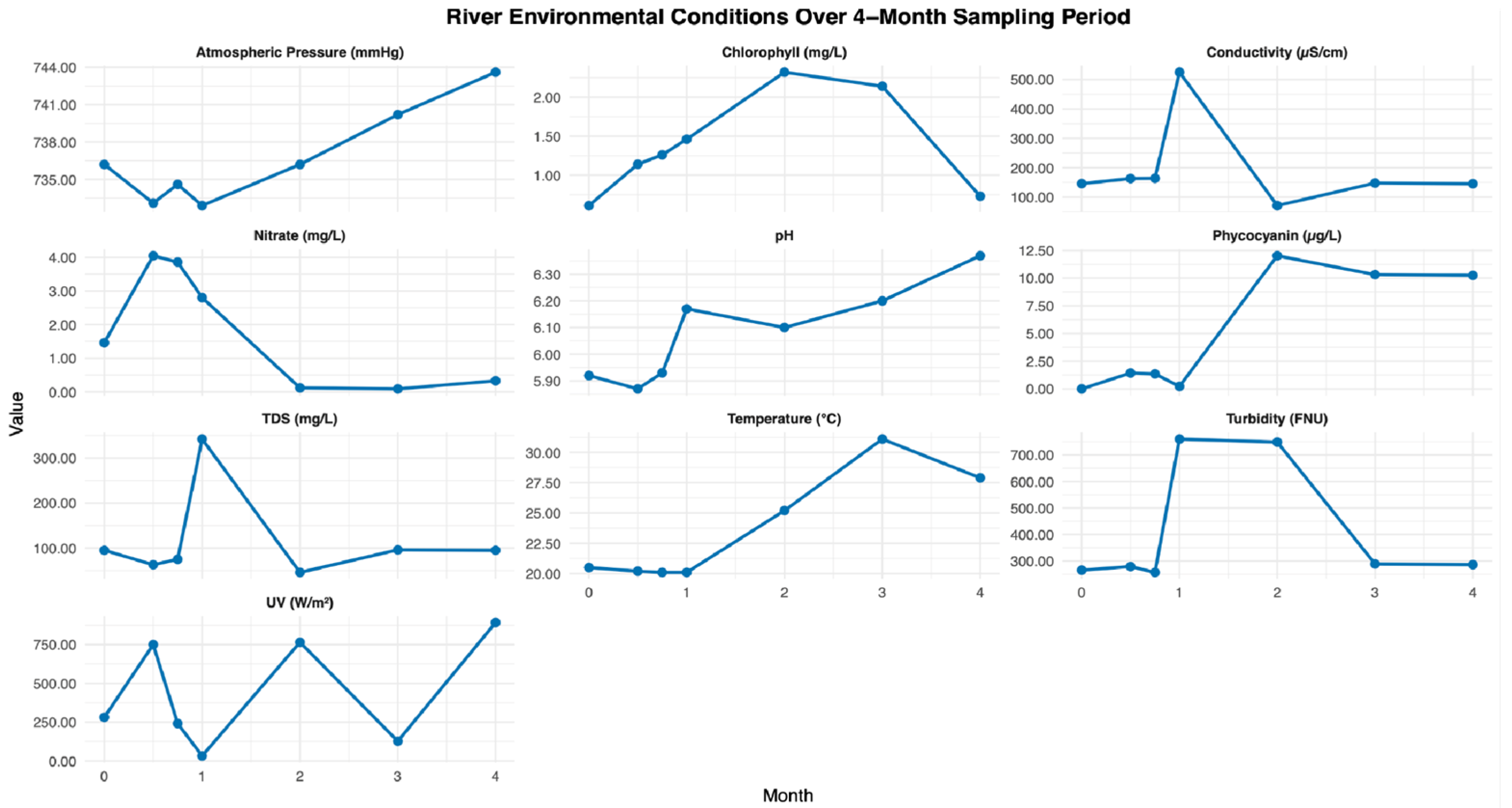
Figure A2.
Environmental conditions at the Third Creek site over the 4-month sampling period. Monthly measurements of atmospheric pressure, chlorophyll, conductivity, nitrate, pH, phycocyanin, total dissolved solids (TDS), temperature, turbidity, and UV radiation are shown. Mean site values are summarized in Table 2.
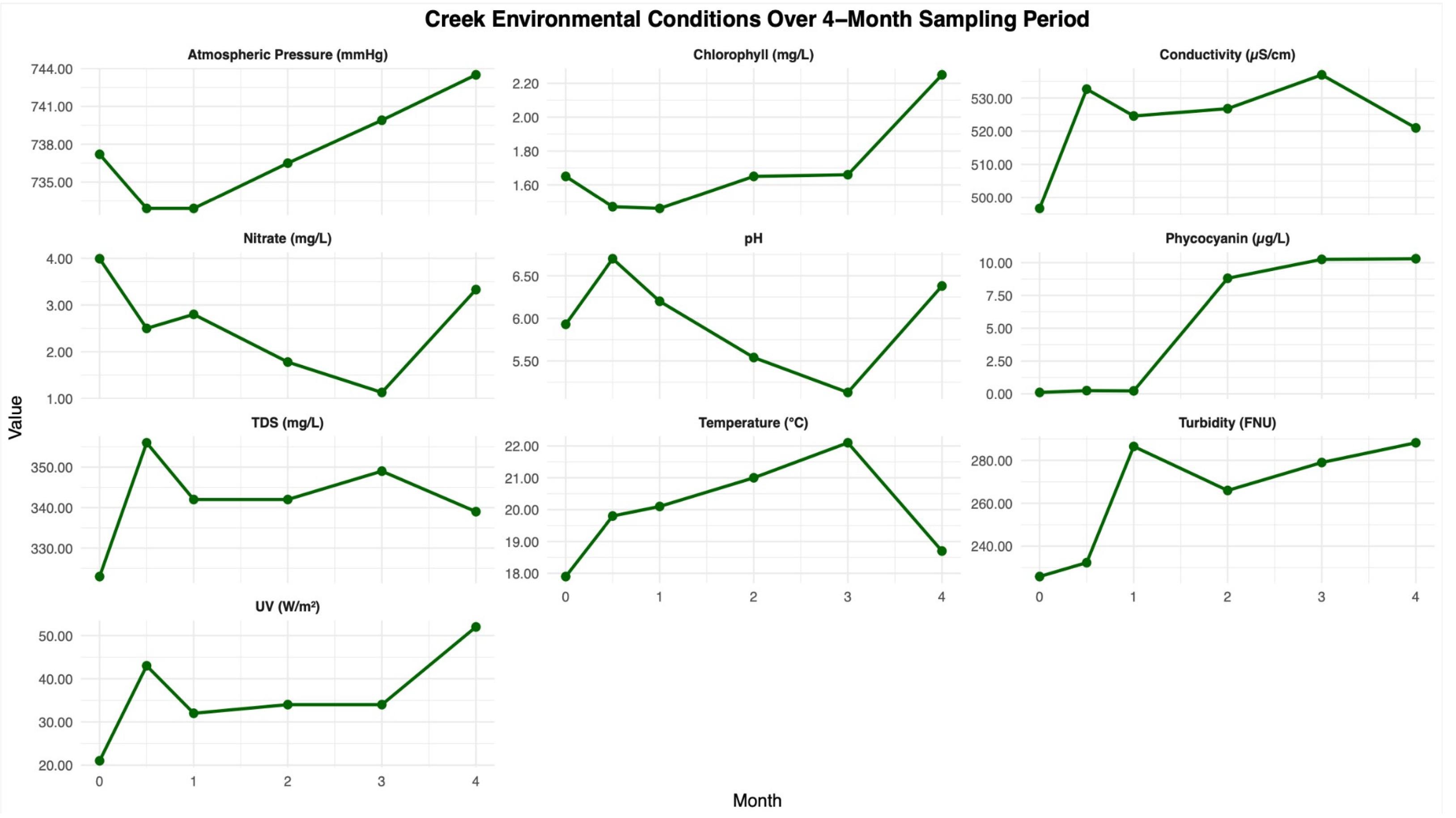
Figure A3.
Environmental conditions at the pond site over the 4-month sampling period. Monthly measurements of atmospheric pressure, chlorophyll, conductivity, nitrate, pH, phycocyanin, total dissolved solids (TDS), temperature, turbidity, and UV radiation are shown. Mean site values are summarized in Table 2.
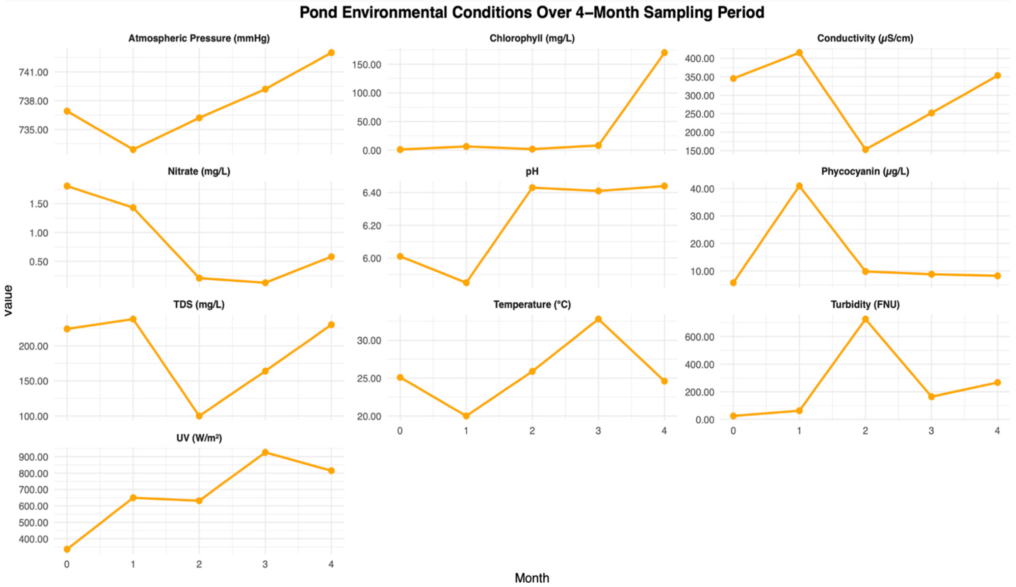
Figure A4.
SEM images of plastic surfaces after 30 days of incubation at the river site. Images show surface roughening, pitting, microfractures, and biofilm accumulation on PHA, PLA, PET, LDPE, and PE (blank control) samples, indicating early-stage surface alteration following environmental exposure.
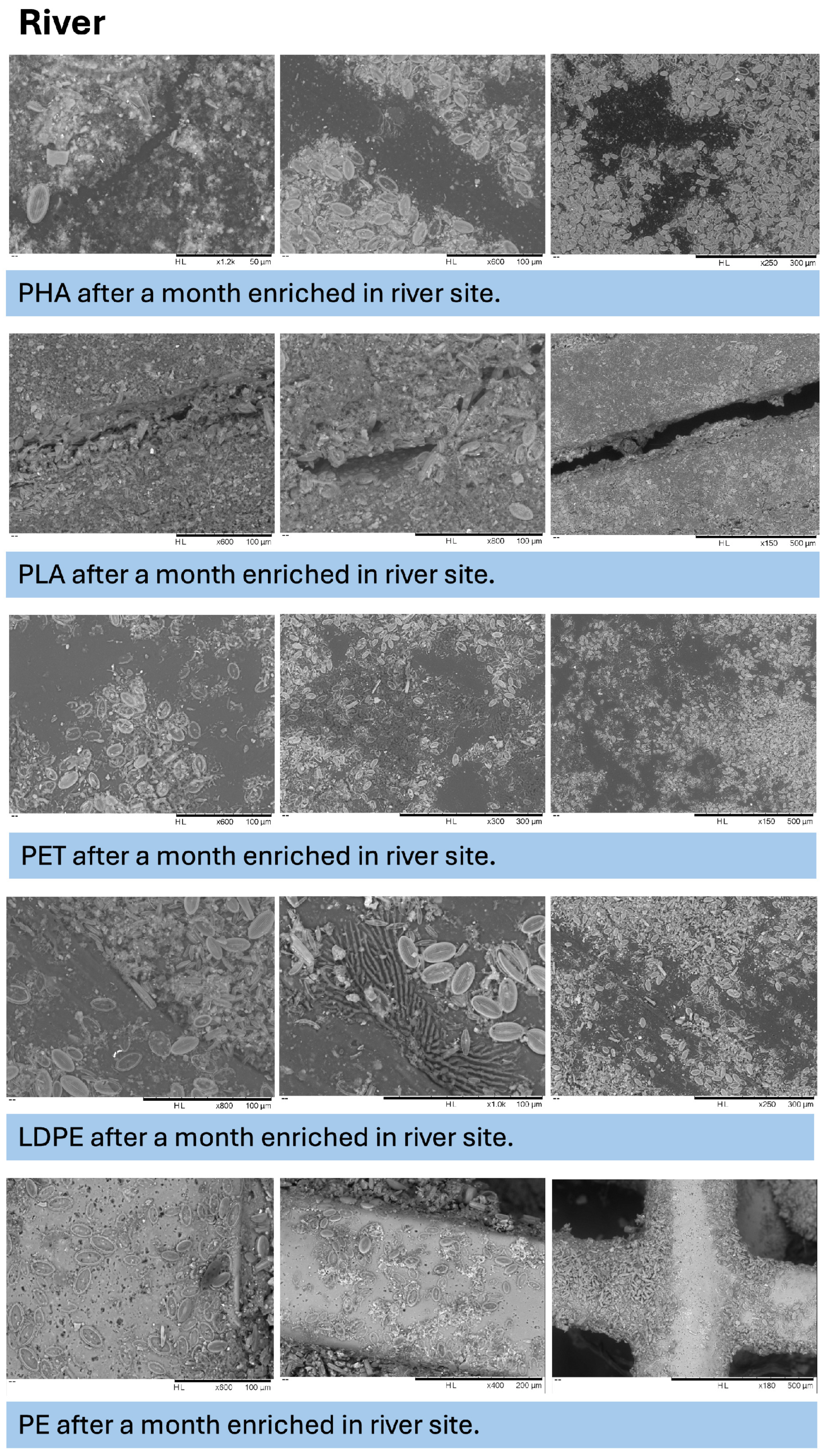
Figure A5.
SEM images of plastic surfaces after 30 days of incubation at the creek site. Images show surface roughening, fissures, and apparent microbial colonization across PHA, PLA, PET, LDPE, and PE (blank control) samples following environmental exposure.
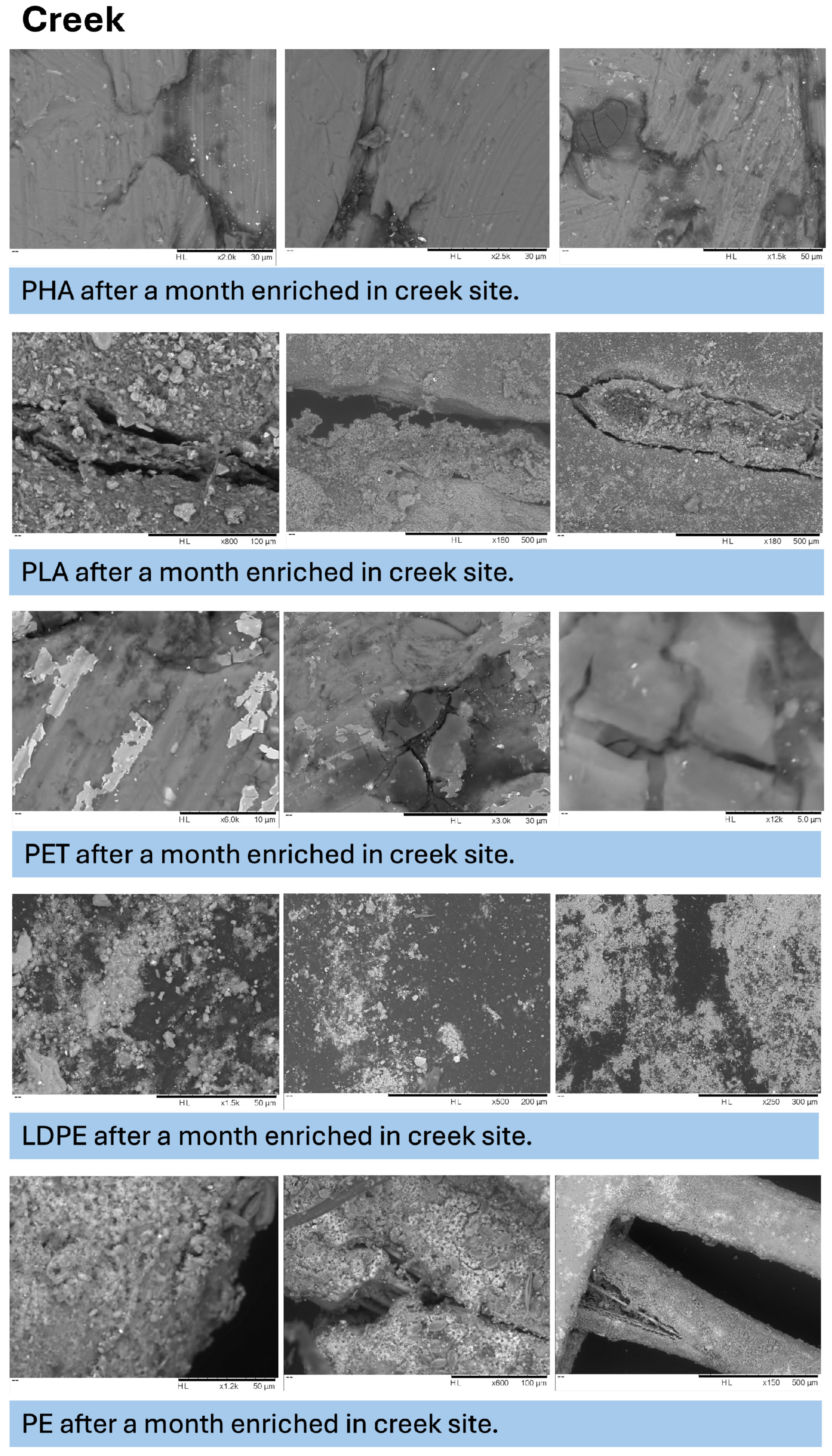
Figure A6.
SEM images of plastic surfaces after 30 days of incubation at the pond site. Images show surface roughening, cracking, and apparent biological accumulation on PHA, PLA, PET, LDPE, and PE (blank control) samples under lentic environmental conditions.
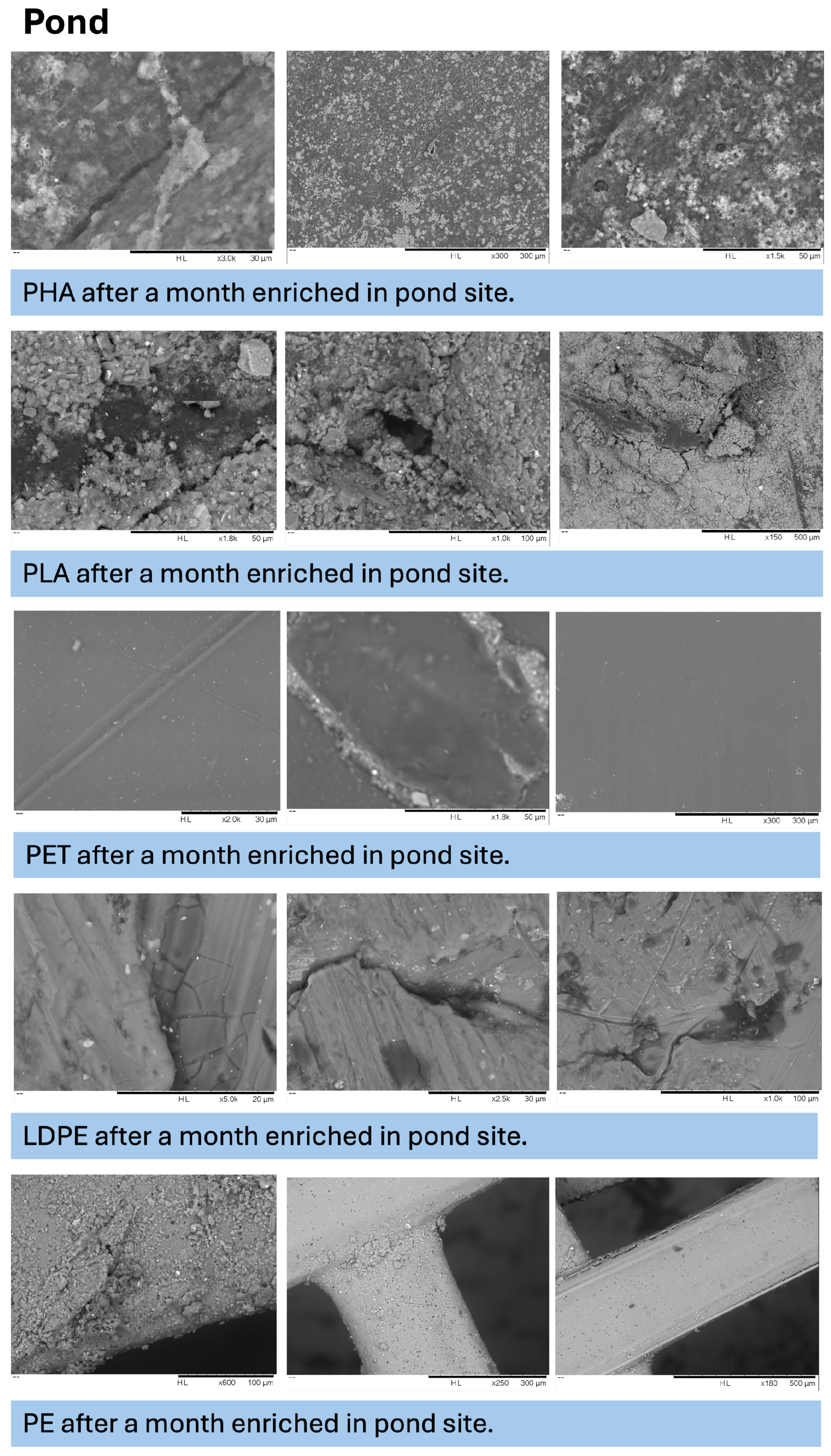
Figure A7.
SEM images of plastic surfaces after 30 days under control conditions. Images illustrate baseline surface morphology of PHA, PLA, PET, LDPE, and PE (blank control) samples in the absence of environmental exposure.
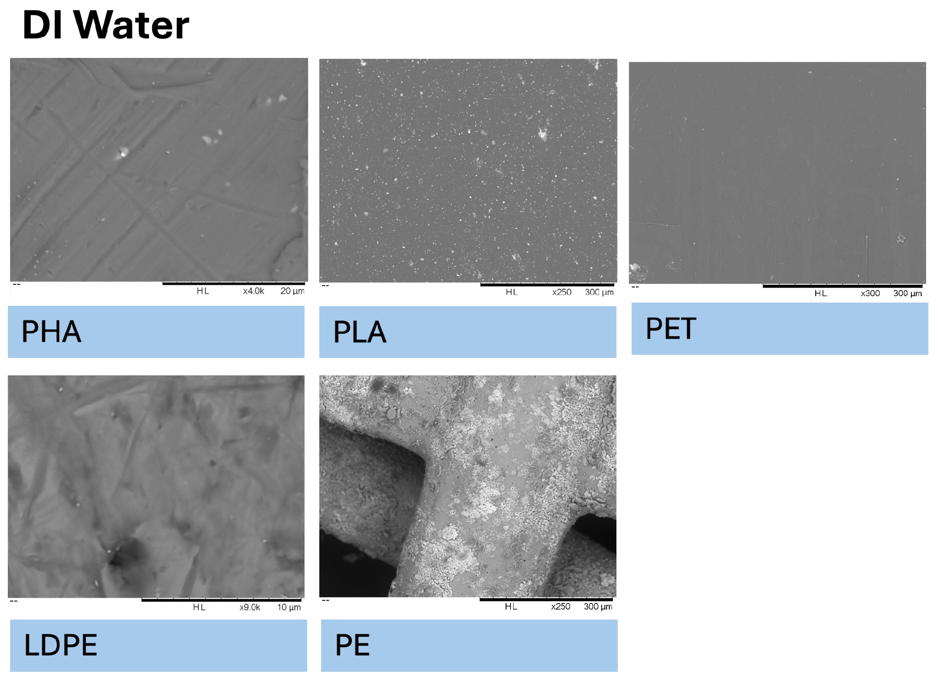
Figure A8.
SEM images of plastic surfaces after 120 days of incubation at the river site. Images show surface morphology changes on PHA, PLA, PET, LDPE, and PE blank control samples under river conditions.
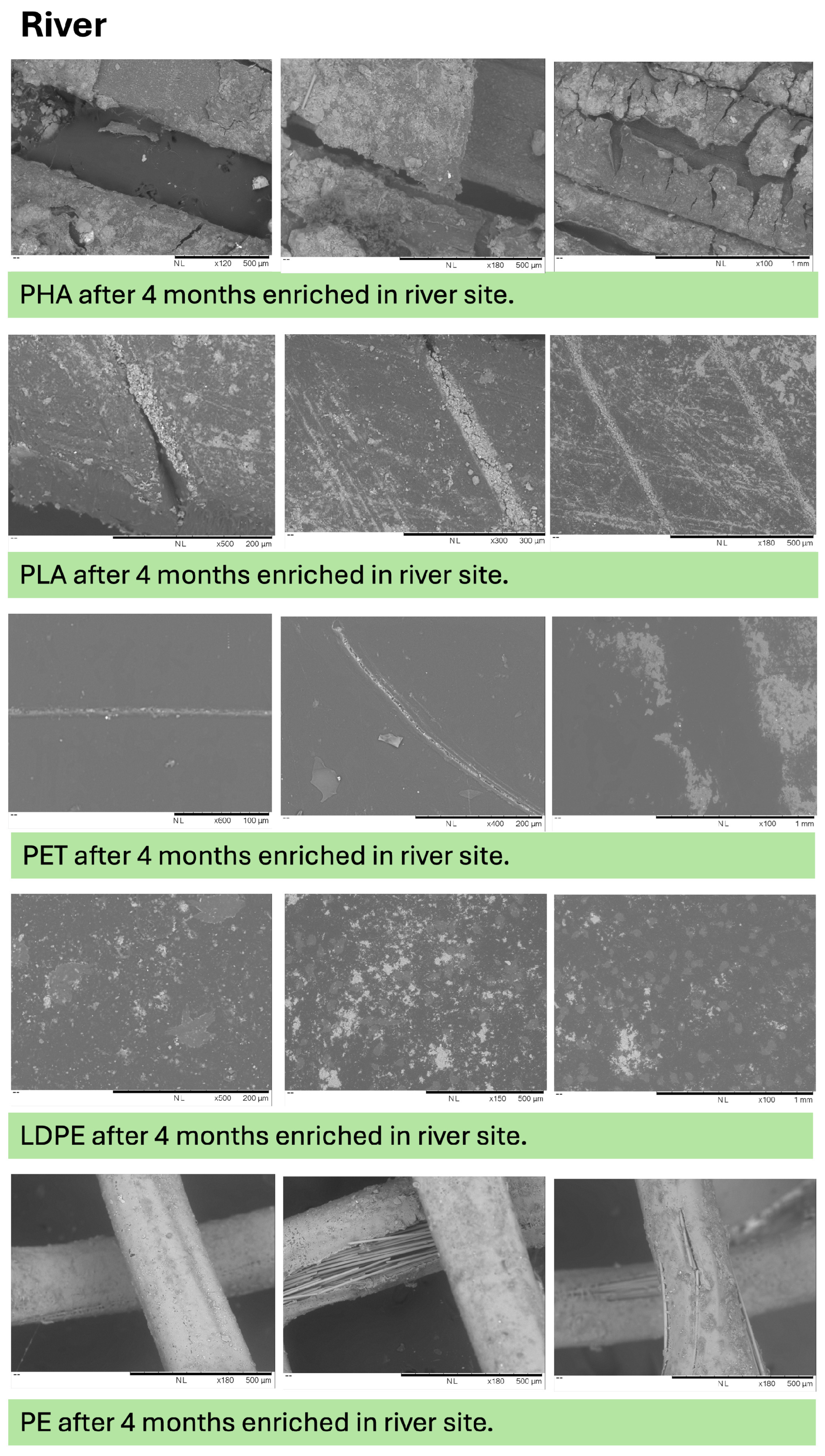
Figure A9.
SEM images of plastic surfaces after 120 days of incubation at the creek site. Images show changes in surface morphology, including pitting and cracking, on PHA, PLA, PET, LDPE, and PE (blank control) samples under creek conditions.
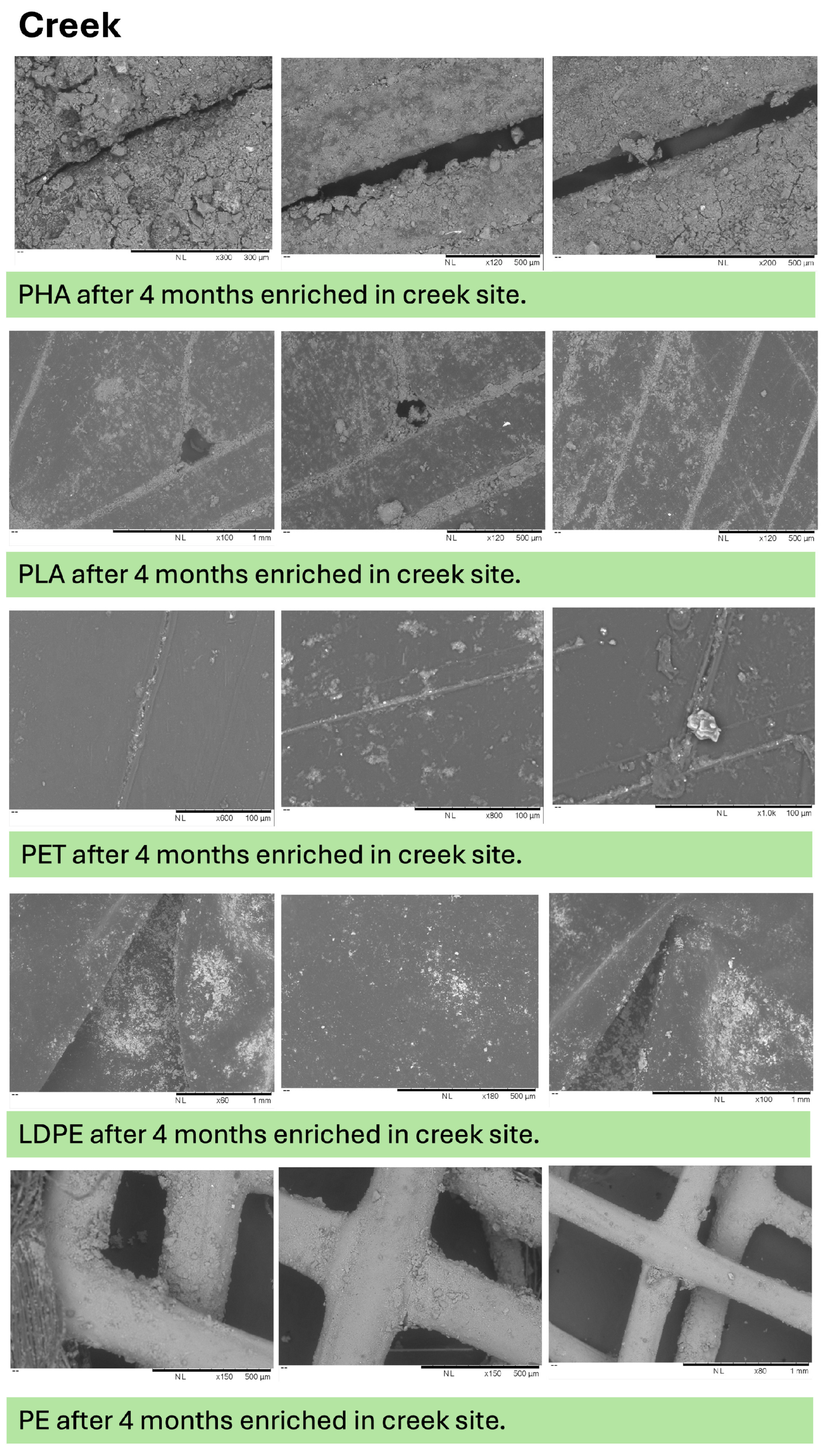
Figure A10.
SEM images of plastic surfaces after 120 days of incubation at the pond site. Images show changes in surface morphology, including pitting and fissuring, on PHA, PLA, PET, LDPE, and PE (blank control) samples under lentic environmental conditions.
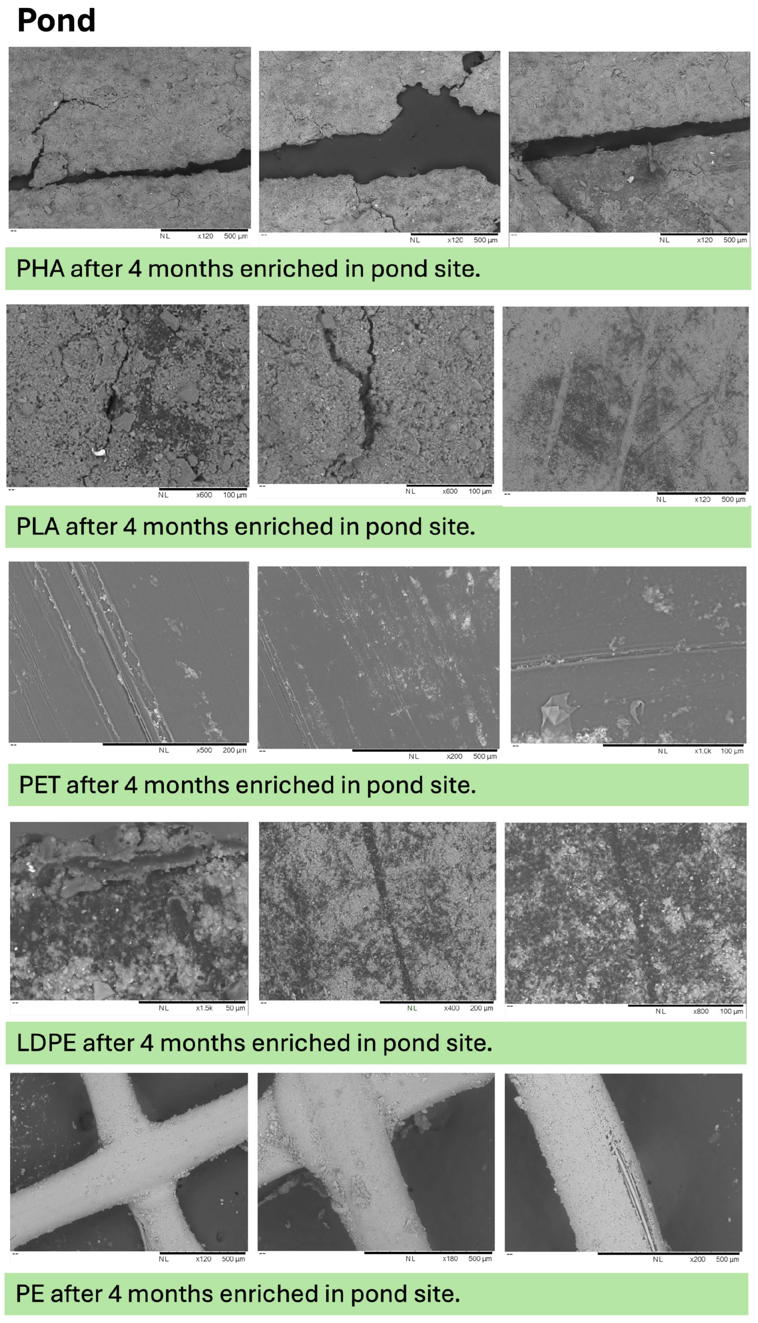
Figure A11.
SEM images of plastic surfaces after 120 days of incubation under control conditions. Micrographs depict baseline surface morphology of PHA, PLA, PET, LDPE, and PE (blank control) samples in the absence of environmental exposure.
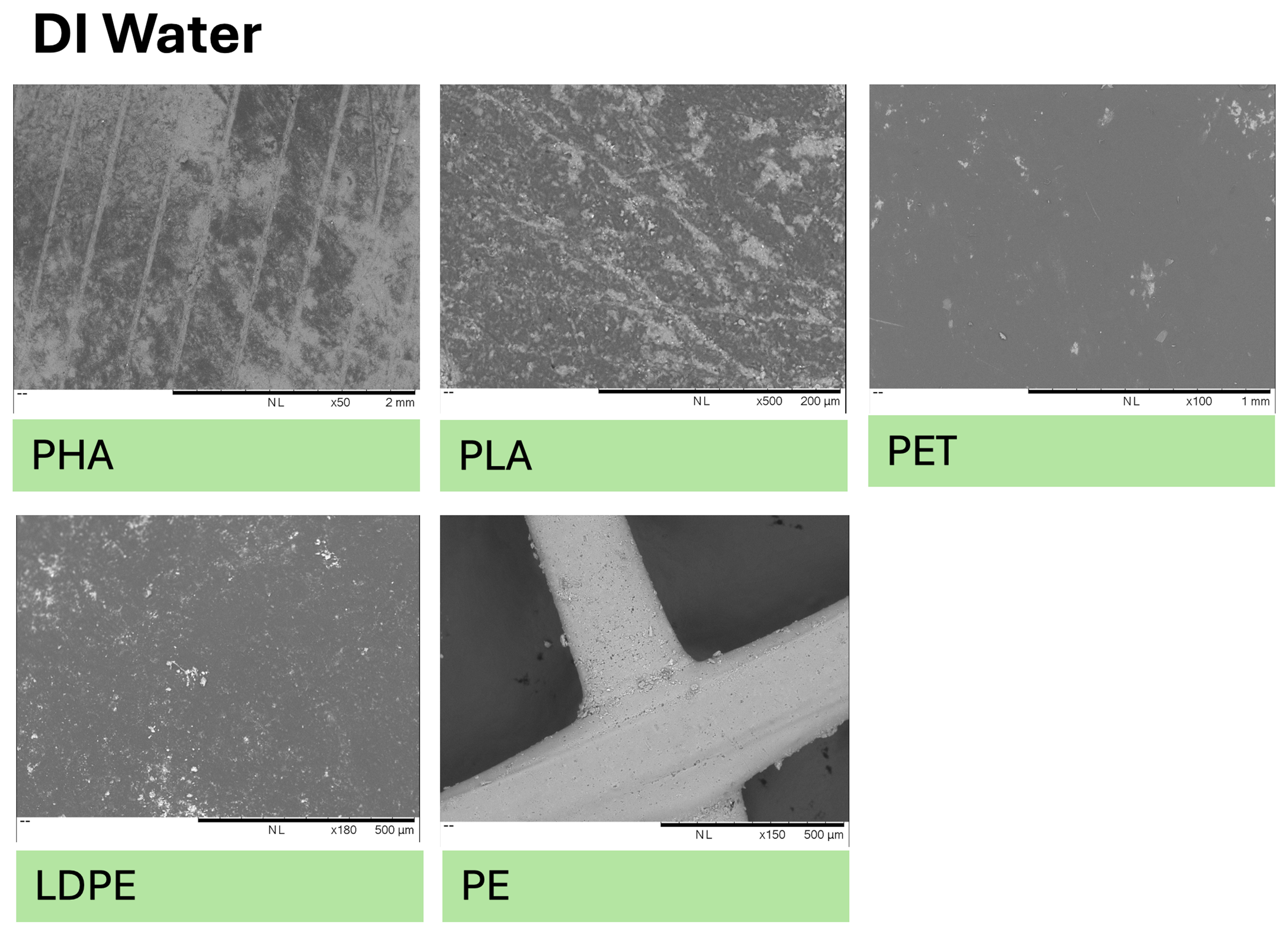
Figure A12.
Shannon diversity (genus level) of microbial communities associated with deployed plastic substrates (Bag Blank, LDPE, PET, PHA, and PLA) across freshwater environments. Panels are faceted by environment (Creek, Pond, and River), and points represent individual samples. Alpha diversity varied among substrates and environments, with substantial overlap among plastics across sites.
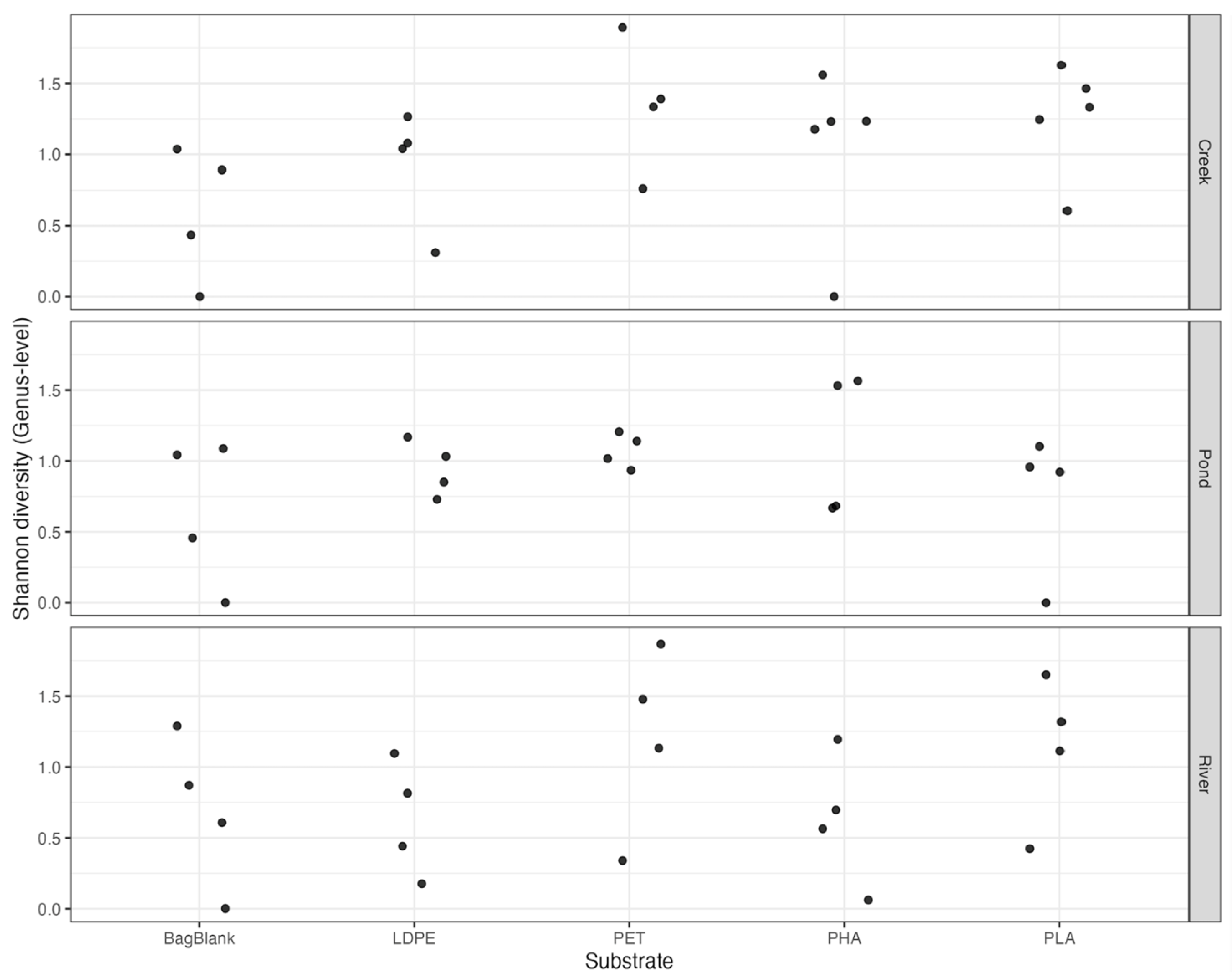
Figure A13.
Principal coordinates analysis (PCoA) of genus-level microbial communities associated with deployed plastic substrates (PET, LDPE, PHA, and PLA). Ordination is based on Bray–Curtis dissimilarity of relative abundance data. Points represent individual plastic samples, colored by polymer type and shaped by environment (Creek, Pond, and River). Axes represent the primary coordinates explaining the greatest variation in community composition.
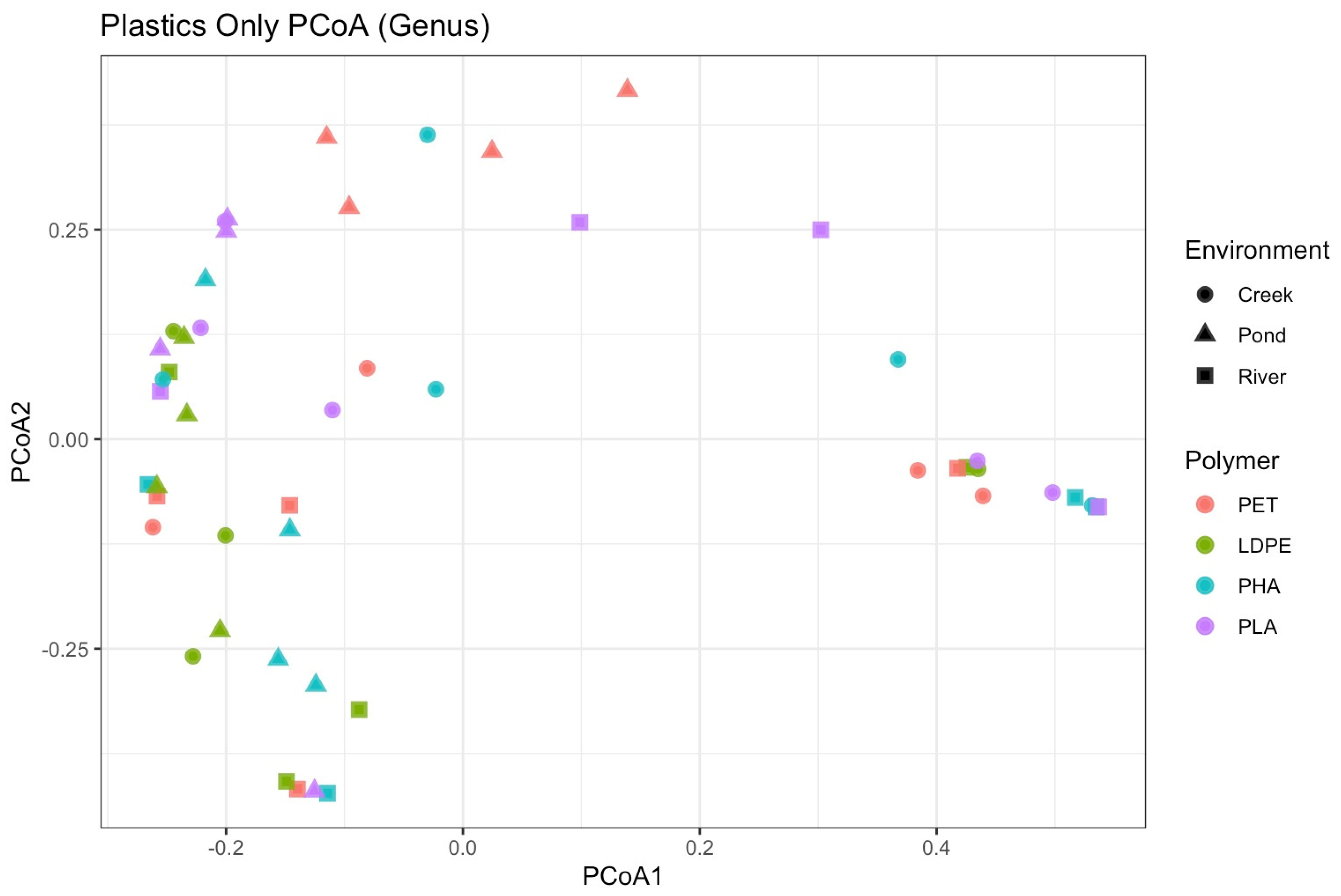
Table A1.
Spectrophotometer (OD600) measurements of bacterial growth for all samples across sites, polymer types, sampling months, and dilution factors.
Table A2.
Genus-level differential abundance results comparing plastics and water samples using DESeq2. Positive log2 fold change values indicate enrichment on plastics; negative values indicate enrichment in water samples. Adjusted p-values (padj) were calculated using the Benjamini–Hochberg correction.
Table A3.
Genus-level differential abundance results comparing LDPE and bag blank samples using DESeq2. Positive log2 fold change values indicate enrichment on LDPE. Adjusted p-values (padj) were calculated using the Benjamini–Hochberg correction. No genera were significantly different after correction for multiple testing (padj < 0.05).
Table A4.
Genus-level differential abundance results comparing PHA and bag blank samples using DESeq2. Positive log2 fold change values indicate enrichment on PHA. Adjusted p-values (padj) were calculated using the Benjamini–Hochberg correction. No genera were significantly different after correction for multiple testing (padj < 0.05).
Table A5.
Genus-level differential abundance results comparing PLA and bag blank samples using DESeq2. Positive log2 fold change values indicate enrichment on PLA. Adjusted p-values (padj) were calculated using the Benjamini–Hochberg correction. No genera were significantly different after correction for multiple testing (padj < 0.05).
Table A6.
Genus-level differential abundance results comparing PET and bag blank samples using DESeq2. Positive log2 fold change values indicate enrichment on PET. Adjusted p-values (padj) were calculated using the Benjamini–Hochberg correction. Nitrobacter was significantly enriched on PET after correction for multiple testing (padj < 0.05).
Table A7.
Genus-level differential abundance results comparing creek and pond water samples using DESeq2. Positive log2 fold change values indicate enrichment in the pond relative to the creek. Adjusted p-values (padj) were calculated using the Benjamini–Hochberg correction. No genera were significantly different after correction for multiple testing (padj < 0.05).
Table A8.
Genus-level differential abundance results comparing creek and river water samples using DESeq2. Positive log2 fold change values indicate enrichment in the river relative to the creek. Adjusted p-values (padj) were calculated using the Benjamini–Hochberg correction. Nitrobacter and Methanobacterium were significantly enriched in river samples after correction for multiple testing (padj < 0.05).
Table A9.
Genus-level differential abundance results comparing river and pond water samples using DESeq2. Positive log2 fold change values indicate enrichment in the river relative to the pond. Adjusted p-values (padj) were calculated using the Benjamini–Hochberg correction. Nitrobacter was significantly enriched in river samples after correction for multiple testing (padj < 0.05).
Table A10.
Summary of bacterial genera reported to be associated with LDPE and evaluation of experimental evidence supporting LDPE degradation. For each genus, reported evidence types (e.g., mass loss, FTIR spectral changes, SEM surface alteration, molecular weight reduction, enzymatic activity) are synthesized, and degradation capacity is interpreted based on findings from prior studies.
Table A11.
Summary of bacterial genera detected in this study and assessment of experimental evidence supporting their involvement in PHA degradation. Reported evidence types are synthesized to evaluate whether each genus demonstrates confirmed extracellular PHA depolymerization or lacks direct degradation evidence based on prior studies.
Table A12.
Summary of bacterial genera associated with polylactic acid (PLA) and evaluation of experimental evidence supporting PLA degradation. Reported evidence types are synthesized to assess degradation capacity based on prior studies.
Table A13.
Summary of bacterial genera detected in this study and assessment of experimental evidence supporting their involvement in PET degradation. Reported evidence types are synthesized to evaluate whether each genus demonstrates confirmed extracellular PET depolymerization or lacks direct degradation evidence based on prior studies.
References
- Li, C.; Busquets, R.; Campos, L.C. Assessment of microplastics in freshwater systems: A review. Sci. Total Environ. 2020, 707, 135578. [Google Scholar] [CrossRef] [PubMed]
- Rangel-Buitrago, N.; Neal, W.; Williams, A. The Plasticene: Time and rocks. Mar. Pollut. Bull. 2022, 185, 114358. [Google Scholar] [CrossRef] [PubMed]
- Eerkes-Medrano, D.; Thompson, R.C.; Aldridge, D.C. Microplastics in freshwater systems: A review of the emerging threats, identification of knowledge gaps and prioritisation of research needs. Water Res. 2015, 75, 63–82. [Google Scholar] [CrossRef]
- Jambeck, J.R.; Geyer, R.; Wilcox, C.; Siegler, T.R.; Perryman, M.; Andrady, A.; Narayan, R.; Law, K.L. Plastic waste inputs from land into the ocean. Science 2015, 347, 768–771. [Google Scholar] [CrossRef]
- Yang, J.; Monnot, M.; Sun, Y.; Asia, L.; Wong-Wah-Chung, P.; Doumenq, P.; Moulin, P. Microplastics in different water samples (seawater, freshwater, and wastewater): Removal efficiency of membrane treatment processes. Water Res. 2023, 232, 119673. [Google Scholar] [CrossRef]
- Azoulay, D.; Villa, P.; Arellano, Y.; Gordon, M.F.; Moon, D.; Miller, K.A.; Thompson, K. Plastic & Health: The Hidden Costs of a Plastic Planet; Center for International Environmental Law: Washington, DC, USA, 2019. [Google Scholar]
- Bridson, J.H.; Gaugler, E.C.; Smith, D.A.; Northcott, G.L.; Gaw, S. Leaching and extraction of additives from plastic pollution to inform environmental risk: A multidisciplinary review of analytical approaches. J. Hazard. Mater. 2021, 414, 125571. [Google Scholar] [CrossRef]
- Bhagat, J.; Zang, L.; Nishimura, N.; Shimada, Y. Zebrafish: An emerging model to study microplastic and nanoplastic toxicity. Sci. Total Environ. 2020, 728, 138707. [Google Scholar] [CrossRef]
- Omura, T.; Isobe, N.; Miura, T.; Ishii, S.; Mori, M.; Ishitani, Y.; Kimura, S.; Hidaka, K.; Komiyama, K.; Suzuki, M.; et al. Microbial decomposition of biodegradable plastics on the deep-sea floor. Nat. Commun. 2024, 15, 568. [Google Scholar] [CrossRef] [PubMed]
- Demirel, B.; Daver, F. The effects on the properties of PET bottles of changes to bottle-base geometry. J. Appl. Polym. Sci. 2009, 114, 3811–3818. [Google Scholar] [CrossRef]
- Perz, V.; Zumstein, M.T.; Sander, M.; Zitzenbacher, S.; Ribitsch, D.; Guebitz, G.M. Biomimetic approach to enhance enzymatic hydrolysis of the synthetic polyester poly(1,4-butylene adipate): Fusing binding modules to esterases. Biomacromolecules 2015, 16, 3889–3896. [Google Scholar] [CrossRef]
- Taniguchi, I.; Yoshida, S.; Hiraga, K.; Miyamoto, K.; Kimura, Y.; Oda, K. Biodegradation of PET: Current status and application aspects. ACS Catal. 2019, 9, 4089–4105. [Google Scholar] [CrossRef]
- Debroas, D.; Domaizon, I.; Humbert, J.-F.; Jardillier, L.; Lepère, C.; Oudart, A.; Taïb, N. Overview of freshwater microbial eukaryotes diversity: A first analysis of publicly available metabarcoding data. FEMS Microbiol. Ecol. 2017, 93, fix023. [Google Scholar] [CrossRef]
- Newton, R.J.; Jones, S.E.; Eiler, A.; McMahon, K.D.; Bertilsson, S. A guide to the natural history of freshwater lake bacteria. Microbiol. Mol. Biol. Rev. 2011, 75, 14–49. [Google Scholar] [CrossRef] [PubMed]
- Chen, M.; Hur, J.; Gu, J.-D.; Yamashita, Y. Microbial degradation of various types of dissolved organic matter in aquatic ecosystems and its influencing factors. Sci. China Earth Sci. 2023, 66, 169–189. [Google Scholar] [CrossRef]
- Thorp, J.H.; Rogers, D.C. Thorp and Covich’s Freshwater Invertebrates: Ecology and General Biology, 4th ed.; Elsevier Science & Technology: Amsterdam, The Netherlands, 2014. [Google Scholar]
- Pérez-García, P.; Sass, K.; Wongwattanarat, S.; Amann, J.; Feuerriegel, G.; Neumann, T.; Bäse, N.; Schmitz, L.S.; Dierkes, R.F.; Gurschke, M.F.; et al. Microbial plastic degradation: Enzymes, pathways, challenges, and perspectives. Microbiol. Mol. Biol. Rev. 2025, 89, e0008724. [Google Scholar] [CrossRef]
- Cai, Z.; Li, M.; Zhu, Z.; Wang, X.; Huang, Y.; Li, T.; Gong, H.; Yan, M. Biological degradation of plastics and microplastics: A recent perspective on associated mechanisms and influencing factors. Microorganisms 2023, 11, 1661. [Google Scholar] [CrossRef]
- Zettler, E.R.; Mincer, T.J.; Amaral-Zettler, L.A. Life in the “Plastisphere”: Microbial communities on plastic marine debris. Environ. Sci. Technol. 2013, 47, 7137–7146. [Google Scholar] [CrossRef] [PubMed]
- Zhai, X.; Zhang, X.-H.; Yu, M. Microbial colonization and degradation of marine microplastics in the plastisphere: A review. Front. Microbiol. 2023, 14, 1127308. [Google Scholar] [CrossRef]
- Blettler, M.C.M.; Abrial, E.; Khan, F.R.; Sivri, N.; Espinola, L.A. Freshwater plastic pollution: Recognizing research biases and identifying knowledge gaps. Water Res. 2018, 143, 416–424. [Google Scholar] [CrossRef]
- Galloway, T.S.; Cole, M.; Lewis, C. Interactions of microplastic debris throughout the marine ecosystem. Nat. Ecol. Evol. 2017, 1, 0116. [Google Scholar] [CrossRef]
- Amaral-Zettler, L.A.; Zettler, E.R.; Mincer, T.J. Ecology of the plastisphere. Nat. Rev. Microbiol. 2020, 18, 139–151. [Google Scholar] [CrossRef] [PubMed]
- Rendueles, O.; Beloin, C.; Latour-Lambert, P.; Ghigo, J.-M. A new biofilm-associated colicin with increased efficiency against biofilm bacteria. ISME J. 2014, 8, 1275–1288. [Google Scholar] [CrossRef] [PubMed]
- Bocci, V.; Galafassi, S.; Levantesi, C.; Crognale, S.; Amalfitano, S.; Congestri, R.; Matturro, B.; Rossetti, S.; Di Pippo, F. Freshwater plastisphere: A review on biodiversity, risks, and biodegradation potential with implications for the aquatic ecosystem health. Front. Microbiol. 2024, 15, 1395401. [Google Scholar] [CrossRef]
- Emmerik, T.; Schwarz, A. Plastic debris in rivers. Wiley Interdiscip. Rev. Water 2020, 7, e1398. [Google Scholar] [CrossRef]
- Oberbeckmann, S.; Osborn, A.M.; Duhaime, M.B. Microbes on a bottle: Substrate, season and geography influence community composition of microbes colonizing marine plastic debris. PLoS ONE 2016, 11, e0159289. [Google Scholar] [CrossRef]
- Di Pippo, F.; Bocci, V.; Amalfitano, S.; Crognale, S.; Levantesi, C.; Pietrelli, L.; Di Lisio, V.; Martinelli, A.; Rossetti, S. Microbial colonization patterns and biodegradation of petrochemical and biodegradable plastics in lake waters: Insights from a field experiment. Front. Microbiol. 2023, 14, 1290441. [Google Scholar] [CrossRef]
- Choi, J.; Kim, H.; Ahn, Y.-R.; Kim, M.; Yu, S.; Kim, N.; Lim, S.Y.; Park, J.-A.; Ha, S.-J.; Lim, K.S.; et al. Recent advances in microbial and enzymatic engineering for the biodegradation of micro- and nanoplastics. RSC Adv. 2024, 14, 9943–9966. [Google Scholar] [CrossRef]
- Boström, K.H.; Simu, K.; Hagström, Å.; Riemann, L. Optimization of DNA extraction for quantitative marine bacterioplankton community analysis. Limnol. Oceanogr. Methods 2004, 2, 365–373. [Google Scholar] [CrossRef]
- Somerville, C.C.; Knight, I.T.; Straube, W.L.; Colwell, R.R. Simple, rapid method for direct isolation of nucleic acids from aquatic environments. Appl. Environ. Microbiol. 1989, 55, 548–554. [Google Scholar] [CrossRef]
- Zhou, J.; Bruns, M.A.; Tiedje, J.M. DNA recovery from soils of diverse composition. Appl. Environ. Microbiol. 1996, 62, 316–322. [Google Scholar] [CrossRef] [PubMed]
- Tennessee Department of Environment and Conservation, Division of Water Resources. Posted Streams, Rivers, and Reservoirs in Tennessee. Available online: https://www.tn.gov/content/dam/tn/health/documents/wr_wq_fish-advisories.pdf (accessed on 5 May 2026).
- Choe, S.; Kim, Y.; Park, G.; Lee, D.H.; Park, J.; Mossisa, A.T.; Lee, S.; Myung, J. Biodegradation of 3D-printed biodegradable/non-biodegradable plastic blends. ACS Appl. Polym. Mater. 2022, 4, 5077–5090. [Google Scholar] [CrossRef]
- McDonnell, G.; Russell, A.D. Antiseptics and disinfectants: Activity, action, and resistance. Clin. Microbiol. Rev. 1999, 12, 147–179. [Google Scholar] [CrossRef] [PubMed]
- Chen, C.-C.; Dai, L.; Ma, L.; Guo, R.-T. Enzymatic degradation of plant biomass and synthetic polymers. Nat. Rev. Chem. 2020, 4, 114–126. [Google Scholar] [CrossRef] [PubMed]
- Lear, G.; Maday, S.D.M.; Gambarini, V.; Northcott, G.; Abbel, R.; Kingsbury, J.M.; Weaver, L.; Wallbank, J.A.; Pantos, O. Microbial abilities to degrade global environmental plastic polymer waste are overstated. Environ. Res. Lett. 2022, 17, 043002. [Google Scholar] [CrossRef]
- Pooja, N.; Chakraborty, I.; Rahman, M.H.; Mazumder, N. An insight on sources and biodegradation of bioplastics: A review. 3 Biotech 2023, 13, 220. [Google Scholar] [CrossRef]
- Alam, O.; Yang, L.; Yanchun, X. Determination of the selected heavy metal and metalloid contents in various types of plastic bags. J. Environ. Health Sci. Eng. 2019, 17, 161–170. [Google Scholar] [CrossRef]
- Gilewski, A.L.; Shrestha, S.; Kahara, S.N.; Stasulli, N.M. Microbial composition on microplastics mediated by stream impairment. Environ. Microbiome 2025, 20, 32. [Google Scholar] [CrossRef] [PubMed]
- Febria, F.A.; Syafrita, A.; Putra, A.; Hidayat, H.; Febrion, C. Degradation of low-density polyethylene by a novel strain of bacteria isolated from the plastisphere of marine ecosystems. Glob. J. Environ. Sci. Manag. 2024, 10, 805–820. [Google Scholar] [CrossRef]
- Relucenti, M.; Familiari, G.; Donfrancesco, O.; Taurino, M.; Li, X.; Chen, R.; Artini, M.; Papa, R.; Selan, L. Microscopy methods for biofilm imaging: Focus on SEM and VP-SEM pros and cons. Biology 2021, 10, 51. [Google Scholar] [CrossRef]
- Chao, Y.; Zhang, T. Optimization of fixation methods for observation of bacterial cell morphology and surface ultrastructures by atomic force microscopy. Appl. Microbiol. Biotechnol. 2011, 92, 381–392. [Google Scholar] [CrossRef]
- Dassanayake, R.P.; Falkenberg, S.M.; Stasko, J.A.; Shircliff, A.L.; Lippolis, J.D.; Briggs, R.E. Identification of a reliable fixative solution to preserve the complex architecture of bacterial biofilms for scanning electron microscopy evaluation. PLoS ONE 2020, 15, e0233973. [Google Scholar] [CrossRef]
- Talbot, M.J.; White, R.G. Methanol fixation of plant tissue for scanning electron microscopy improves preservation of tissue morphology and dimensions. Plant Methods 2013, 9, 36. [Google Scholar] [CrossRef] [PubMed]
- Fischer, E.R.; Hansen, B.T.; Nair, V.; Hoyt, F.H.; Schwartz, C.L.; Dorward, D.W. Scanning electron microscopy. Curr. Protoc. 2024, 4, e1034. [Google Scholar] [CrossRef] [PubMed]
- Alhede, M.; Qvortrup, K.; Liebrechts, R.; Høiby, N.; Givskov, M.; Bjarnsholt, T. Combination of microscopic techniques reveals a comprehensive visual impression of biofilm structure and composition. FEMS Immunol. Med. Microbiol. 2012, 65, 335–342. [Google Scholar] [CrossRef] [PubMed]
- Ali, R.; El-Boubbou, K.; Boudjelal, M. An easy, fast and inexpensive method of preparing a biological specimen for scanning electron microscopy (SEM). MethodsX 2021, 8, 101521. [Google Scholar] [CrossRef]
- Ru, J.; Huo, Y.; Yang, Y. Microbial degradation and valorization of plastic wastes. Front. Microbiol. 2020, 11, 442. [Google Scholar] [CrossRef]
- Stevenson, E.M.; Buckling, A.; Cole, M.; Murray, A. Culturing the plastisphere: Comparing methods to isolate culturable bacteria colonising microplastics. Front. Microbiol. 2023, 14, 1259287. [Google Scholar] [CrossRef] [PubMed]
- Lathrop, S.K.; Cooper, K.G.; Binder, K.A.; Starr, T.; Mampilli, V.; Detweiler, C.S.; Steele-Mortimer, O. Salmonella Typhimurium infection of human monocyte-derived macrophages. Curr. Protoc. Microbiol. 2018, 50, e56. [Google Scholar] [CrossRef]
- Beal, J.; Farny, N.G.; Haddock-Angelli, T.; Selvarajah, V.; Baldwin, G.S.; Buckley-Taylor, R.; Gershater, M.; Kiga, D.; Marken, J.; Sanchania, V.; et al. Robust estimation of bacterial cell count from optical density. Commun. Biol. 2020, 3, 512. [Google Scholar] [CrossRef]
- Tsuchiya, K.; Cao, Y.-Y.; Kurokawa, M.; Ashino, K.; Yomo, T.; Ying, B.-W. A decay effect of the growth rate associated with genome reduction in Escherichia coli. BMC Microbiol. 2018, 18, 101. [Google Scholar] [CrossRef]
- Jarerat, A.; Tokiwa, Y.; Tanaka, H. Production of poly(l-lactide)-degrading enzyme by Amycolatopsis orientalis for biological recycling of poly(l-lactide). Appl. Microbiol. Biotechnol. 2006, 72, 726–731. [Google Scholar] [CrossRef]
- Elbing, K.L.; Brent, R. Recipes and tools for culture of Escherichia coli. Curr. Protoc. Mol. Biol. 2019, 125, e83. [Google Scholar] [CrossRef]
- Yoshida, S.; Hiraga, K.; Takehana, T.; Taniguchi, I.; Yamaji, H.; Maeda, Y.; Toyohara, K.; Miyamoto, K.; Kimura, Y.; Oda, K. Response to comment on “A bacterium that degrades and assimilates poly(ethylene terephthalate)”. Science 2016, 353, 759. [Google Scholar] [CrossRef] [PubMed]
- QIAGEN. QIAamp PowerFecal Pro DNA Kit: Quick-Start Protocol (HB-2559-002); QIAGEN: Hilden, Germany, 2019. [Google Scholar]
- Illumina, Inc. 16S Metagenomic Sequencing Library Preparation: Preparing 16S Ribosomal RNA Gene Amplicons for the Illumina MiSeq System, Part #15044223 Rev. B; Illumina, Inc.: San Diego, CA, USA, 2019. [Google Scholar]
- Reis, S.N.; Aldridge, D.C. Biofilm development on macroplastics along a freshwater-estuarine gradient with attention to diatom assemblages. ACS ES&T Water 2024, 4, 5522–5530. [Google Scholar] [CrossRef]
- Jiang, C.; Almuhtaram, H.; McKie, M.J.; Andrews, R.C. Assessment of biofilm growth on microplastics in freshwaters using a passive flow-through system. Toxics 2023, 11, 987. [Google Scholar] [CrossRef] [PubMed]
- Restrepo-Flórez, J.-M.; Bassi, A.; Thompson, M.R. Microbial degradation and deterioration of polyethylene—A review. Int. Biodeterior. Biodegrad. 2014, 88, 83–90. [Google Scholar] [CrossRef]
- Raddadi, N.; Fava, F. Biodegradation of oil-based plastics in the environment: Existing knowledge and needs of research and innovation. Sci. Total Environ. 2019, 679, 148–158. [Google Scholar] [CrossRef]
- Shah, A.A.; Hasan, F.; Hameed, A.; Ahmed, S. Biological degradation of plastics: A comprehensive review. Biotechnol. Adv. 2008, 26, 246–265. [Google Scholar] [CrossRef]
- Lyshtva, P.; Voronova, V.; Barbir, J.; Leal Filho, W.; Kröger, S.D.; Witt, G.; Miksch, L.; Saborowski, R.; Gutow, L.; Frank, C.; et al. Degradation of a poly(3-hydroxybutyrate-co-3-hydroxyvalerate) (PHBV) compound in different environments. Heliyon 2024, 10, e24770. [Google Scholar] [CrossRef] [PubMed]
- Bocci, V.; De Vivo, M.; Alfano, S.; Rossetti, S.; Di Pippo, F.; Pietrelli, L.; Martinelli, A. Biodegradation in freshwater: Comparison between compostable plastics and their biopolymer matrices. Polymers 2025, 17, 2236. [Google Scholar] [CrossRef] [PubMed]
- Mori-Bazzano, L.; Nguyen, N.H.A.; Sevcu, A.; Riha, J.; Fu, T.; Slaveykova, V.I.; Ibelings, B.W. Plastic degradation in Lake Geneva: Influence of depth, seasonal shifts, and bacterial community dynamics. Sci. Total Environ. 2025, 983, 179615. [Google Scholar] [CrossRef] [PubMed]
- Gong, M.; Yang, G.; Zhuang, L.; Zeng, E.Y. Microbial biofilm formation and community structure on low-density polyethylene microparticles in lake water microcosms. Environ. Pollut. 2019, 252, 94–102. [Google Scholar] [CrossRef]
- Mistry, A.N.; Kachenchart, B.; Wongthanaroj, A.; Somwangthanaroj, A.; Luepromchai, E. Rapid biodegradation of high-molecular-weight semi-crystalline polylactic acid at ambient temperature via enzymatic and alkaline hydrolysis by a defined bacterial consortium. Polym. Degrad. Stab. 2022, 202, 110051. [Google Scholar] [CrossRef]
- Yoshida, S.; Hiraga, K.; Takehana, T.; Taniguchi, I.; Yamaji, H.; Maeda, Y.; Toyohara, K.; Miyamoto, K.; Kimura, Y.; Oda, K. A bacterium that degrades and assimilates poly(ethylene terephthalate). Science 2016, 351, 1196–1199. [Google Scholar] [CrossRef]
- Gewert, B.; Plassmann, M.M.; MacLeod, M. Pathways for degradation of plastic polymers floating in the marine environment. Environ. Sci. Process. Impacts 2015, 17, 1513–1521. [Google Scholar] [CrossRef]
- Hossain, M.R.; Jiang, M.; Wei, Q.; Leff, L.G. Microplastic surface properties affect bacterial colonization in freshwater. J. Basic Microbiol. 2019, 59, 54–61. [Google Scholar] [CrossRef]
- Thauer, R.K.; Kaster, A.-K.; Seedorf, H.; Buckel, W.; Hedderich, R. Methanogenic archaea: Ecologically relevant differences in energy conservation. Nat. Rev. Microbiol. 2008, 6, 579–591. [Google Scholar] [CrossRef]
- Carvalho, C.C.C.R.; Costa, S.S.; Fernandes, P.; Couto, I.; Viveiros, M. Membrane transport systems and the biodegradation potential and pathogenicity of genus Rhodococcus. Front. Physiol. 2014, 5, 133. [Google Scholar] [CrossRef] [PubMed]
- Alam, K.; Mazumder, A.; Sikdar, S.; Zhao, Y.-M.; Hao, J.; Song, C.; Wang, Y.; Sarkar, R.; Islam, S.; Zhang, Y.; et al. Streptomyces: The biofactory of secondary metabolites. Front. Microbiol. 2022, 13, 968053. [Google Scholar] [CrossRef] [PubMed]
- Danso, D.; Chow, J.; Streit, W.R. Plastics: Environmental and biotechnological perspectives on microbial degradation. Appl. Environ. Microbiol. 2019, 85, e01095-19. [Google Scholar] [CrossRef]
- Ghatge, S.; Yang, Y.; Ahn, J.-H.; Hur, H.-G. Biodegradation of polyethylene: A brief review. Appl. Biol. Chem. 2020, 63, 27. [Google Scholar] [CrossRef]
- Crump, B.C.; Adams, H.E.; Hobbie, J.E.; Kling, G.W. Biogeography of bacterioplankton in lakes and streams of an Arctic tundra catchment. Ecology 2007, 88, 1365–1378. [Google Scholar] [CrossRef]
- Liu, M.; Han, X.; Tong, J.; Zhu, H.; Bai, X. Mutual environmental drivers of the community composition, functional attributes and co-occurrence patterns of bacterioplankton in the composite aquatic ecosystem of Taihu watershed in China. FEMS Microbiol. Ecol. 2020, 96, fiaa137. [Google Scholar] [CrossRef]
- Niño-García, J.P.; Ruiz-González, C.; del Giorgio, P.A. Interactions between hydrology and water chemistry shape bacterioplankton biogeography across boreal freshwater networks. ISME J. 2016, 10, 1755–1766. [Google Scholar] [CrossRef]
- Stadler, M.; del Giorgio, P.A. Terrestrial connectivity, upstream aquatic history and seasonality shape bacterial community assembly within a large boreal aquatic network. ISME J. 2022, 16, 937–947. [Google Scholar] [CrossRef]
- Bai, C.; Cai, J.; Zhou, L.; Jiang, X.; Hu, Y.; Dai, J.; Shao, K.; Tang, X.; Yang, X.; Gao, G. Geographic patterns of bacterioplankton among lakes of the middle and lower reaches of the Yangtze River Basin, China. Appl. Environ. Microbiol. 2020, 86, e02423-19. [Google Scholar] [CrossRef]
- Savio, D.; Sinclair, L.; Ijaz, U.Z.; Parajka, J.; Reischer, G.H.; Stadler, P.; Blaschke, A.P.; Blöschl, G.; Mach, R.L.; Kirschner, A.K.T.; et al. Bacterial diversity along a 2600 km river continuum. Environ. Microbiol. 2015, 17, 4994–5007. [Google Scholar] [CrossRef] [PubMed]
- Wurtsbaugh, W.A.; Paerl, H.W.; Dodds, W.K. Nutrients, eutrophication and harmful algal blooms along the freshwater to marine continuum. Wiley Interdiscip. Rev. Water 2019, 6, e1373. [Google Scholar] [CrossRef]
- Jones, I.D.; Smol, J.P. Wetzel’s Limnology: Lake and River Ecosystems, 4th ed.; Elsevier Academic Press: Amsterdam, The Netherlands, 2024. [Google Scholar]
- Read, D.S.; Gweon, H.S.; Bowes, M.J.; Newbold, L.K.; Field, D.; Bailey, M.J.; Griffiths, R.I. Catchment-scale biogeography of riverine bacterioplankton. ISME J. 2015, 9, 516–526. [Google Scholar] [CrossRef] [PubMed]
- Boyandin, A.N.; Prudnikova, S.V.; Karpov, V.A.; Ivonin, V.N.; Đỗ, N.L.; Nguyễn, T.H.; Lê, T.M.H.; Filichev, N.L.; Levin, A.L.; Filipenko, M.L.; et al. Microbial degradation of polyhydroxyalkanoates in tropical soils. Int. Biodeterior. Biodegrad. 2013, 83, 77–84. [Google Scholar] [CrossRef]
- Rong, Z.; Ding, Z.-H.; Wu, Y.-H.; Xu, X.-W. Degradation of low-density polyethylene by the bacterium Rhodococcus sp. C-2 isolated from seawater. Sci. Total Environ. 2024, 907, 167993. [Google Scholar] [CrossRef]
- Maleki Rad, M.; Moghimi, H.; Azin, E. Biodegradation of thermo-oxidative pretreated low-density polyethylene (LDPE) and polyvinyl chloride (PVC) microplastics by Achromobacter denitrificans Ebl13. Mar. Pollut. Bull. 2022, 181, 113830. [Google Scholar] [CrossRef] [PubMed]
- Atanasova, N.; Paunova-Krasteva, T.; Stoitsova, S.; Radchenkova, N.; Boyadzhieva, I.; Petrov, K.; Kambourova, M. Degradation of poly(ε-caprolactone) by a thermophilic community and Brevibacillus thermoruber strain 7 isolated from a Bulgarian hot spring. Biomolecules 2021, 11, 1488. [Google Scholar] [CrossRef]
- Mohan, M.; Abedien, Z.U.; Kaparaju, P. Thermophilic anaerobic digestion of polylactic acid, polyethylene and polypropylene microplastics: Effect of inoculum–substrate ratio and microbiome. Biodegradation 2025, 36, 95. [Google Scholar] [CrossRef]
- Abraham, J.; Ghosh, E.; Mukherjee, P.; Gajendiran, A. Microbial degradation of low-density polyethylene. Environ. Prog. Sustain. Energy 2017, 36, 147–154. [Google Scholar] [CrossRef]
- Oliveira, J.; Almeida, P.L.; Sobral, R.G.; Lourenço, N.D.; Gaudêncio, S.P. Marine-derived actinomycetes: Biodegradation of plastics and formation of PHA bioplastics—A circular bioeconomy approach. Mar. Drugs 2022, 20, 760. [Google Scholar] [CrossRef] [PubMed]
- Jebashalomi, V.; Emmanuel Charles, P.; Rajaram, R. Microbial degradation of low-density polyethylene (LDPE) and polystyrene using Bacillus cereus (OR268710) isolated from a plastic-polluted tropical coastal environment. Sci. Total Environ. 2024, 924, 171580. [Google Scholar] [CrossRef]
- Mazaheri, H.; Nazeri, S. Biodegradation and detoxification of low-density polyethylene (LDPE) by Stenotrophomonas sp. and an Alcaligenaceae bacterium. Bull. Environ. Contam. Toxicol. 2024, 112, 19. [Google Scholar] [CrossRef]
- Alamer, N.J.; Aldayel, M.F.; Khalifa, A. Isolation and characterization of Brucella spp., low-density polyethylene (LDPE) plastic-degrading bacteria in the Al-Ahsa region, Saudi Arabia. Appl. Sci. 2023, 13, 4629. [Google Scholar] [CrossRef]
- Singleton, S.L.; Davis, E.W.; Arnold, H.K.; Daniels, A.M.Y.; Brander, S.M.; Parsons, R.J.; Sharpton, T.J.; Giovannoni, S.J. Identification of rare microbial colonizers of plastic materials incubated in a coral reef environment. Front. Microbiol. 2023, 14, 1259014. [Google Scholar] [CrossRef]
- Ma, Y.; Wang, J.; Liu, Y.; Wang, X.; Zhang, B.; Zhang, W.; Chen, T.; Liu, G.; Xue, L.; Cui, X. Nocardioides: “Specialists” for hard-to-degrade pollutants in the environment. Molecules 2023, 28, 7433. [Google Scholar] [CrossRef]
- Muenmee, S.; Chiemchaisri, W.; Chiemchaisri, C. Enhancement of biodegradation of plastic wastes via methane oxidation in semi-aerobic landfill. Int. Biodeterior. Biodegrad. 2016, 113, 244–255. [Google Scholar] [CrossRef]
- Zhang, H.; Lu, Y.; Wu, H.; Liu, Q.; Sun, W. Effect of an Acinetobacter pittobacter on low-density polyethylene. Environ. Sci. Pollut. Res. Int. 2023, 30, 10495–10504. [Google Scholar] [CrossRef]
- Szabó, I.; Al-Omari, J.; Dąbrowska, A.; Szerdahelyi, S.G.; Sebők, R.; Szabó, G.; Micsinai, A.; Kaszab, E.; Szoboszlay, S.; Kriszt, B. Long-term study of the bacterial colonization of polypropylene microplastics in a freshwater lake by optical and molecular methods. J. Mol. Liq. 2024, 393, 123637. [Google Scholar] [CrossRef]
- Zhong, R.; Li, M.; Zhu, Z.; Fang, X.; Wang, X.; Gong, H.; Yan, M. Bacterial degradation of polyethylene and polypropylene microplastics in a mangrove ecosystem. Chemosphere 2024, 369, 143908. [Google Scholar] [CrossRef] [PubMed]
- Muangchinda, C.; Pinyakong, O. Enrichment of LDPE-degrading bacterial consortia: Community succession and enhanced degradation efficiency through various pretreatment methods. Sci. Rep. 2024, 14, 28795. [Google Scholar] [CrossRef] [PubMed]
- Bardají, D.K.R.; Furlan, J.P.R.; Stehling, E.G. Isolation of a polyethylene-degrading Paenibacillus sp. from a landfill in Brazil. Arch. Microbiol. 2019, 201, 699–704. [Google Scholar] [CrossRef]
- Kämpfer, P.; Glaeser, S.P.; Busse, H.-J.; Eisenberg, T.; Scholz, H.C. Falsochrobactrum ovis gen. nov., sp. nov., isolated from a sheep. Int. J. Syst. Evol. Microbiol. 2013, 63, 3841–3847. [Google Scholar] [CrossRef]
- Ezaki, T.; Kawamura, Y.; Li, N.; Li, Z.-Y.; Zhao, L.; Shu, S. Proposal of the genera Anaerococcus gen. nov., Peptoniphilus gen. nov. and Gallicola gen. nov. for members of the genus Peptostreptococcus. Int. J. Syst. Evol. Microbiol. 2001, 51, 1521–1528. [Google Scholar] [CrossRef]
- Park, W.J.; Hwangbo, M.; Chu, K.-H. Plastisphere and microorganisms involved in polyurethane biodegradation. Sci. Total Environ. 2023, 886, 163932. [Google Scholar] [CrossRef]
- Busse, H.-J. Review of the taxonomy of the genus Arthrobacter, emendation of the genus Arthrobacter sensu lato, proposal to reclassify selected species of the genus Arthrobacter in the novel genera Glutamicibacter gen. nov., Paeniglutamicibacter gen. nov., Pseudoglutamicibacter gen. nov., Paenarthrobacter gen. nov. and Pseudarthrobacter gen. nov., and emended description of Arthrobacter roseus. Int. J. Syst. Evol. Microbiol. 2016, 66, 9–37. [Google Scholar] [CrossRef]
- Stackebrandt, E.; Goebel, B.M. Taxonomic note: A place for DNA–DNA reassociation and 16S rRNA sequence analysis in the present species definition in bacteriology. Int. J. Syst. Bacteriol. 1994, 44, 846–849. [Google Scholar] [CrossRef]
- Greene, A.C.; Euzéby, J.P.; Tindall, B.J.; Patel, B.K.C. Proposal of Frondihabitans gen. nov. to replace the illegitimate genus name Frondicola Zhang et al. 2007. Int. J. Syst. Evol. Microbiol. 2009, 59, 447–448. [Google Scholar] [CrossRef] [PubMed][Green Version]
- Carlier, A.; Cnockaert, M.; Fehr, L.; Vandamme, P.; Eberl, L. Draft genome and description of Orrella dioscoreae gen. nov., sp. nov., a new species of Alcaligenaceae isolated from leaf acumens of Dioscorea sansibarensis. Syst. Appl. Microbiol. 2017, 40, 11–21. [Google Scholar] [CrossRef] [PubMed]
- Jeon, Y.; Jin, H.; Kong, Y.; Cha, H.-G.; Lee, B.W.; Yu, K.; Yi, B.; Kim, H.T.; Joo, J.C.; Yang, Y.-H.; et al. Poly(3-hydroxybutyrate) degradation by Bacillus infantis sp. isolated from soil and identification of phaZ- and bdhA-expressing PHB depolymerase. J. Microbiol. Biotechnol. 2023, 33, 1076–1083. [Google Scholar] [CrossRef] [PubMed]
- Di, Y.; Xia, H.; Jiao, Y.; Zhang, X.; Fang, Q.; Li, F.; Chen, S. Biodegradation of polyhydroxybutyrate by Pseudomonas sp. DSDY0501 and purification and characterization of polyhydroxybutyrate depolymerase. 3 Biotech 2019, 9, 359. [Google Scholar] [CrossRef]
- Jeszeová, L.; Puškárová, A.; Bučková, M.; Kraková, L.; Grivalský, T.; Danko, M.; Mosnáčková, K.; Chmela, Š.; Pangallo, D. Microbial communities responsible for the degradation of poly(lactic acid)/poly(3-hydroxybutyrate) blend mulches in soil burial respirometric tests. World J. Microbiol. Biotechnol. 2018, 34, 101. [Google Scholar] [CrossRef]
- Raghuwanshi, S.; Agarwal, T.; Yadav, A.; Zaidi, M.; Shouche, Y.; Goel, R. Selection of poly(R)-3-hydroxybutyric acid-utilising bacteria by enrichment, optimisation and compatibility testing for consortia development. Chem. Ecol. 2016, 32, 583–597. [Google Scholar] [CrossRef]
- Karimnezhad, H.; Rahimpour, F.; Samadi, S. Bioconversion of food waste to poly(3-hydroxybutyrate-co-3-hydroxyvalerate) biopolymer using Stenotrophomonas geniculata: Study of operating parameters and process optimization. Biodegradation 2025, 36, 57. [Google Scholar] [CrossRef]
- Jiménez, N.; Richnow, H.H.; Vogt, C.; Treude, T.; Krüger, M. Methanogenic hydrocarbon degradation: Evidence from field and laboratory studies. J. Mol. Microbiol. Biotechnol. 2016, 26, 227–242. [Google Scholar] [CrossRef]
- Patel, P.D.; Patel, R.B.; Munshi, N.S. Exploring the poly(3-hydroxybutyrate-co-3-hydroxyvalerate) production potential of total petroleum hydrocarbon-degrading bacteria using oily sludge waste as feedstock. ACS EST Eng. 2024, 4, 1622–1634. [Google Scholar] [CrossRef]
- Celli, J. The intracellular life cycle of Brucella spp. Microbiol. Spectr. 2019, 7, BAI-0006-2019. [Google Scholar] [CrossRef]
- Albuquerque, L.; Simões, C.; Nobre, M.F.; Pino, N.M.; Battista, J.R.; Silva, M.T.; Rainey, F.A.; da Costa, M.S. Truepera radiovictrix gen. nov., sp. nov., a new radiation-resistant species and the proposal of Trueperaceae fam. nov. FEMS Microbiol. Lett. 2005, 247, 161–169. [Google Scholar] [CrossRef]
- Mabrouk, M.E.S.; El-Badan, D.E.S.; Ghanem, N.B.; Sabry, S.A. Degradation of polyesters by a novel marine Nocardiopsis aegyptia sp. nov.: Application of Plackett–Burman experimental design for the improvement of PHB depolymerase activity. J. Gen. Appl. Microbiol. 2005, 51, 151–158. [Google Scholar] [CrossRef]
- Suzuki, M.; Tachibana, Y.; Takizawa, R.; Morikawa, T.; Takeno, H.; Kasuya, K. A novel poly(3-hydroxybutyrate)-degrading actinobacterium isolated from the plastisphere formed on marine plastic debris. Polym. Degrad. Stab. 2021, 183, 109461. [Google Scholar] [CrossRef]
- Al-Khattaf, F.S.; Al-Ansari, M.M.; Maruthamuthu, M.K.; Dyona, L.; Agastian, P. Polyhydroxybutyrate degradation by biocatalyst of municipal sludge water and degradation efficacy in a sequencing batch biofilm reactor. Environ. Res. 2022, 204, 112336. [Google Scholar] [CrossRef] [PubMed]
- Yokota, A.; Hamada, T.; Uchino, Y. Proposal of Pseudorhodobacter ferrugineus gen. nov., comb. nov., for a non-photosynthetic marine bacterium, Agrobacterium ferrugineum, related to the genus Rhodobacter. J. Gen. Appl. Microbiol. 2002, 48, 309–319. [Google Scholar] [CrossRef] [PubMed]
- Kämpfer, P.; Martin, E.; Lodders, N.; Jäckel, U. Transfer of Defluviibacter lusatiensis to the genus Aquamicrobium as Aquamicrobium lusatiense comb. nov. and description of Aquamicrobium aerolatum sp. nov. Int. J. Syst. Evol. Microbiol. 2009, 59, 2468–2470. [Google Scholar] [CrossRef]
- Vigneswari, S.; Vijaya, S.; Majid, M.I.A.; Sudesh, K.; Sipaut, C.S.; Azizan, M.N.M.; Amirul, A.A. Enhanced production of poly(3-hydroxybutyrate-co-4-hydroxybutyrate) copolymer with manipulated variables and its properties. J. Ind. Microbiol. Biotechnol. 2009, 36, 547–556. [Google Scholar] [CrossRef]
- Cattaneo, C.R.; Rodríguez, Y.; Rene, E.R.; García-Depraect, O.; Muñoz, R. Biogas bioconversion into poly(3-hydroxybutyrate) by a mixed microbial culture in a novel Taylor flow bioreactor. Waste Manag. 2022, 150, 364–372. [Google Scholar] [CrossRef]
- Takeuchi, M.; Weiss, N.; Schumann, P.; Yokota, A. Leucobacter komagatae gen. nov., sp. nov., a new aerobic, Gram-positive, nonsporulating rod with 2,4-diaminobutyric acid in the cell wall. Int. J. Syst. Bacteriol. 1996, 46, 967–971. [Google Scholar] [CrossRef]
- Rodríguez, J.; Andersson, A.; Björn, E.; Timonen, S.; Brugel, S.; Skrobonja, A.; Rowe, O. Inputs of terrestrial dissolved organic matter enhance bacterial production and methylmercury formation in oxic coastal water. Front. Microbiol. 2022, 13, 809166. [Google Scholar] [CrossRef]
- Jarerat, A.; Pranamuda, H.; Tokiwa, Y. Poly(L-lactide)-degrading activity in various actinomycetes. Macromol. Biosci. 2002, 2, 420–428. [Google Scholar] [CrossRef]
- Tokiwa, Y.; Calabia, B.P. Biodegradability and biodegradation of poly(lactide). Appl. Microbiol. Biotechnol. 2006, 72, 244–251. [Google Scholar] [CrossRef]
- Kawashima, N.; Tokuda, J.; Yagi, T.; Takahashi, K. Isolation of a Nocardiopsis chromatogenes strain that degrades PLA (polylactic acid) in pig waste-based compost. Arch. Microbiol. 2022, 204, 599. [Google Scholar] [CrossRef] [PubMed]
- Bonifer, K.S.; Wen, X.; Hasim, S.; Phillips, E.K.; Dunlap, R.N.; Gann, E.R.; DeBruyn, J.M.; Reynolds, T.B. Bacillus pumilus B12 degrades polylactic acid and degradation is affected by changing nutrient conditions. Front. Microbiol. 2019, 10, 2548. [Google Scholar] [CrossRef] [PubMed]
- Jeon, H.J.; Kim, M.N. Biodegradation of poly(L-lactide) (PLA) exposed to UV irradiation by a mesophilic bacterium. Int. Biodeterior. Biodegrad. 2013, 85, 289–293. [Google Scholar] [CrossRef]
- Pourhajibagher, M.; Pourakbari, B.; Bahador, A. Contribution of antimicrobial photo-sonodynamic therapy in wound healing: An in vivo effect of curcumin–nisin-based poly(L-lactic acid) nanoparticles on Acinetobacter baumannii biofilms. BMC Microbiol. 2022, 22, 28. [Google Scholar] [CrossRef]
- Mao, Z.; Yu, C.; Xin, L. Enhancement of phenol biodegradation by Pseudochrobactrum sp. through ultraviolet-induced mutation. Int. J. Mol. Sci. 2015, 16, 7320–7333. [Google Scholar] [CrossRef]
- Zhang, M.; Li, Q.; Nie, R.; Bai, X.; Hu, G.; Pan, K.; Liu, H.; Zhu, Q.; Yan, X.; Hong, Q. Unveiling the highly efficient degradation mechanism of strobilurin fungicides by strain Hyphomicrobium sp. DY-1. Int. Biodeterior. Biodegrad. 2024, 187, 105722. [Google Scholar] [CrossRef]
- Guo, W.; Duan, J.; Shi, Z.; Yu, X.; Shao, Z. Biodegradation of PET by the membrane-anchored PET esterase from the marine bacterium Rhodococcus pyridinivorans P23. Commun. Biol. 2023, 6, 1090. [Google Scholar] [CrossRef] [PubMed]
- Krainara, S.; Mistry, A.N.; Malee, C.; Chavananikul, C.; Pinyakong, O.; Assavalapsakul, W.; Jitpraphai, S.M.; Kachenchart, B.; Luepromchai, E. Development of a plastic-waste treatment process by combining deep eutectic solvent pretreatment and bioaugmentation with a plastic-degrading bacterial consortium. J. Hazard. Mater. 2023, 460, 132507. [Google Scholar] [CrossRef]
- Verschoor, J.-A.; Croese, M.R.J.; Lakemeier, S.E.; Mugge, A.; Burgers, C.M.C.; Innocenti, P.; Willemse, J.; Crooijmans, M.E.; van Wezel, G.P.; Ram, A.F.J.; et al. Polyester degradation by soil bacteria: Identification of conserved BHETase enzymes in Streptomyces. Commun. Biol. 2024, 7, 725. [Google Scholar] [CrossRef]
- Roberts, C.; Edwards, S.; Vague, M.; León-Zayas, R.; Scheffer, H.; Chan, G.; Swartz, N.A.; Mellies, J.L. Environmental consortium containing Pseudomonas and Bacillus species synergistically degrades polyethylene terephthalate plastic. mSphere 2020, 5, e01151-20. [Google Scholar] [CrossRef]
- Din, S.U.; Satti, S.M.K.; Uddin, S.; Mankar, S.V.; Ceylan, E.; Hasan, F.; Khan, S.; Badshah, M.; Beldüz, A.O.; Canakci, S.; et al. Purification and characterization of a cutinase-like enzyme with activity on polyethylene terephthalate (PET) from a newly isolated bacterium, Stenotrophomonas maltophilia PRS8, at a mesophilic temperature. Appl. Sci. 2023, 13, 3686. [Google Scholar] [CrossRef]
- Singh, S.; Thakur, R.S.; Manickam, N. Insights into the molecular mechanism of plasticizer biodegradation in Dietzia kunjamensis IITR165 and Brucella intermedia IITR166 isolated from a solid-waste dumpsite. J. Appl. Microbiol. 2023, 134, lxad231. [Google Scholar] [CrossRef]
- Zhao, S.; Liu, R.; Wang, J.; Lv, S.; Zhang, B.; Dong, C.; Shao, Z. Biodegradation of polyethylene terephthalate (PET) by diverse marine bacteria in deep-sea sediments. Environ. Microbiol. 2023, 25, 2719–2731. [Google Scholar] [CrossRef]
- Carr, C.M.; Clarke, D.J.; Dobson, A.D.W. Microbial polyethylene terephthalate hydrolases: Current and future perspectives. Front. Microbiol. 2020, 11, 571265. [Google Scholar] [CrossRef] [PubMed]
- Ramasamy, S.; Mathiyalagan, P.; Chandran, P. Characterization and optimization of EPS-producing and diesel oil-degrading Ochrobactrum anthropi MP3 isolated from refinery wastewater. Pet. Sci. 2014, 11, 439–445. [Google Scholar] [CrossRef]
- Esmaeili, A.; Pourbabaee, A.A.; Alikhani, H.A.; Shabani, F.; Esmaeili, E. Biodegradation of low-density polyethylene (LDPE) by mixed culture of Lysinibacillus xylanilyticus and Aspergillus niger in soil. PLoS ONE 2013, 8, e71720. [Google Scholar] [CrossRef]
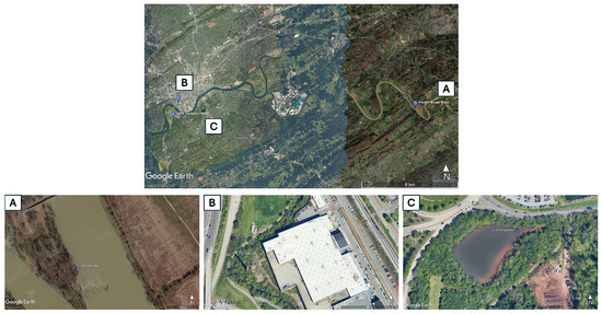
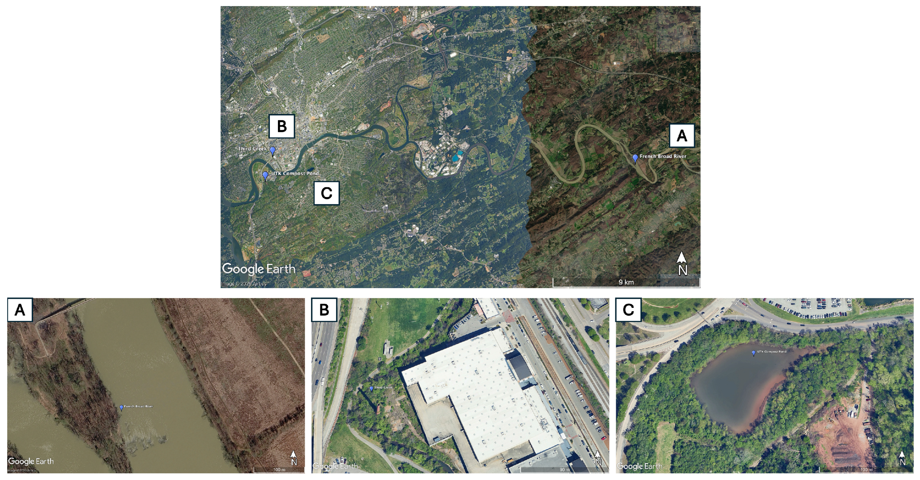
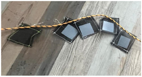
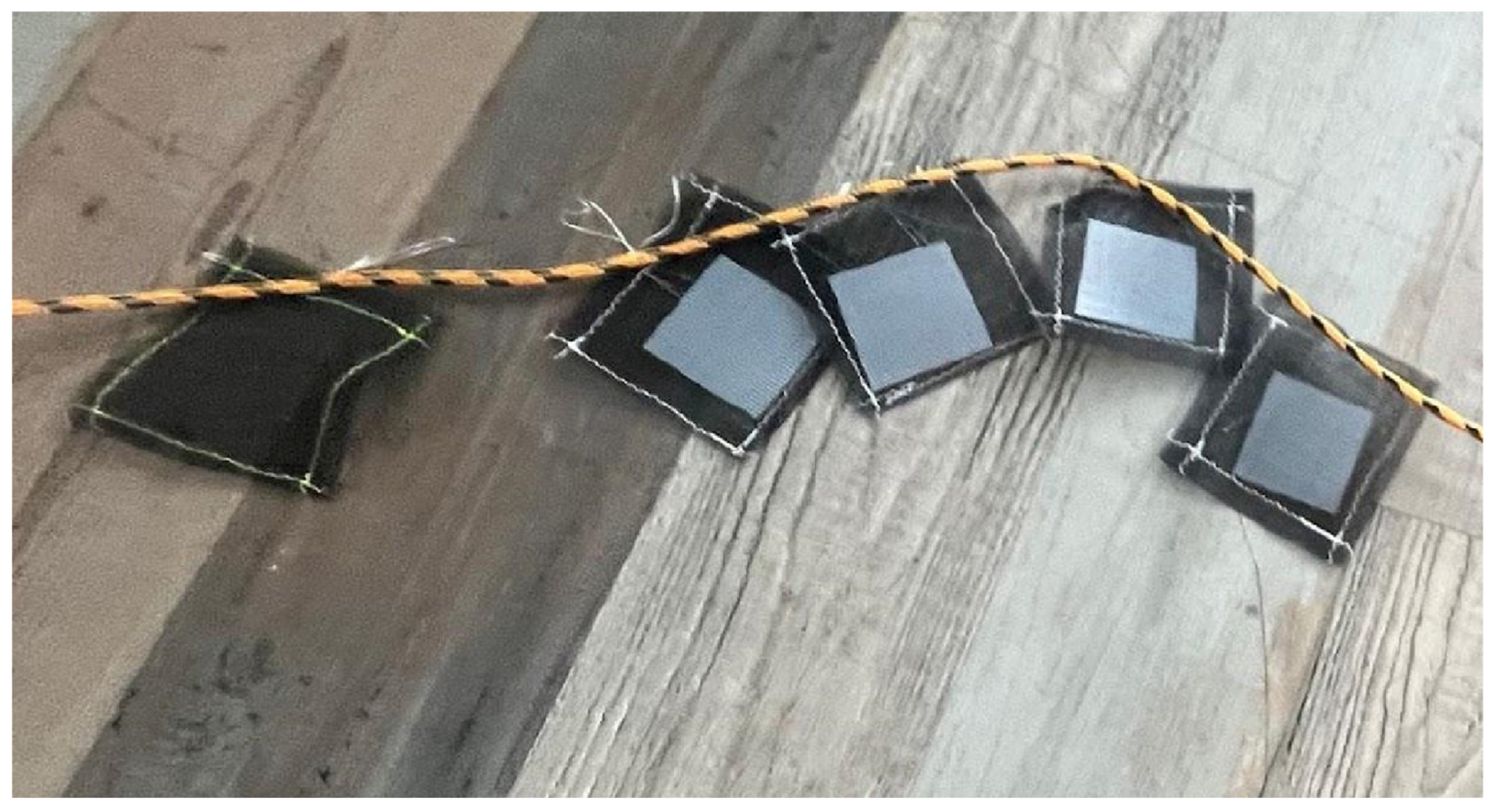
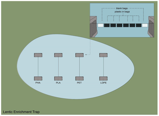
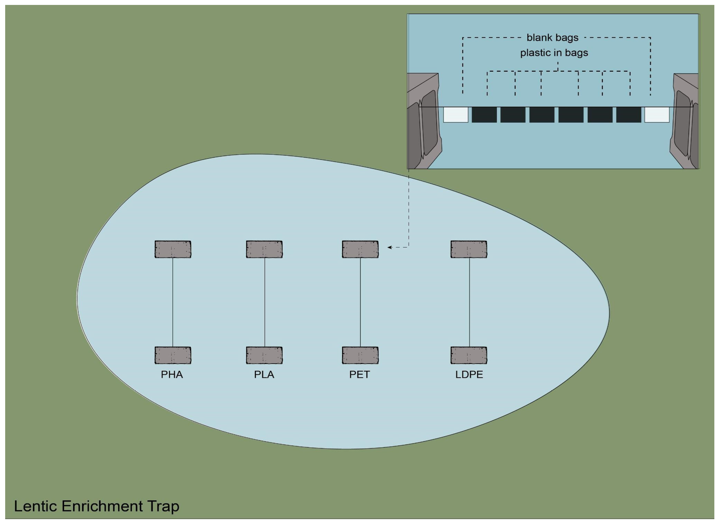
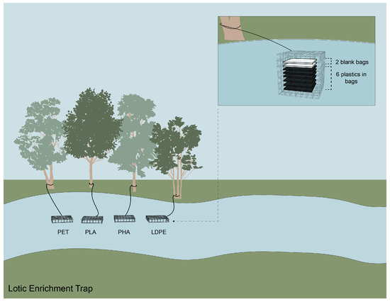
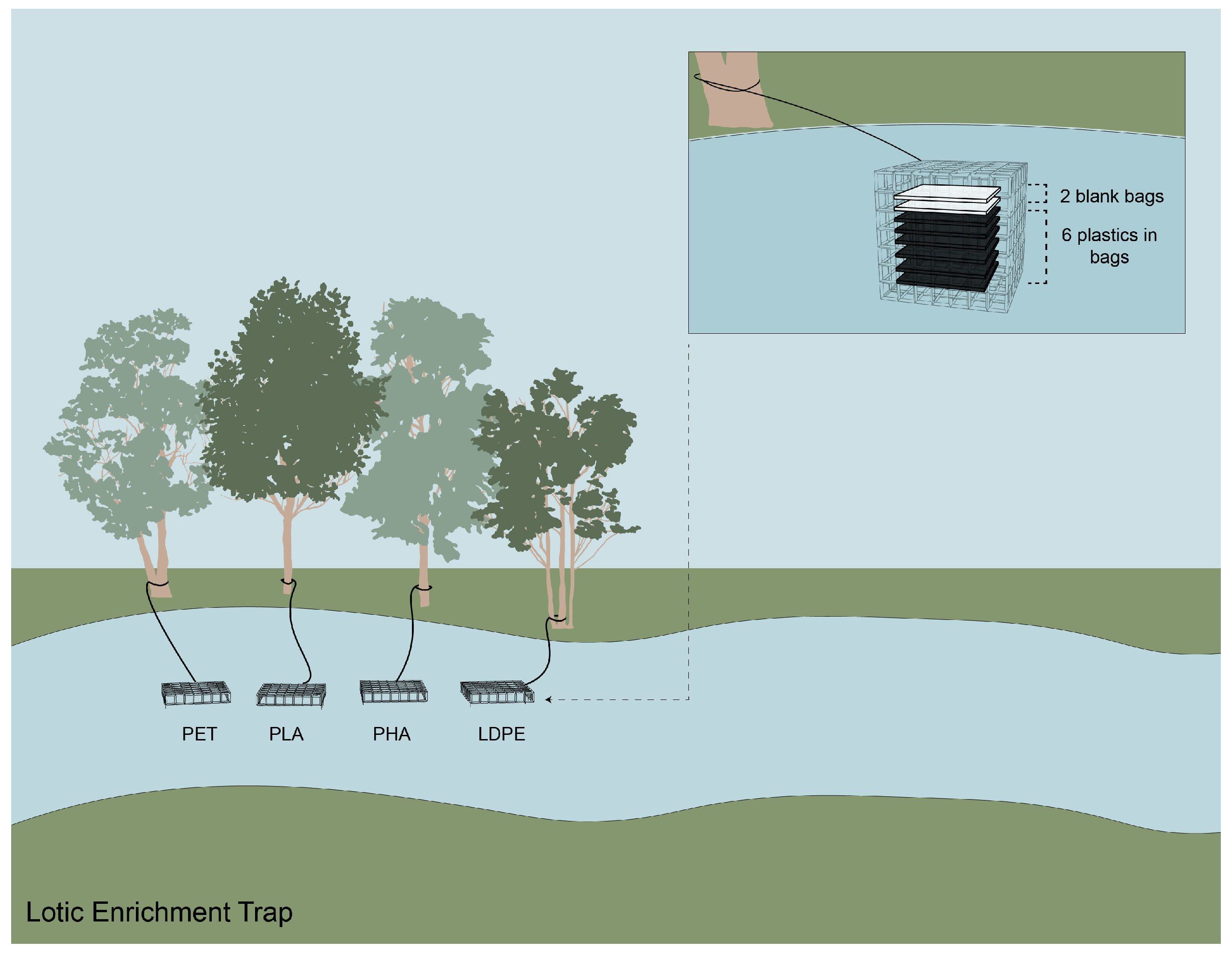
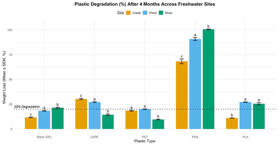
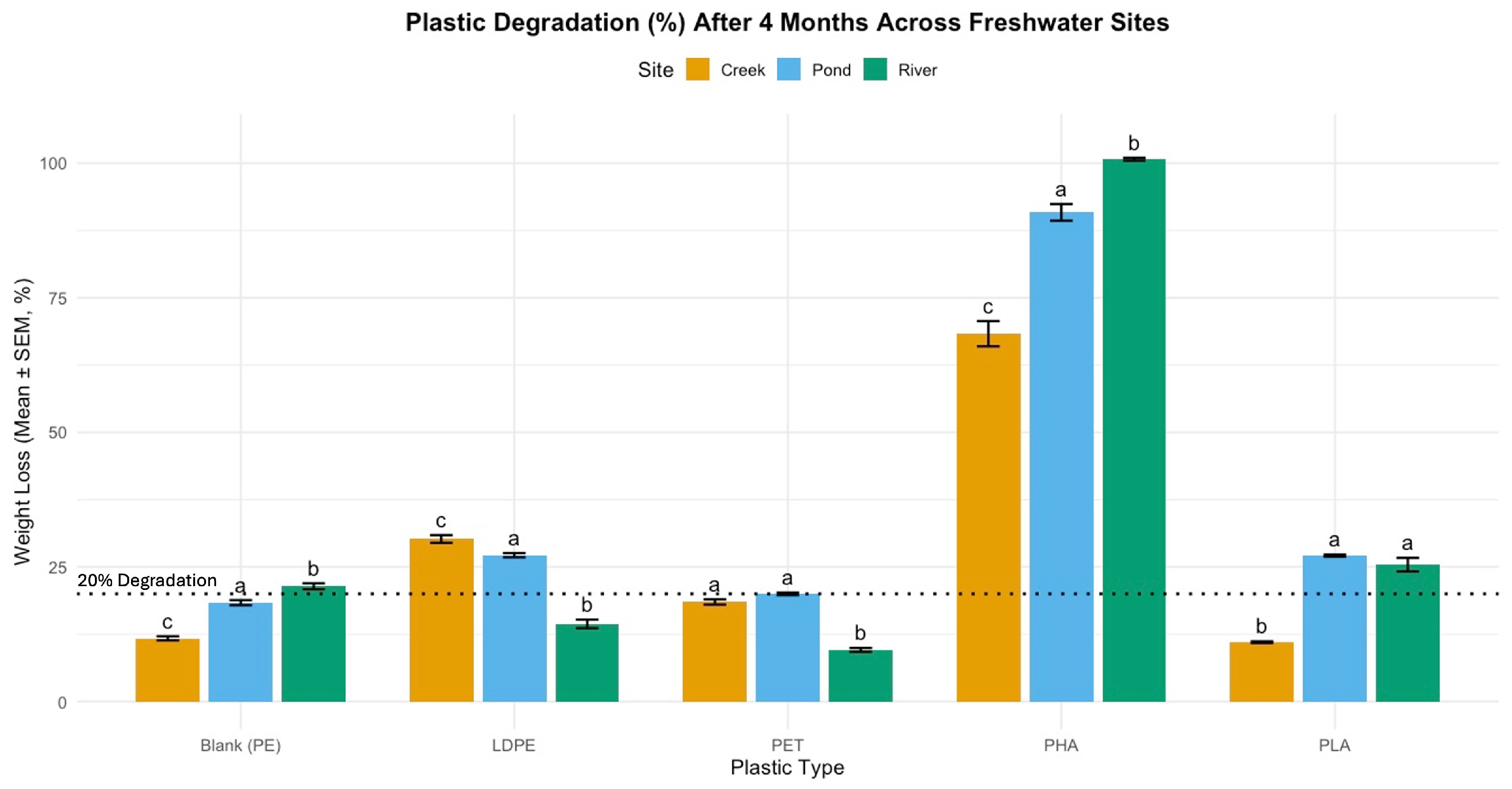
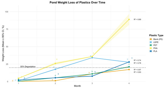
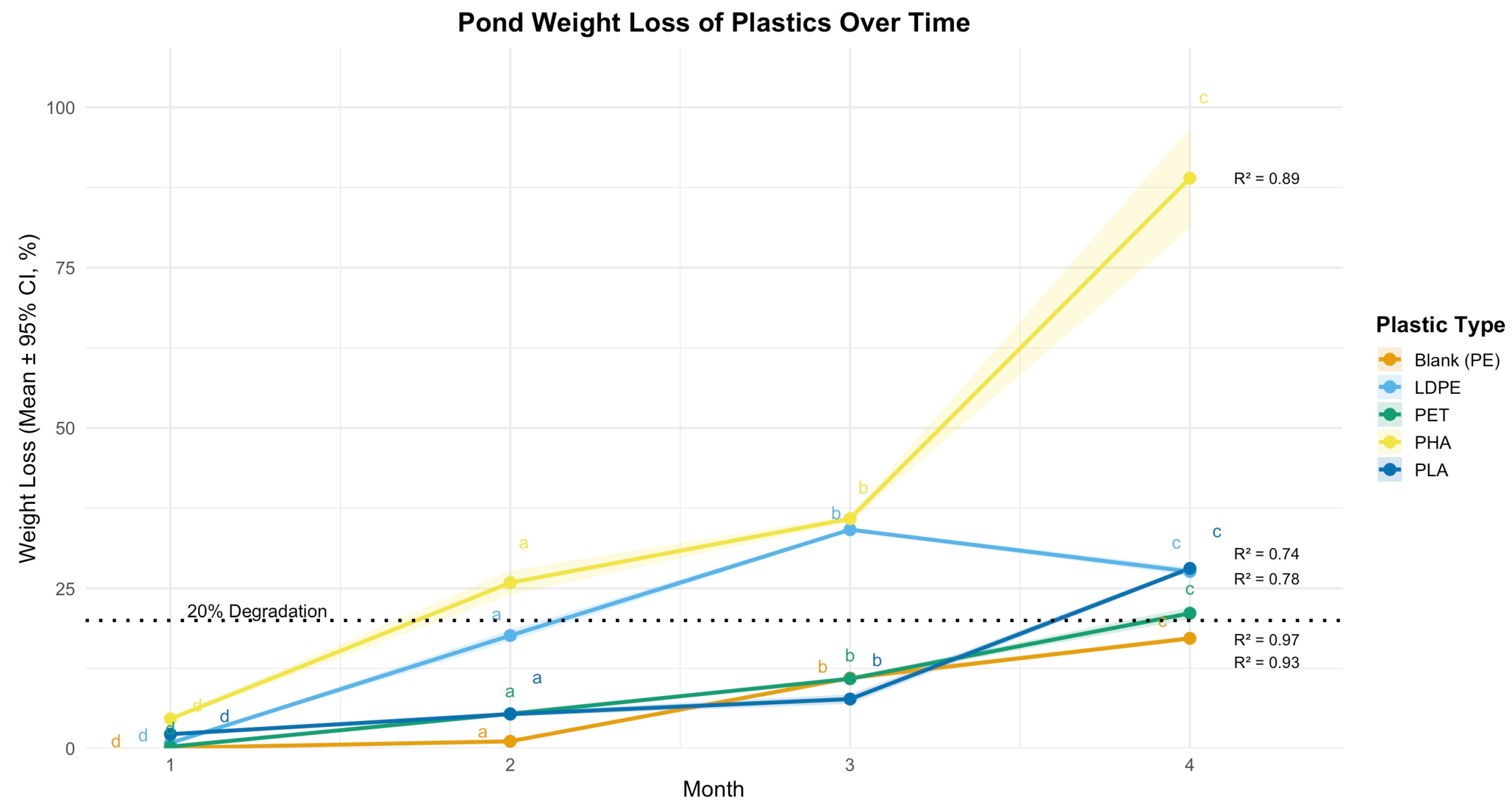
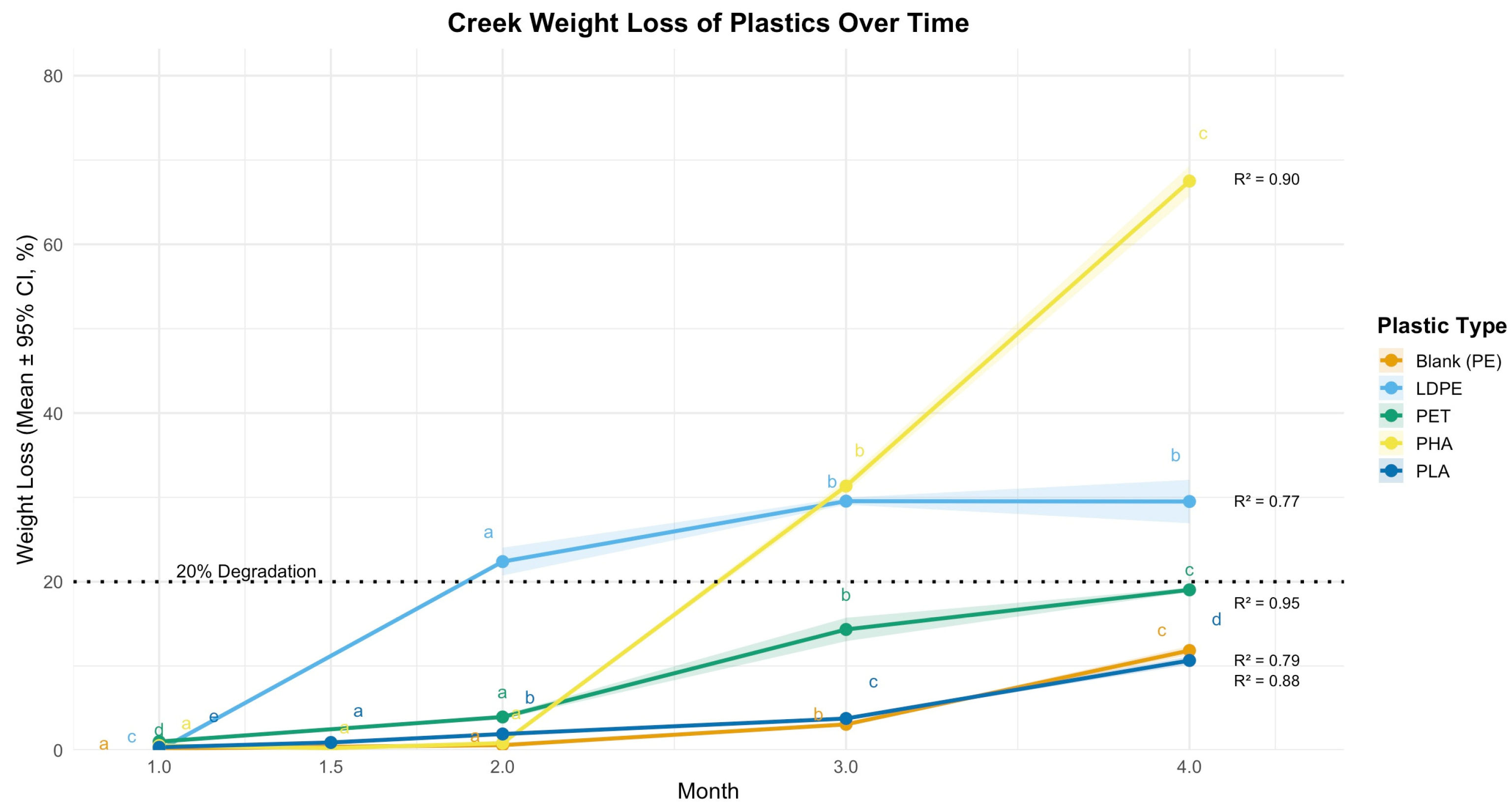
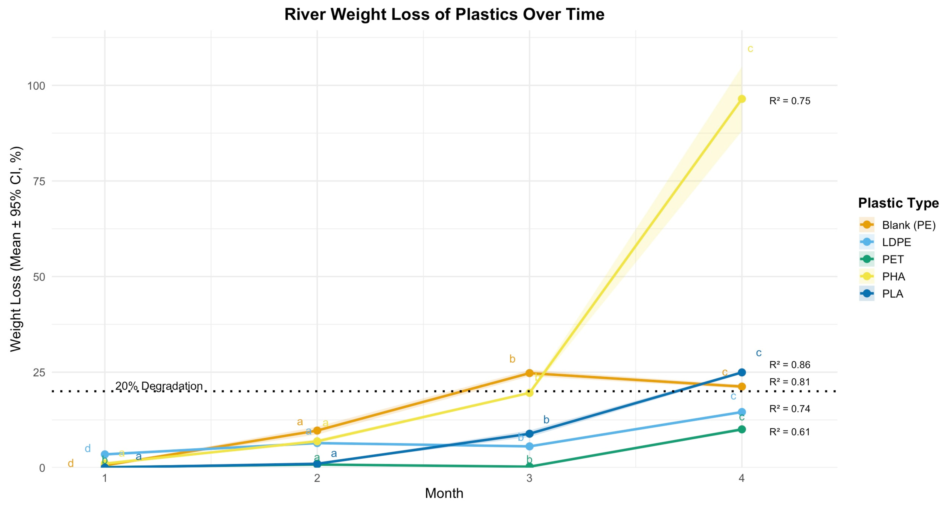
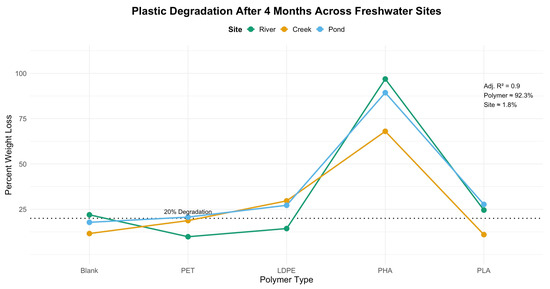
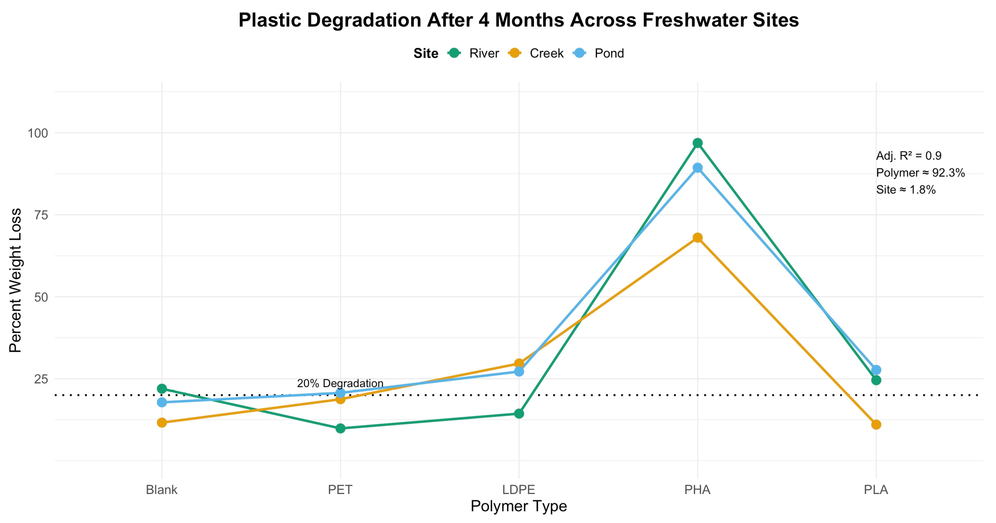
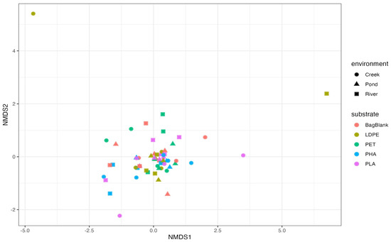
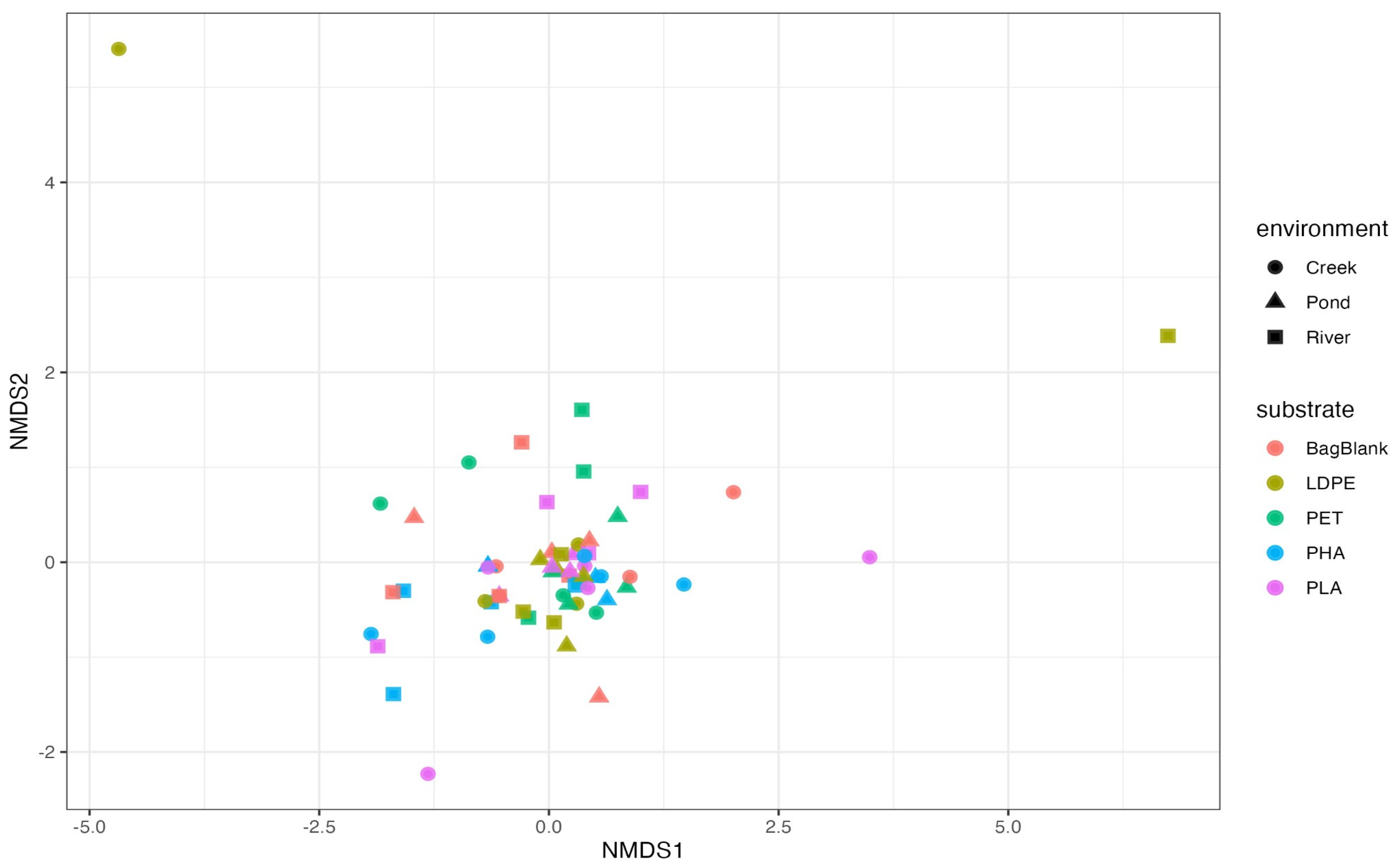
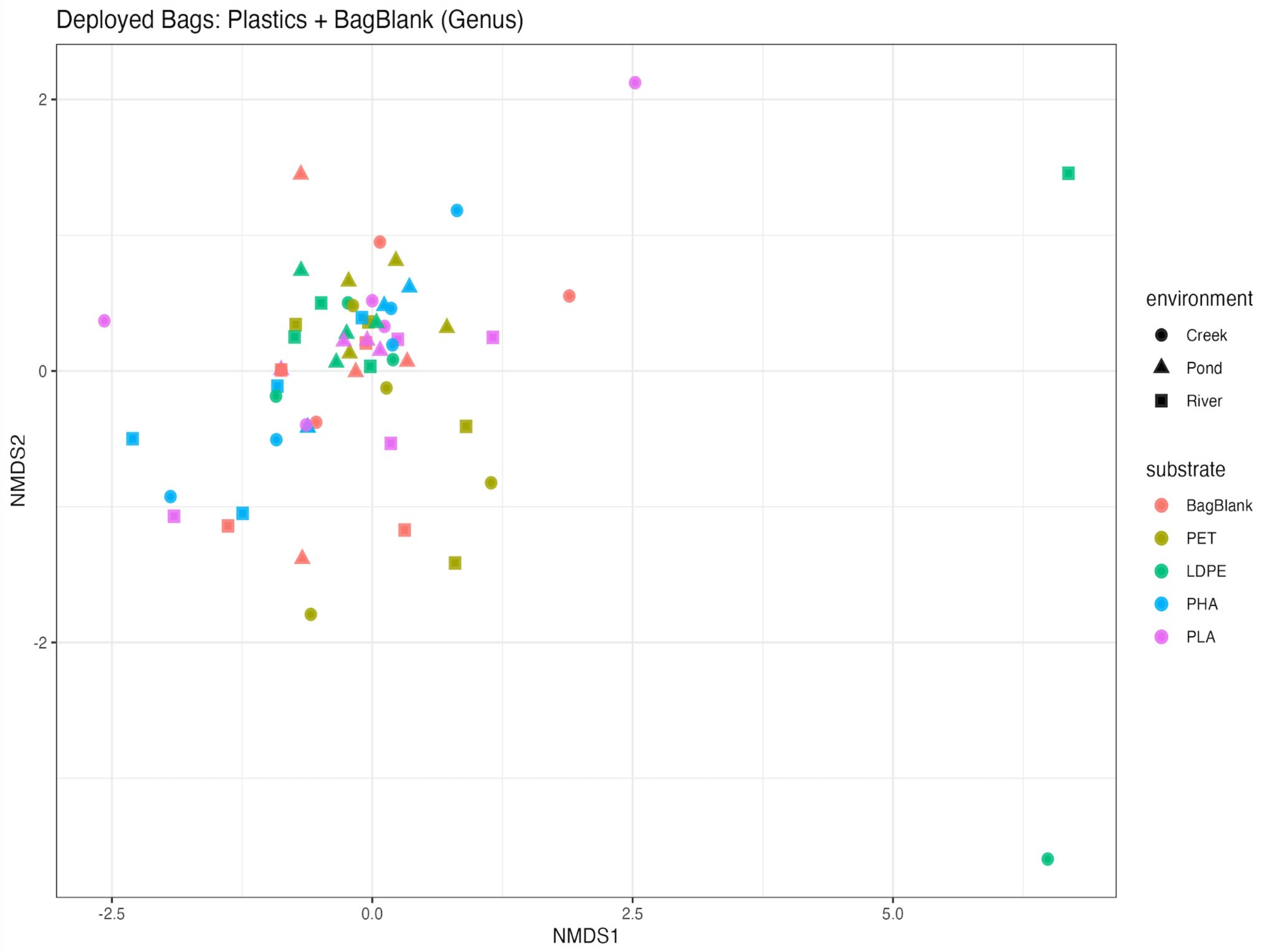
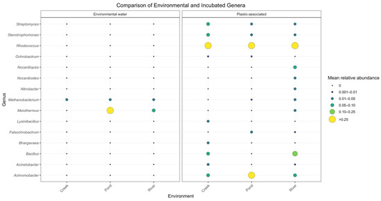
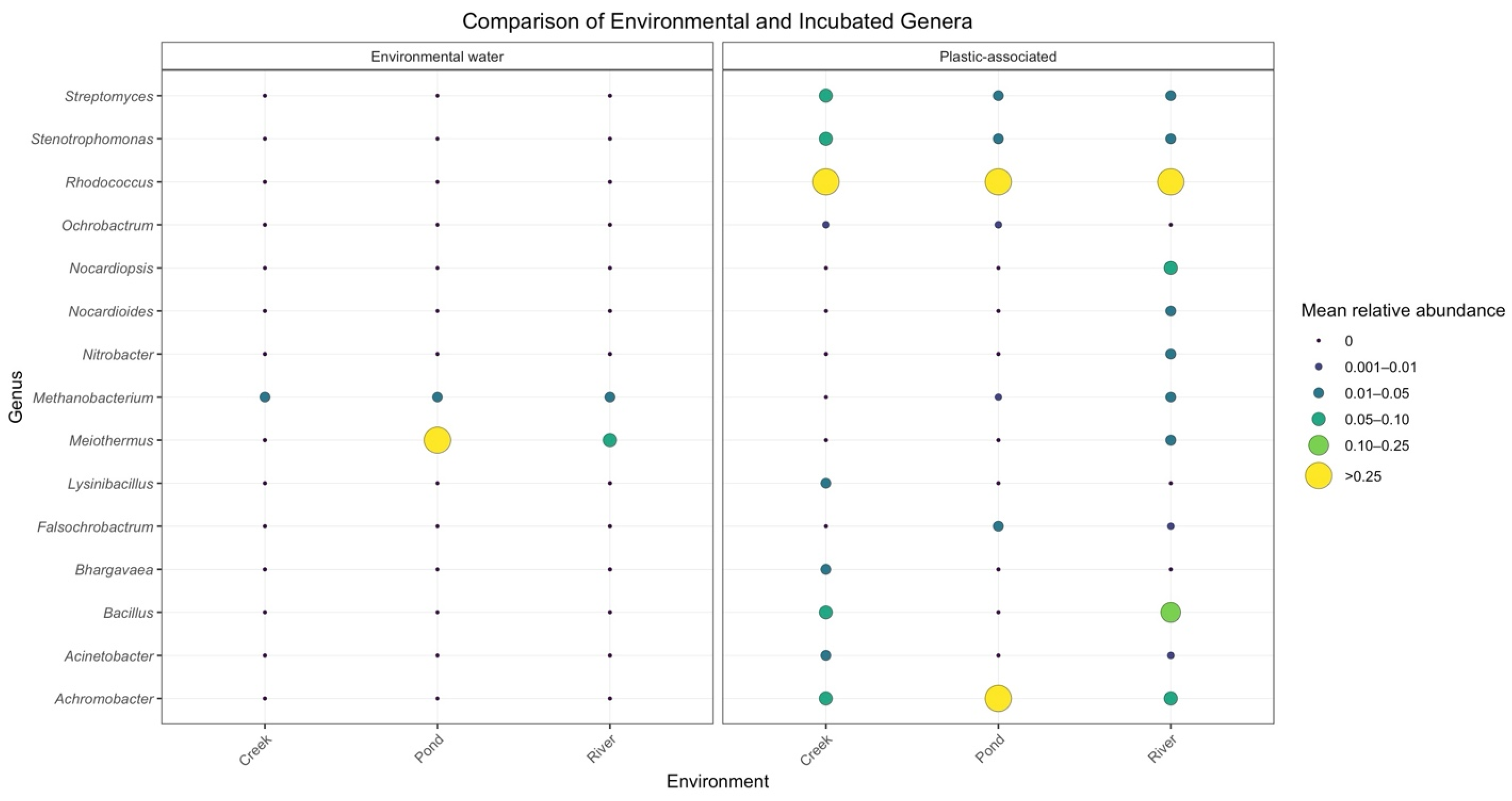
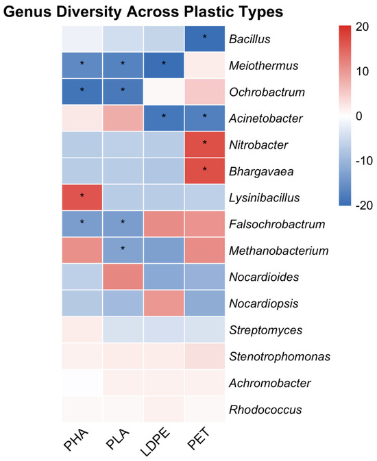
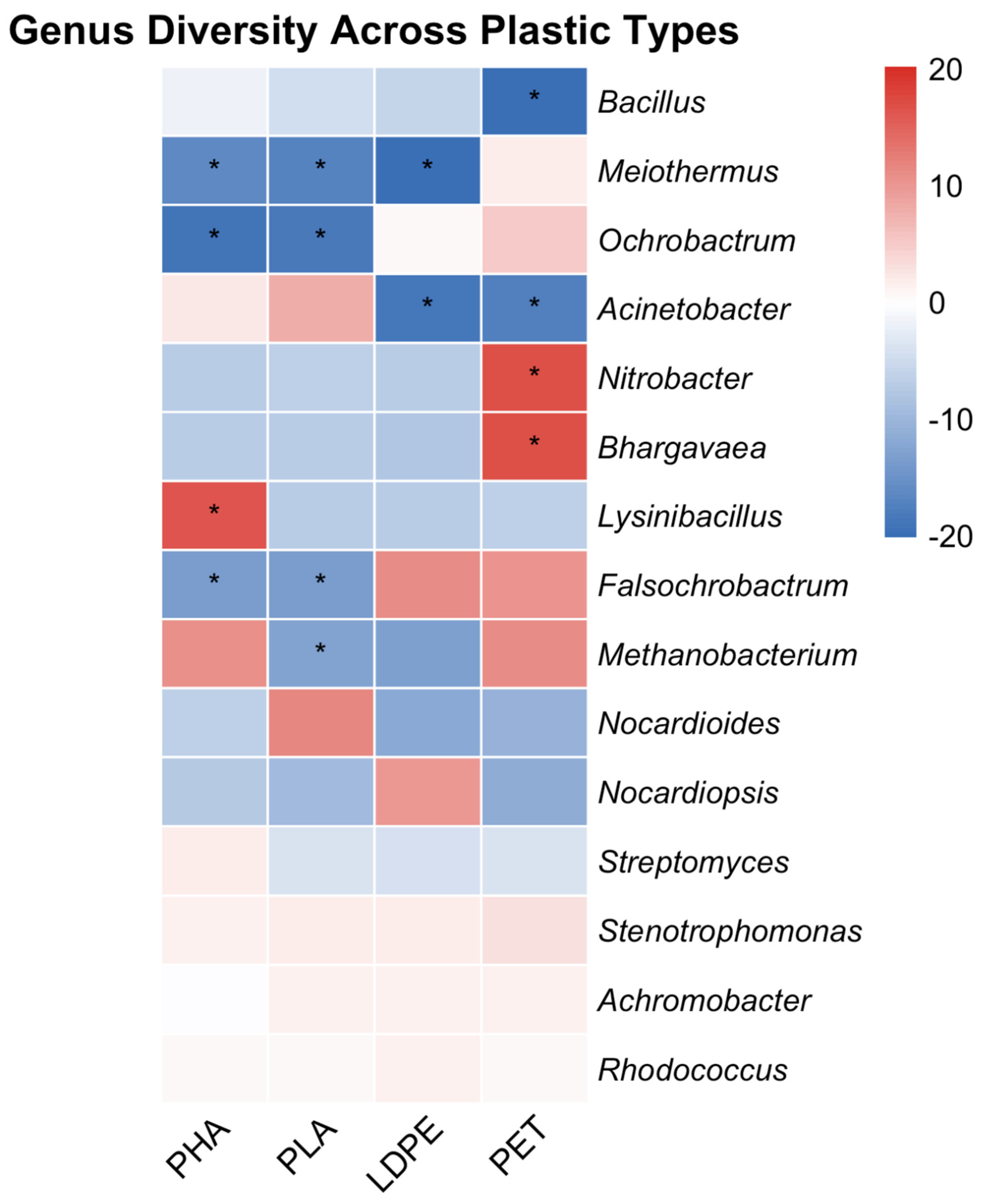
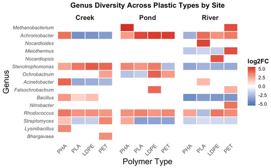
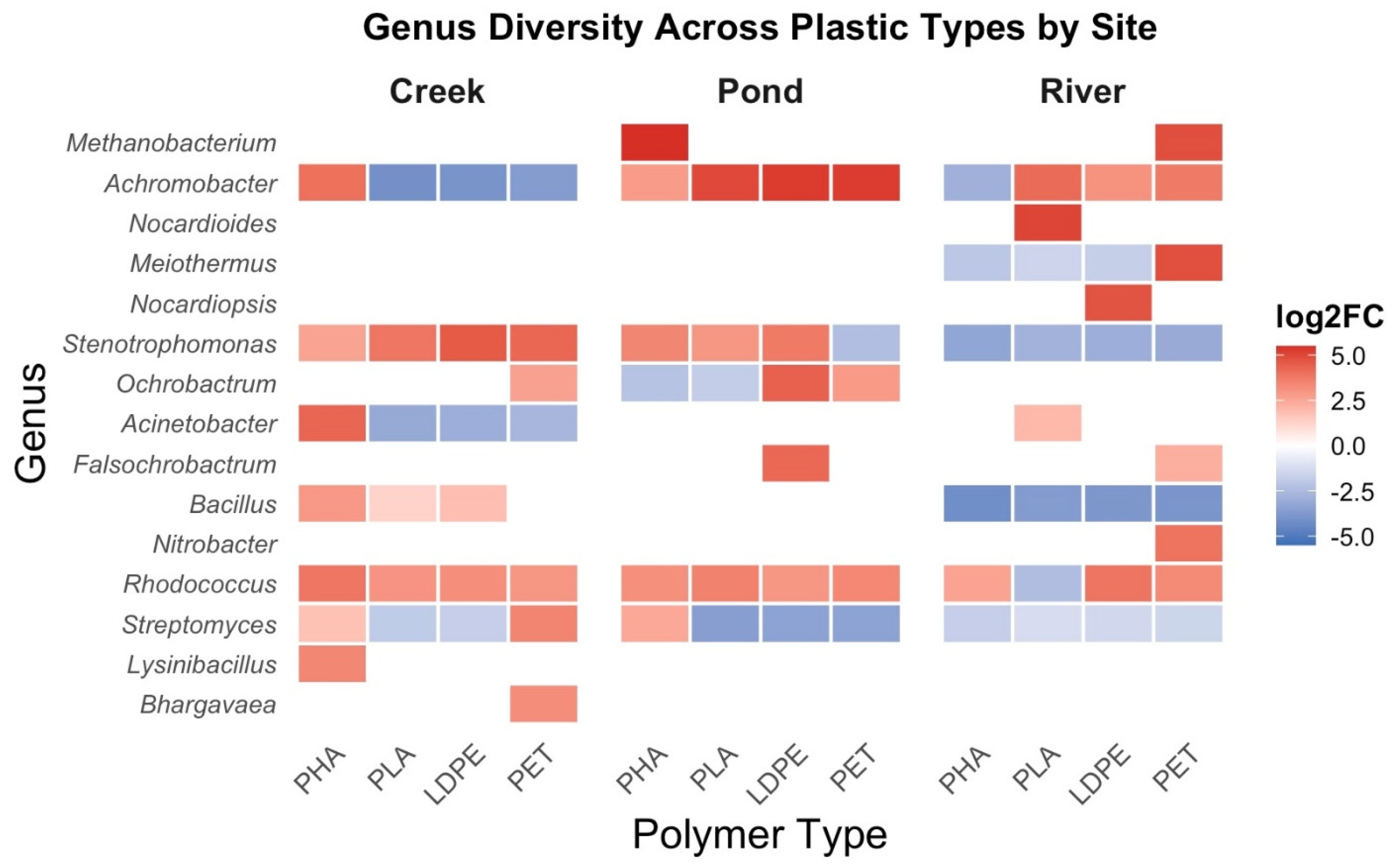
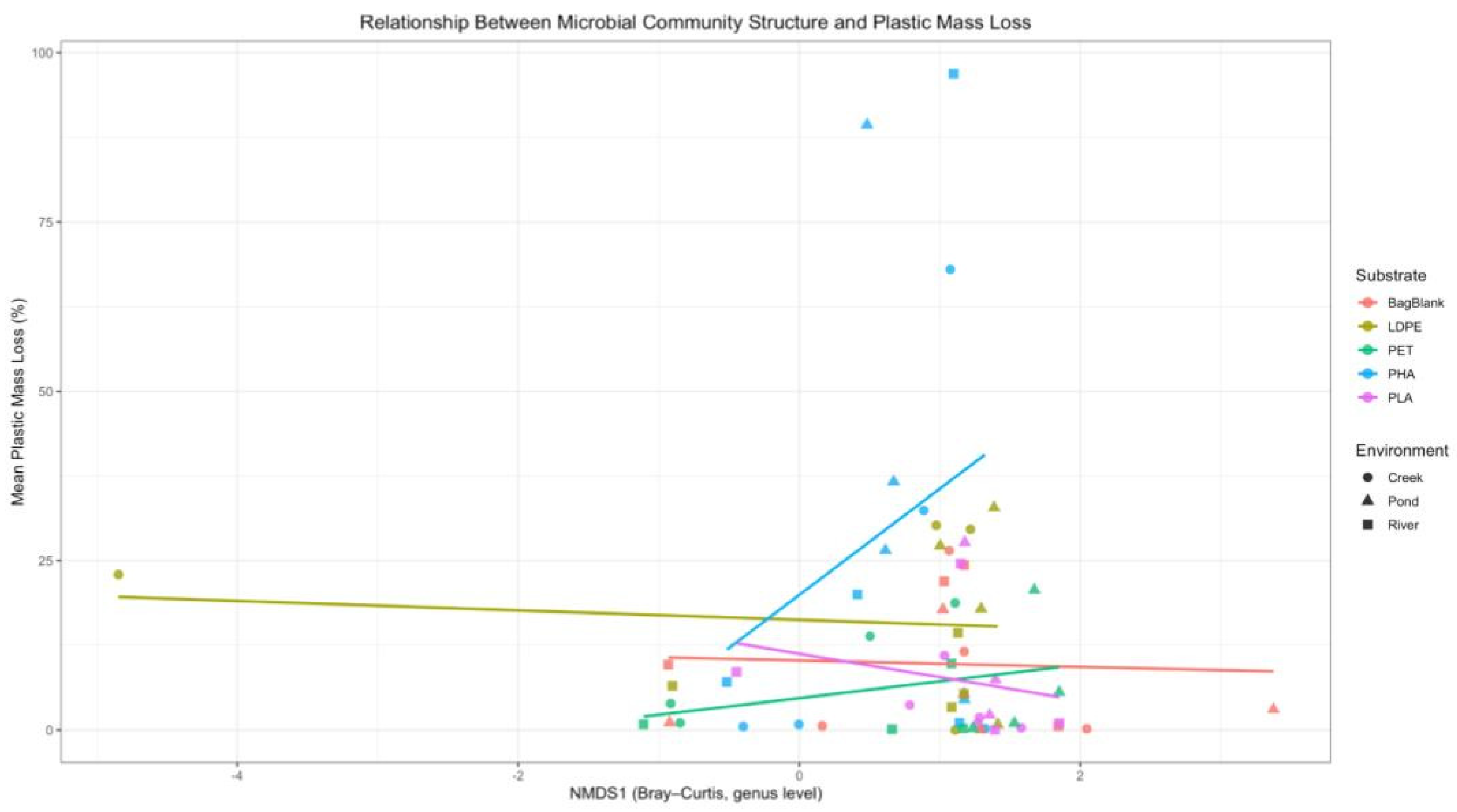

Disclaimer/Publisher’s Note: The statements, opinions and data contained in all publications are solely those of the individual author(s) and contributor(s) and not of MDPI and/or the editor(s). MDPI and/or the editor(s) disclaim responsibility for any injury to people or property resulting from any ideas, methods, instructions or products referred to in the content. |
© 2026 by the authors. Licensee MDPI, Basel, Switzerland. This article is an open access article distributed under the terms and conditions of the Creative Commons Attribution (CC BY) license.