


Abstract
In recent decades, plastic consumption has risen across various industries and everyday products, leading to greater plastic use and the generation of waste, which results in the leaching of micro- and nanoplastics into the environment. This review summarizes recent analytical methods for the detection of nanoplastics (NPs) in several marine matrices, divided into three main stages: extraction, separation, and identification. The literature reviewed indicates that chemical and enzymatic digestion are the most commonly used procedures for the extraction step. For the separation step, flotation, filtration, and centrifugation are the most used techniques. Finally, two groups of techniques may be used for the identification step. The first category consists of methods used for qualitative identification, with spectroscopic methods such as Raman and FTIR being the most frequently used. The second category comprises those used for the quantitative analysis of NPs, where fluorescence-based methods and nanoparticle tracking analysis are increasingly used for this assessment. Despite these advances, significant challenges remain, such as matrix interferences caused by salinity and organic matter, low environmental concentrations of NPs, and the lack of standardized protocols. This review highlights the need for standardized protocols, validated reference materials, and integrated multi-technique approaches to improve the comparability of nanoplastics measurements in marine environments.
1. Introduction
1.1. From Plastics to Nanoplastics
Plastics have gained popularity in recent decades, mainly due to their unique properties (e.g., durability and versatility) and their high production rate [1]. Plastics have become an integral part of society, increasing from 1.5 million metric tons in the mid-twentieth century to 359 million metric tons in 2018. However, the disproportionate use of this material has led to problems related to its use, which pose a threat to the environment and to human health [2]. There are more than 300 types of plastics, although only about 60 are commonly used in industry. Fossil fuels are the main source of the raw materials used to make plastics, accounting for about 99% of them, while only 1% is derived from biomass [3]. These fundamental materials include high-density polyethylene (HDPE), low-density polyethylene (LDPE), polyethylene terephthalate (PET), polypropylene (PP), polystyrene (PS) [4], polyvinyl chloride (PVC), and polyethylene (PE) [5].
Many plastics share the characteristic of being used only once; therefore, they are also known as single-use plastics (SUPs) [6]. The production of SUPs starts with the extraction of oil and natural gas to generate PP granulate, which is thermoformed to synthesize two main types of plastic polymers [7]: thermoplastic and thermoset polymers. On the one hand, thermoplastic polymers can be moulded multiple times and are viable for recycling [3]. On the other hand, thermoset polymers are petrochemical byproducts that are not recyclable [3].
There is a huge variability in SUPs production around the world due to the different industrial development of each country. China, the United States, and Europe are known to have the highest SUP production. China is the largest agricultural producer; therefore, the amount of plastic used for mulching crops has increased fivefold over the last decade [6]. For Europe and the USA, the main use of plastics refers to packaging consumption in many different fields, such as grocery bags, clothing or food packaging, leading to increased plastic use by people, not just by industries [6]. Other countries, such as South Africa, have contributed significantly to plastic pollution in oceans due to their extensive coastlines [6].
According to Liu et al. [8], the main use of plastics is in the shopping bag industry. This leads to an increase in the amount of plastic debris generated that should be correctly managed. To achieve this purpose, there are different end-of-life (EoL) options. EoL options are defined as different pathways for managing plastic waste to either recirculate it into a new product or break it down and degrade it, causing the least environmental pollution possible [9]. Several studies report recycling, incineration, and landfilling as routes to manage plastic waste [7]. First, recycling is recognized as part of the circular economy as it converts waste products into new, valuable products [10]. Second, incineration transforms waste materials into gases and ashes by combusting them [10]. Finally, landfilling is considered an end-of-life option, although it has many drawbacks, such as mismanagement, which can lead to unmanaged waste accumulations [10]. Despite these EoL pathways, a huge amount of plastic ends up in soil, oceans, rivers, and lakes, causing a contamination problem that has increased in recent decades. Chen et al. [6] reported some of these environmental impacts: SUPs contamination in soil leads to decreased water use efficiency, nutrient uptake, and biomass accumulation. Regarding air pollution, research has been limited; however, SUPs can become effective carriers for air pollutants that may be transported long distances by wind [11].
SUPs have become more popular in different applications, increasing the probability of ending up in rivers, lakes or the marine environment, where the SUP contamination has more significant effects [12]. Rivers and lakes have low relative density, making plastic waste buoyant in water [6]; this waste can then be transported into oceans, where SUPs concentration is higher than in any other environment [12], with an average of 2.76 items/m3, reaching up to 4.98 items/m3 in the Atlantic Ocean [13]. This concentration may result in the destruction and degradation of ocean water properties, which also affects aquatic flora and fauna [14]. Studies on plastic ingestion by marine species have shown that micro- and nanoplastics are the most common type of SUPs found in these animals [6]. SUPs can fragment into large pieces, leading to the formation of macroplastics (>25 mm). These macroplastics may further break down into smaller pieces, giving rise to mesoplastics (5–25 mm), which can eventually degrade into microplastics (1 µm–5 mm). Ultimately, microplastics may degrade further into nanoparticles (<1 µm), commonly referred to as nanoplastics (NPs) [15] (Figure 1).
Figure 1.
Fragmentation of single-use plastics (SUPs).
NPs are characterized by heterogeneous morphologies, including spheroids, fragments, flakes or cylinders, which influence their interaction with the environment [16]. NPs are composed of a wide variety of polymers, but the most common are polyethylene (PE), polyvinyl chloride (PVC), polystyrene (PS), polyethylene terephthalate (PET), polyamide (PA), polypropylene (PP), poly(methyl methacrylate (PMMA), and polycarbonate (PC) [17,18]. Due to their small size, nanoplastics (NPs) exhibit a high surface-to-volume ratio, which makes them effective carriers of other pollutants and facilitates their adsorption and absorption by a wide range of aquatic organisms [16]. Inside these organisms, some biomolecules may encapsulate NPs, developing an eco-corona layer [19]. This may affect the physicochemical properties and fate of NPs, increasing the probability of uptake by marine organisms [20]. As particle size decreases, particle movement transitions from being dominated by gravitational forces to being governed by intermolecular forces and Brownian motion, resulting in colloidal behavior [21]. This allows nanoparticles (NPs) to remain in the water column longer than other plastic debris [16]. Colloidal stability may be influenced by two important factors: particle size and particle charge. On the one hand, particle size may increase through the sorption of different species, causing particles to again exhibit motion governed by gravitational forces [21]. Particle charge is pH-dependent and is affected when the pH approaches the point of zero charge or when the ionic strength increases [21]. In both cases, electrostatic repulsion between particles decreases [21] while humic acid increases this repulsion [16]. In addition, NPs can interact with natural and anthropogenic colloids due to their high surface reactivity, inducing heterogeneous aggregation (heteroaggregation) depending on the environmental conditions. The final behavior and fate of these heteroaggregates depends on their form, composition and size, which is difficult to study because of the wide variety of natural colloids [16].
NPs can be classified as primary nanoplastics (PNPs) and secondary nanoplastics (SNPs) according to their origin. On the one hand, PNPs are those intentionally engineered at the nanometre scale for specific functions in different industries such as pharmaceuticals, electronics, cosmetics and personal care products [22]. PS and PE are the most common polymers found in PNPs; PS nanoparticles are used as model particles to study the toxicity, fate and transport of nanoplastics colloids [23]. On the other hand, SNPs originate when larger plastic pieces are broken by different processes such as photooxidation, weathering, abrasion, hydrolysis or biodegradation. This releases NPs into the environment in various forms, such as plastic debris from agriculture, car tires or textile microfibers [23]. These types of NPs are more difficult to detect and potentially more harmful at the cellular level [24]. SNPs have more heterogeneous and irregular shapes and surfaces than PNPs due to weathering processes (exposure to water, oxygen or light) and can be composed of different polymers such as PE, PS, PET and PVC. SNPs can form associations with microorganisms or exudates more easily than PNPs, leading to the formation of eco-corona layers as mentioned before [23]. Recent evidence indicates that secondary nanoplastics (SNPs) are likely to dominate environmental nanoplastic contamination because they are continuously generated through the degradation of larger plastic items. In contrast, primary nanoplastics (PNPs) are released directly from industrial and commercial applications and are generally considered a less abundant environmental source [25]. Supporting this view, a recent field investigation in the North Atlantic Ocean estimated the presence of more than one million tonnes of nanoplastics, highlighting the major contribution of secondary formation processes to marine nanoplastic pollution [26].
As mentioned above, aquatic environments are the main transport pathway for NPs. In this context, NPs may reach these environments following different paths [27]. For example, NPs present in the atmosphere may settle in aquatic and terrestrial environments through rain, snowfall or wind, while plastics from soil may be moved by surface runoff into oceans [28]. When NPs reach oceans, they can travel long distances, becoming ubiquitous waste products whose concentration has increased in recent decades. This distribution is influenced not only by NPs properties and interaction with the marine environment, but also by the proximity to urban centers, waste management, population density and sewage overflow [29]. The reason for this may be poorer waste management or weaker regulations of these areas. Since NPs are ubiquitous, they can reach different organisms including animals or humans, causing health problems related to their toxicity and pollutant properties [30].
1.2. Environmental Relevance and Global Concerns
NPs are emerging pollutants that may cause environmental stress and affect the behavior of marine organisms [31]. Bianco et al. [32] studied the effects of PS-NPs as environmental stressors by investigating their degradation and reactions with radicals commonly found in aquatic environments, such as hydroxyl radicals (-OH). The results suggested that the products derived from NPs degradation (carboxylic acids and toxic compounds such as benzene and phenol) can absorb light and may therefore interfere with the natural photochemical equilibrium in these environments [32]. According to Wu et al. [33], the surface of NPs changes when they react with hydroxyl radicals or are exposed to light, which may affect particle aggregation [33]. All the experiments mentioned above were conducted in aquatic environments, where many organisms may also be affected by the ingestion of these modified particles.
As the degradation of the plastics continues and reaches aquatic environments, NPs could be transported both vertically and horizontally throughout these environments, but their fate depends on their behavior as colloids or aggregation with organic matter [34]. The vertical transport of these plastics is facilitated by different mechanisms, such as aggregation with organic matter or biofouling, finally reaching the ocean sediments, where NPs accumulate [35]. In this context, marine sediments are recognised as a major sink for nanoplastics, thus acting as long-term reservoirs where these NPs may persist and accumulate [36]. Once these NPs are deposited, they aggregate with organic matter and form eco-coronas [37]. In addition to these interactions, the strong Brownian motion promotes settling processes by enhancing the aggregation and precipitation of NPs, forming the accumulation hot spots of NPs in sediments [38]. Despite this, it is important to notice that depending on the density of NPs, their fate may be different. While low-density polymers can float and be transported through the surface of marine environments, other polymers, such as PET or PVC, which possess higher densities, tend to settle in the aquatic sediments [39]. Importantly, sediments not only act as passive sinks of NPs but also as dynamic compartments due to several processes, such as currents or storms, which can resuspend previously deposited NPs, reintroducing them into the water column [40]. Owing to sedimentation, combined with the small size and surface properties of NPs, their bioavailability increases, facilitating their ingestion by aquatic organisms [34].
Many marine animal species, such as mussels, fish, rotifers, and seaweed, can absorb and accumulate NPs, causing adverse effects on them. Due to their size, NPs can enter the blood circulation and cross the cell membrane, potentially causing cellular damage [41]. Marine bacteria are affected by NPs; interactions with positively charged NPs can impair biofilm formation, potentially reducing their survival capacity and increasing oxidative stress [22]. Gonçalves and Bebianno [22] and Zaki and Aris [42] have shown in their respective reviews an increase in the mortality rate of rotifers and reduced reproductive capacity derived from the ingestion of NPs, although these consequences depend on both polymer type and size. NPs can provoke health problems in the phylum Echinodermata, acting as environmental stressors during embryonic stages, causing skeletal malformations and undeveloped embryos [22]. NPs accumulation in echinoderms also affects phagocytes, inducing lysosomal damage and apoptosis [42]. Crustaceans are another group of animals affected by NPs ingestion: NPs can leach chemical additives that affect these animals [42] and can diminish the organisms’ swimming speed [22] and increase the production of reactive oxygen species (ROS), causing oxidative damage and inhibition of their antioxidant defence system [43]. Another problem caused by NPs ingestion and accumulation is the reduction of fecundity and increased embryo malformations [22]. In this regard, Timilsina et al. [43] have reported that NPs may be transferred from parents to their offspring, affecting embryonic development and growth. Finally, NPs accumulation in the alimentary tract of crustaceans is recognized as a cause of mortality of these species [44]. Molluscs, such as mussels, oysters, or clams, are also affected by nanoplastic ingestion through the trophic chain. Effects of NPs on molluscs’ health have been studied recently, and the main problems include increased oxidative stress [45]. Although these effects depend on dose levels, NPs ingestion may lead to long-term starvation, affecting the neurological and immunological functions of molluscs [42]. The immune system of molluscs is compromised by reduced lysosomal membrane stability. The reproductive system of molluscs is affected by NPs ingestion, damaging the gametes and jeopardizing embryonic development [22]. Gong et al. [41] have reported that NPs can enter the bloodstream of fish and be transported to different tissues, causing important problems to the organism, such as increasing oxidative stress and neurotoxicity [42], decreasing locomotion (motor dysfunction), alteration of the lipid metabolism, and, therefore, an energy imbalance [41]. A lower cell viability was shown in the study of Gonçalves and Bebianno [22]: NPs cause changes in molecular signalling pathways and affect mRNA transcription, inducing a higher rate of mutations.
Although NPs may cause damage at different trophic levels, these effects depend on the dose level of NPs inside the organisms. Studies were conducted to assess this dose, and the results showed that for organisms, such as crustaceans, rotifers, bivalves and fish, the physiological damage occurred when the dose level exceeded tens of µg/L [46]. Yang et al. [46] calculated predicted no-effect concentrations of 72 µg/L for marine systems, suggesting that exposures above this threshold may provoke significant damage in these organisms.
Marine flora is also affected by nanoplastic contamination. Ge et al. [47] reported that an increase in the concentration of NPs causes higher negative effects on marine algae. As mentioned before, oxidative stress is a common form of damage caused by NP contamination in animals, and this is also a problem in marine flora. Algal growth is inhibited by this contamination [48]. Moreover, NPs may increase the toxic effects of other pollutants, such as titanium dioxide, which enhances lipid peroxidation and modifies the activity of antioxidant enzymes [49]. Photosynthesis and cell viability are also compromised by NPs. On the one hand, pigment and lipid composition are damaged, leading to cell wall and pigmentation issues, which may inactivate photosystems and thus inhibit photosynthesis [22]. On the other hand, cell viability is affected by NPs through adhesion and internalization, which leads to a massive accumulation of these nanoplastics inside algae [50], reducing their mitochondrial homeostasis [22] and growth efficiency [50]. As for marine fauna, the harmful effects of NPs on marine flora depend on the dose level. For example, Slaveykova et al. [51] reported that EC50 (effective concentration 50%) values of 83–132 mg/L of PMMA-NPs could induce significant stress on photosynthetic efficiency.
All the above-mentioned harmful effects lead to a need for global measures to manage plastic waste to diminish contamination and, therefore, MPs and NPs pollution [52]. Several objectives are common across the four continents: reduce plastic use, improve different ways of recycling plastic waste, along with increasing different control procedures to manage this waste and limit plastic use in industrial products. Some of these measures taken on each continent are explained below.
Beginning with Europe, the European Commission aims to properly differentiate harmful MPs and NPs from all those materials that are not considered a risk for the environment or human health. Those harmful polymers should be analyzed by different processes to assess how long they could be in the environment without causing damage [53]. To achieve this purpose, the European Union (EU) and the Socioeconomic Analysis Committee (SEAC) have proposed different maximum time periods depending on the use of plastics. An example of this is fragrance encapsulation, where the disposal time range is between 5 and 8 years. Additionally, the European Commission also aims to improve and develop more scientific research to evaluate the toxic capacity of NPs [53].
Next, in the United States, there are different agencies that enforce laws to deal with NP pollution. For example, the US Environmental Protection Agency (USEPA) aims to fund projects whose purpose is to diminish the amount of trash that enters the environment and to promote its removal. Two of these projects funded by USEPA are the Safe Drinking Water Act (SDWA), which restricts the level of pollutants permissible in water from anthropogenic and natural sources, and the Resource Conservation and Recovery Act (RCRA), whose goals are to develop a proper way of managing plastic waste and disposal [54]. Another example is the Food and Drug Administration (FDA). This agency deals with plastic use in industries like food, drugs, medical devices, and cosmetics. The FDA estimates an acceptable concentration of contaminants in food packaging of less than 1.5 µg/person/day [54].
Following this, Asian countries have also developed different policies to manage plastic waste. For example, India has implemented the Plastic Waste Management Amendment Rules that make producers, brand owners, and importers responsible for the use and management of plastics. In addition, these rules have banned 19 categories of SUPs and established criteria for degradable plastics in order to avoid MP and NP pollution [55]. Finally, African countries have implemented different methods, although most of them are focused on the later stages of the plastic product life cycle and establishing taxes for plastic products. Examples of these are: Cape Verde, which has restricted the production of MPs and NPs and Southern African countries, which have banned the use of plastic bags. Despite these measures, NPs pollution in Africa remains a major challenge because there is no rule implemented for the manufacturing or distribution of plastics [56].
Despite these laws and measures to deal with plastic waste and, therefore, NPs contamination, numerous studies have focused on NPs effects on individual organisms, but some evidence indicates that NPs contamination can be transferred through the trophic chain, inducing bioaccumulation in superior organisms and health problems as mentioned above. Through these food webs, NPs may reach humans, jeopardizing human health [57].
1.3. Potential Impact on Human Health
NPs can enter the environment and the food chain, eventually reaching humans via consumption of contaminated food (meat, packaged poultry, fish, molluscs) [58]. NPs can follow several exposure routes to enter the human body. First, oral intake is the most common and represents a continuous exposure because NPs are found in many food and drinking products, such as food packaging or plastic water bottles [58]. Second, humans can also be exposed to NPs through the food chain: NPs can be absorbed by lower organisms that will be ingested by higher animals in the trophic chain, eventually reaching humans and causing health problems by bioaccumulation [59]. Finally, inhalation is another route for NPs. NPs have been found to carry chemical pollutants in the atmosphere that may enter the human body through the respiratory system. Besides these routes, it was found that NPs could enter humans through dermal exposure, and when their size is smaller than 40 nm, they are able to pass through the epidermal barrier [59].
On the one hand, once inside the human body, NPs can produce different effects in different organs and systems owing to their toxicity [60]. Yee et al. [61] reported that NPs can be found in the gastrointestinal (GI) system through ingestion. NPs can permeate the gut epithelium; therefore, they may be modified by the interaction with other molecules such as lipids, ions, water, proteins and carbohydrates. For example, they can be encompassed by proteins, developing an eco-corona layer that facilitates NPs translocation through the body [61]. NPs can also affect other organs, such as the lungs, by air inhalation. In this system, they can carry pathogens and parasite vectors. Besides, the alveolar barrier is thin enough to allow NPs to permeate, allowing them to reach the blood system and, thus, the entire human body [61].
On the other hand, at a cellular level, Molina and Benedé [62] reported that the ingestion of NPs through food is a particular concern for humans because they can damage different cell processes, such as apoptosis through the mitochondrial pathway. In the epithelial intestinal cells, PS-NPs can interact and aggregate with mucin, inducing apoptosis of these cells and increasing the production of reactive oxygen species (ROS), which causes macrophage stress, eventually promoting apoptosis [62]. Although experimental studies on human cells are scarce, Xu et al. [63] have determined that ROS increases as PS-NPs increase in a concentration of 0.1–10 µg/mL. Finally, recent studies have shown that NPs’ toxicity may also affect the metabolic homeostasis by influencing signalling systems and vesicle transport and distribution. NPs may be internalized and alter the epithelium of different tissues, promoting allergic diseases [61]. For example, several in vitro studies have found that PS-NP particles in lung cells reduced phagocytosis by alveolar macrophages. Another example is PE-NPs, which increase the production of some pro-inflammatory factors such as TNFα or IL-1, and pro-osteoclastic factors, inducing a higher rate of bone resorption [61]. Research has been done on different human cells to assess the cytotoxic effect of NPs in human cells. For example, Ma et al. [64] evaluated the effects of exposure to NPs on cardiomyocyte function; the results showed that a reduction in viability and contractile dysfunction was found in those cells exposed to PS-NPs at a dose of 0.1 µg/L [64]. Another example was carried out by Huang et al. [65] in nasal epithelial cells; in these cells, the dose of NPs necessary to produce damage was much higher than in cardiomyocytes, reaching 10–1250 µg/mL. In summary, NPs may harm different cells and organs inside the human body, but these effects will depend on the dose level and location.
Nanoplastics (NPs) are an emerging environmental concern with potential impacts on human and animal health. Therefore, it is necessary to develop effective methods for their detection to mitigate their potential effects. However, there are several challenges associated with detecting NPs, which make it difficult to obtain reliable results. One of the main issues is that NPs are present at very low concentrations and are mixed with organic matter, mineral particles, and other colloids such as salts [66]. For this reason, multiple steps are required in the detection process to remove interferences from coexisting compounds and to concentrate the nanoplastics, thereby improving detection accuracy. Given these drawbacks, this review aims to evaluate the existing approaches for the detection of nanoplastics.
1.4. Objectives of This Review
Given the concerns regarding the use and environmental and human health impacts of NPs, it is essential to assess the prevalence of nanoplastics in the marine environment and associated fauna. Hence, the aim of this review is to compile the methodologies published in the literature on different methods for detecting NPs in marine environments and organisms. These methods will be detailed: extraction, separation and identification.
To this end, a search was carried out for several studies on two platforms: SCOPUS (https://www.scopus.com/ (accessed on 15 April 2024)) and Google Scholar (https://scholar.google.es/ (accessed on 15 April 2024)) from 2020 to the present, to find the latest advances in this matter, as well as a comparison between these methods. SCOPUS focuses on high-quality literature, with over 25,000 peer-reviewed journals indexed. Thus, it ensures academic standards. Google Scholar, on the other hand, has a much broader reach, as it does not apply prior quality filters, so it includes documents of varying quality, such as grey literature. In this way, the present review covers a wider body of scientific literature. There are several reasons for this:
- To reduce the publication bias. Studies with negative or non-significant results are not published in traditional journals.
- To ensure completeness of available evidence. Grey literature represents between 50–80% of the total scientific literature, although this depends on the specific study area.
- Access to the newest research. Conference papers or technical reports usually contain more up-to-date information since they do not need to undergo editorial processes.
- Diversity of methodological perspectives. Grey literature includes case studies, program evaluations and other approaches that may not match scientific criteria of the traditional academic journals.
- Evidence of practical implementation. These search engines include reports from government, international organizations, and the private sector that describe the real-world applications of the theoretical knowledge.
- Methodological transparency. Including grey literature strengthens the credibility of the findings and allows other researchers to evaluate the comprehensiveness of the work.
2. Detection of NPs in Marine Samples
In this section, four fundamental steps for the detection of nanoplastics (NPs) in marine samples will be described (Figure 2). First, the sampling methods will be presented (Section 2.1) to ensure an adequate and representative collection from aquatic environments and biota. Then, methods for the extraction of NPs (Section 2.2) from the collected material, including chemical and enzymatic approaches, will be explained, followed by an overview of the main separation procedures to isolate these particles (Section 2.3). Finally, identification techniques for the qualitative and quantitative characterization of NPs (Section 2.4) will be detailed.
Figure 2.
General outline of the NP analysis process.
2.1. Sampling Methods
Plastics can be found in different matrices; therefore, it is necessary to collect the samples where the detection studies will be conducted. It is important to decide the sampling method for this kind of analysis [67]. There are three different types of samples to be analyzed in marine environments: aquatic samples, sediment samples and biota samples (Figure 3).
Figure 3.
Overview of sampling methods for water, sediments and biota samples in marine environments.
2.1.1. Water Samples
Water samples may be collected by several methods depending on whether the target is surface water or the water column [68,69]. In addition to their size, other variables and difficulties may hinder the collection of NPs. One of these difficulties is the physicochemical characteristics of NPs, because they vary more at the nanoscale than at the microscale in terms of strength, reactivity, conductivity and surface area. Another difficulty is the ability of NPs to form aggregates (heteroaggregates), which modifies their mobility and transport. Finally, the unintentional contamination of samples from the researchers’ clothing, air or equipment devices used to conduct the analysis [70] also represents a problem in NP analysis.
For MP analysis, there are some sampling methods that have been standardized for both surface water and water column: nets and sieves have been used for sampling due to their ease of use. The main nets used for this purpose are Floating Neuston and trawling Manta nets [69,71,72]. These nets have also been considered for standardization in NP analysis, but as the mesh size decreases, they are easily obstructed by particulate organic matter [69]. In addition, both of them are typically made of nylon (polyamide), polyester and in some cases they may contain PE or PP, which may cause overestimation in NPs analysis [73].
Other methods used in this step of the analysis are bulk techniques, which allow the collection of the entire volume of the sample, making these methods suitable for MP and NP analysis [72]. In these techniques, there are several devices used to collect water: buckets or water bottle samplers, which are commonly used for the analysis of NPs despite the small volume collected. Examples of instruments used for sampling are Niskin bottles, Rosette devices, and an Integrating Water Sampler (IWS). Niskin bottles allow the acquisition of water from different depths (water column), but, as they are usually made of plastic, they may be a potential source of contamination for the analysis. Rosette devices are another example of instruments used for sampling. They are made up of 6–24 Niskin bottles on a rosette frame and are usually used for taking samples of different sequential depths. Finally, the IWS is a sampling instrument that can be controlled according to a specific pre-selected depth, but it can also be a potential source of contamination for the analysis as it is made of acrylic glass (i.e., Plexiglass) [69].
It is important to highlight that other water samples, such as sea ice samples, can be collected by some of the devices mentioned above, like Niskin bottles [26], but can also be sampled by onboard pumps found in research vessels [74], although this method collects more and smaller particles than nanoplastics, possibly leading to interferences during the analysis [75].
Currently, no standardized sampling method has been implemented. Despite this, there have been several developments in this direction. Water surface microlayer (SML) sampling consists of several glass plates that are immersed in water and later vertically drawn out. There is an important disadvantage regarding this method, since the small volume of the area causes poor representativeness [69].
2.1.2. Sediment Samples
Sediment samples may be gathered by different methods, which use different devices that are mainly composed of stainless steel. The first method of sampling marine sediments is by using grab samplers, which are widely used because of their simplicity and suitability for different sediments [76]. These samplers are formed of two hinged buckets that will close around the sample, although they may disturb the structure of the sample during the collection [35]. To avoid this disturbance, box corers are an alternative method. These devices can also be used to sample sediments from marine environments because this method collects a block of the sediment, preserving its layering structure, which is important to study the distribution of NPs along different depth profiles of the sediments [35,77].
Another method to conduct this step of the process is by using core samplers. This technique can extract the internal piece of the sample and maintain its layers [35,78] by using freeze corers, gravity corers, or piston corers [35]. Lastly, manual tools can also be used, but they present higher contamination risks, and the sampling process is less consistent across different sampling periods [35].
2.1.3. Biota Samples
For the analysis of NPs found in biota samples, the sampling method depends on the animal type. On the one hand, invertebrates are gathered using a net whose characteristics depend on the animal’s living environment and behavior. In case of benthic macroinvertebrates, three net sampling methods can be used: the hand net method (which consists of collecting samples with a nylon net), the D-frame dip net method (which uses a stainless-steel corer that can penetrate different types of substrates) and litter bags (which are made of plastic, meaning that they could cause interferences in NPs analysis) [79]. Other invertebrates, such as nektonic and planktonic invertebrates, are usually collected by Manta nets (mesh size > 300 µm) and Bongo nets (mesh size > 500 µm) [80,81].
On the other hand, vertebrates are usually fished in different ways [68,72]. Crustaceans and bivalves are usually gathered from the field by hand [80]. Fish species can be collected using different devices depending on the habitat (surface, midwater and benthic) [80]: a beach seine is used for both flowing and standing water, but it is limited by depth; alternatively, trawls can be used for larger water volumes, although fish can be injured [82]. Finally, many of these marine species can be found in markets where they can also be captured, although the sampling method remains unknown [80]. Once animals have been collected, they are transported to the laboratory to be washed, weighed and dissected for analysis [83].
To study the presence and quantity of NPs in marine matrices, the following sections of this review summarize different procedures that have been used from 2020 to the present for the detection of NPs.
2.2. Extraction Methods
The step following sample gathering is the extraction of NPs. Water, sediments and biota samples also contain other components besides NPs, such as inorganic and complex organic substances, which may form homo- and heteroaggregates with NPs. This is the reason why a pretreatment step to eliminate these components is necessary. This step is known as extraction [17]. There are several methods to extract NPs from these samples, but the most common is biomass digestion to degrade organic materials. For an overview of extraction methods, see Figure 4.
Figure 4.
Overview of extraction methods used for NPs analysis in aquatic and biota samples.
2.2.1. Chemical Digestion
The first digestion procedure described in this review is chemical digestion, which eliminates organic and inorganic substances to extract NPs. There are three different types of digestion depending on the reagent used: alkali, oxidative and acid digestion.
Alkali Digestion
Alkali digestion has been used in numerous studies of different matrix samples, such as sediments and animal tissues [84]. The main chemical reagents used in this procedure were potassium hydroxide (KOH) [85] and sodium hydroxide (NaOH) [86]. Both KOH and NaOH have shown high digestion efficiency, especially at high temperatures and concentrations [87]. X.-X. Zhou et al. [83] examined the effects of different reagent concentrations used to digest animal tissues. Their results showed that all tissue samples treated with NaOH and TMAH (tetramethylammonium hydroxide) turned yellow except for 15 M NaOH. These results suggest that the colour change of the samples could interfere with the following steps. This problem could be solved by using KOH instead of NaOH for alkali digestion. Potassium hydroxide has some advantages when compared to other reagents because it is less harmful to NPs than other chemicals, such as NaOH, nitric acid (HNO3), or hydrochloric acid (HCl) [88].
Alkali digestion is fast, inexpensive and has high recovery efficiency, but it can change nanoplastic properties, leading to interferences with the following steps of NPs detection [17]. Additionally, it has been reported that alkali digestion can partially degrade some NPs like PET, PE, PVC, and PC [87]. Despite this, digestion processes seem to be a suitable pretreatment method for NPs analysis.
Oxidative Digestion
Another type of chemical digestion to extract NPs from marine samples is oxidative digestion. In this technique, hydrogen peroxide (H2O2) is used as the main reagent. This chemical is usually used in several solutions with different strengths (10–35%) at different temperatures, ranging from 20 to 100 °C [89]. Dellisanti et al. [88] reported that using a solution of 10% KOH is effective for digesting soft tissues of marine biota. Despite this, some researchers, such as Fraissinet et al. [90], slightly change the protocol to optimize this digestion. They prepared a solution with both reagents of alkaline and oxidative digestion (KOH and H2O2, respectively), but there was no significant difference compared to the previous studies.
Although H2O2 does not affect NPs composition [89,91], it can lead to foam formation, which may cause sample losses [88]. Studies on the digestion conditions of organic matter using H2O2 reported that a solution of 30% H2O2 at 70 °C could reduce the size of polypropylene particles [87].
Oxidative digestion has been used in water samples as an extraction method pretreatment, but it can lead to loss of NP particles, which will result in an underestimation of nanoplastic concentrations in this matrix [92]. Despite this, it has been reported that this procedure may achieve a recovery rate of 70–88% [93].
Acid Digestion
The last type of chemical digestion discussed in this review is acid digestion. The main reagents in this technique are nitric acid (HNO3) and hydrochloric acid (HCl) [94] to remove organic matter from animal tissues [84,92]. Research has been conducted with both chemicals. On one hand, Lai et al. [92,95] reported that digesting water samples with HNO3 1 M followed by hydrofluoric acid (HF) 1 M allows the removal of organic silicon particles from samples. On the other hand, Cai et al. [17] reported that a two-step digestion of animal tissues with 0.01 M HCl (50 °C, 2 h) and 5% TMAH solution (60 °C, 1 h) yielded a high recovery rate. This type of chemical digestion showed higher efficiency than other kinds of digestion [88] and high recovery efficiency [17]. On the contrary, acid digestion can cause potential damage to NPs compounds, such as deformation, discoloration [88], and destabilization of particles, leading to the formation of agglomerations [17].
Chemical digestion (alkali, oxidative and acid digestion) presents both advantages and disadvantages when using them. Therefore, it could be necessary to standardize a protocol for this extraction process that provides a high recovery rate and does not damage NPs characteristics and properties.
2.2.2. Enzymatic Digestion
Another method for degrading organic matter in marine samples is enzymatic digestion [84]. Using enzymes for this purpose does not cause any damage to NPs, but it is likely that many enzymes would be needed, which may increase the cost of studies [88]. Common proteases such as proteinase K [96], alcalase and Corolase 7089, and other enzymes such as lipase are used for biomass digestion. For example, Vinay Kumar et al. [97] performed a sequential treatment consisting of sodium dodecyl sulphate (SDS) as surfactant and several enzymes: protease, lipase, cellulase and chitinase. These agents are commonly used together, resulting in different combinations that provide different recovery rates. Chang et al. [98] have studied the digestion efficiency of the following pairs of enzymes in two different freeze-dried matrices (oyster and fish): (1) alcalase/lipase, (2) proteinase K/lipase and (3) Corolase 7089/lipase. The results of this experiment showed that the highest digestion rate was in those matrices treated with Corolase 7089 and lipase, followed by proteinase K and lipase, and finally, alcalase with lipase.
Performing enzymatic digestion with proteinase K presents some benefits since it can prevent NPs aggregate formation, but it needs multiple digestion steps, which increases the cost of the process [17].
The use of enzymes for extracting nanoplastics does not significantly affect their chemical properties and structure because the function of proteases directly affects proteins. Despite this, it is necessary to use more than one enzyme to target different kinds of proteins depending on the sample matrix. For example, cellulase is used to degrade the seaweed remaining on the oyster cells [98] in addition to the other proteases specifically needed to carry out the digestion. This increases the cost of the study; therefore, it may not be possible to establish this process as a routine procedure [88].
Comparatively, enzymatic digestion represents a more suitable process to extract NPs from different matrices than chemical digestion. The reason for this is that enzymes do not affect the components and properties of NPs, although it entails a higher cost [92]. Despite this, both chemical and enzymatic digestion can be used together to perform the extraction step of NP analysis to expand the range of biomass that can be degraded [88].
2.2.3. SPE-Extraction
Other studies have used solid-phase extraction methods to extract NPs from marine samples. This extraction method consists of four fundamental steps: conditioning (preparing the cartridge with an organic solvent and then a compatible solvent), loading (addition of the liquid sample where the compounds of interest are found and will bind the solid phase), washing (to remove the unwanted matrix impurities) and finally elution (a strong solvent is used to break the bonding between the components of interest and the cartridge, allowing the purification and concentration of the compound) [99]. Although this method may seem suitable for NPs analysis, it often suffers from insufficient adsorption capacity and poor selectivity for NPs [100].
To solve this problem, several SPE-derived methods have been developed, such as magnetic solid-phase extraction (MSPE), which may achieve high recoveries of 94–99%. This method uses magnetic or magnetizable materials as adsorbent matrices that can bind the compound of interest. Once these are adsorbed, they can be easily separated by using an external magnetic field [101].
2.3. Separation Methods
After the extraction step to eliminate organic and inorganic substances from different samples, an isolation step to obtain NPs is needed. For this purpose, in this section, several separation methods will be explained (Figure 5).
Figure 5.
Summary of the main methods used for NPs separation from environmental samples.
2.3.1. Main Procedures
This separation step can be done using different methods that can be combined. Density separation (flotation), filtration and centrifugation are the most common processes used for this purpose.
Flotation
The first method is flotation. This technique is based on the density difference between the solution and the NPs, so they can float on the solution. To achieve this separation, this method aims to increase the density of the liquid beyond the density of NPs, allowing them to float [89,102]. As the aim of this method is to increase the density of the solution, several reagents are needed to do so. The most common reagents used are sodium chloride (NaCl), zinc chloride (ZnCl2) [88,89,94,102,103], sodium iodide (NaI) [88,89,94] and sodium tungstate (NaWO4) [88]. To achieve this, researchers reported that using a 1.5 g/cm3 unsaturated NaI solution successfully separated NPs [94]. Other studies revealed that, in the case of using ZnCl2, it is important to transfer the sample to a metal sieve that contains this reagent in a density gradient [103] ranging between 1.2 and 1.5 g/mL [88].
Although flotation seems suitable for NPs separation, it has some limitations. On one hand, the existence of high-density plastics may lead to loss of NP during this step [88]. On the other hand, the small size of NPs and surface fouling can change the density of the particles. For this reason, Yu et al. [94] studied another pathway for density separation, which consisted of using olive oil due to the hydrophobicity of plastics, but this method showed a lower recovery rate compared to the other methods.
In highly saline marine samples, density-based separation may become less effective due to the presence of dissolved salts and natural colloids that interfere with particle isolation [104]. For example, seawater samples collected from estuarine environments often require additional desalting or filtration steps before analysis [105].
Filtration
To achieve the isolation of nanoplastics, several filtration processes can be used. One of them is membrane filtration. This method works as a size range barrier for NPs by retaining larger particles than the pore size of the membrane, letting the smaller particles pass [106]; so as the pore size decreases, the removal of NPs increases [107]. For nanoplastic removal, nano-ultrafiltration membranes are used with a pore size of 0.22 µm, achieving over 92% efficiency [108]. Despite its simplicity, this method presents some drawbacks since other nanoparticles can also be retained by electrostatic charge, causing membrane pore blockage [108].
Another filtration process is the sequential filtration. This technique uses several membranes with different pore sizes, building a chain of different filters. In this type of filtration, the samples pass through a filter chain where the pores are smaller from the first membrane to the last one. The pore diameter depends on the experimental study; for example, Pereira et al. [109] used pore sizes ranging from 50 µm to 1 µm for microplastics. Another example of sequential filtration was carried out by Fraissinet et al. [90]; they performed a six-step filtration where the pore diameter started from 2.2 µm to 0.02 µm for NPs analysis. This sequential filtration can be combined with vacuum filtration, which can also be used with one or two different pore sizes [85,86,89,110,111].
Finally, granular filtration and ultrafiltration can also be used as separation methods. On the one hand, granular filtration uses granular media (activated carbon or sand) to retain NPs, but it is limited by the NPs’ shape and size [106]. Zhang et al. [112] studied the removal effectiveness of granular filtration in MP/NP separation, and they reported that this type of filtration seems suitable for this analysis, achieving high removal percentages. Despite this, there is a critical size range (10–20 µm) at which removal efficiency decreases. On the other hand, ultrafiltration has been recently used to separate nanoplastics thanks to its ability to separate and preconcentrate particles at the same time by isolating them from the solution by applying hydrostatic force [108]. As for granular filtration, ultrafiltration is also limited by the NPs’ size: for particles smaller than 50 nm, removal efficiency decreases [108]. Despite this, ultrafiltration combined with pumps could be more suitable for NPs analysis because a large water volume is pumped through a crossflow ultrafilter; this method can separate nanoplastics smaller than 50 nm [69] because the membranes used in this method present pore sizes in the range of 1–100 nm [113].
Although these filtration techniques have been used to separate and isolate nanoplastics from aquatic matrices, they are limited by the size of nanoplastics, possibly leading to loss of samples.
Centrifugation
The last method discussed in this section is centrifugation [107]. Centrifugation is a technique for separating particles and substances under the influence of centrifugal force [114]. This separation is based on the density, shape and viscosity of the substances and the speed of the rotor of the centrifuge [114]. Ultracentrifugation is based on this separation method [115], but ultracentrifuges can spin at speeds of up to 150,000 rpm [115,116]. Ultracentrifugation has been used in several experiments [17] to study the best conditions for NPs removal [108]. For nanoplastic analysis, a centrifugal force on the order of 105× g is needed to effectively separate small NPs [115]. Keerthana et al. [108] reported that at centrifugation speeds above 10,000 rpm, over 90% of NPs are removed from the sample.
Although centrifugation seems suitable for NPs separation analysis, it can affect plastic particles, causing deformation or compression. Also, this pre-treatment technique should be followed by the filtration step [102]. Finally, a novel centrifugation method was developed for NPs separation: continuous flow ultracentrifugation (CFC). This is a new technique for separating NPs that consists of two centrifuges operating sequentially at different rotational speeds (the first centrifuge operates at 4000 rpm and the second at 17,000 rpm) based on size and density [36,69,71,117].
2.3.2. Other Methods
Other techniques to separate these types of plastics are Field Flow Fractionation (FFF), electrophoresis and chromatography.
Field Flow Fractionation (FFF)
Field flow fractionation is a separation technique that uses a parallelepipedal channel [118] and applies a perpendicular force on particles in flow. This method pumps a fluid suspension through an FFF channel under a force field, which promotes separation of the particles by stratification [119]. This stratification is based on differences in particle mobility under the force field. This leads to the formation of different solute layers that will be displaced by a longitudinal flow [119]. FFF has become one of the most useful techniques for NPs separation because it is able to separate particles across the entire nanometer range and separate nanoplastics from biota samples [120]. Depending on their characteristics (size, density, or shape), particles are separated in the sample without using a stationary phase. This technique can be applied to polydisperse samples, so it is suitable for nanoplastic separation [120].
Asymmetric field flow fractionation (AF4) is a FFF technique that seems to be a promising tool for NPs analysis [121]. In AF4, the sample is introduced into a channel equipped with a semi-permeable membrane, where its motion is driven by the flow of the eluent [121]. The separation of the sample is achieved by a liquid flow applied perpendicular [115] to the channel, also known as cross-flow [118]. In this technique, separation depends on the diffusivity of particles (density, hydrodynamic size and shape) [121]. Despite this, for large-scale applications or samples with a high concentration of NPs, significant adjustments in sample preparation and instrumentation are required [121]. AF4 can be coupled with detectors to provide more information about NPs. Typically, AF4 has been coupled with UV-vis and light scattering detectors [118,121,122,123]. One example is dynamic light scattering (DLS) [118]. DLS has been used in combination with AF4 to acquire information about the hydrodynamic size of the analytes because both techniques are able to measure this characteristic; despite this, DLS is usually used in batch, on the bulk sample [118]. Another example of a detector used for NP analysis in combination with AF4 is multiangle light scattering (MALS), which provides dimensional information because it determines different parameters such as radius of gyration [118]. Although AF4 has many advantages in NPs analysis, it has two important drawbacks: the sample dilution increases, limiting the mass sensitivity, and the membrane pore size limits the molar mass range of the analytes [116].
Despite this, high ionic strength in seawater reduces electrostatic repulsion, alters colloidal stability, and introduces strong matrix effects that compromise size- and density-based separations in FFF, leading to possible interference in NPs analysis [124].
Electrophoresis
Another method that can be used to separate NPs from samples is electrophoresis. This technique uses the charges on the NPs’ surface to trap them as they pass through an electric field [115]. The movement speed of a particle is determined by the intensity of the electrical field, the charge of the particle and the friction coefficient [115,125]. Gel electrophoresis is one of the most common electrophoresis techniques, but as it is not plastic-specific, it does not seem suitable for NPs analysis [115].
Capillary electrophoresis (CE) has emerged as a suitable technique for NPs analysis [126]. Research has been done to demonstrate the effectiveness of CE for separating NPs. Adelantado et al. [126] reported CE as a suitable technique for this because it considers surface charge density for separating NPs by particle diameter. Despite this, CE needs careful regulation of the surface charge using a surfactant, which may interfere with the characterization of NPs [115]. Additionally, high-volume samples are difficult to separate by CE, making this technique more suitable for laboratory studies or small samples.
Chromatography
Finally, chromatography is another technique used for NPs separation. The main principle of chromatography is the differential distribution of mixture components between two phases: the stationary phase (solid or liquid) and the mobile phase (liquid or gas). Each component interacts with both phases, but in a different way, leading to the separation of the components depending on their interaction [127].
There are many chromatography variants depending on the stationary phase and/or mobile phase [127]. For NP separation, the main types of chromatography that have been used for this purpose are high-performance liquid chromatography (HPLC), hydrodynamic chromatography (HDC) and low-pressure size exclusion chromatography (SEC) [45,116,128,129,130]. First, HPLC is a method used to isolate, identify and quantify elements of a mixture [131]. This technique uses a liquid solvent as the mobile phase and high pressure to move this phase through the stationary phase, usually packed in a column. The separation of the components depends on their affinity with the stationary phase [131,132]. This method has been used for NPs analysis, but the separation efficiency reported was low when compared to other methods because the common pore diameter used is about 40 nm (larger than the NP diameter) [115]. Recently, research has been conducted to use HPLC for the characterization of nanoparticles with a size range of 1–40 nm, making it more suitable for NPs. Despite this, more studies are needed because HPLC is limited by the fragmentation and rough surfaces; nevertheless, HPLC can be coupled to a mass spectrometer to identify NPs [115].
Second, HDC is a chromatographic method more suitable for particles in the range of 5 nm to 1200 nm [115]. This technique operates under laminar flow conditions by creating a parabolic velocity profile within the column [133]. In HDC, larger particles are excluded from the slow-moving fluid near the walls, remaining in the center of the channel where the movement is faster; while the smaller particles stay near the walls [134]. HDC has both the advantages of exclusion chromatography and fluid dynamics to separate some NPs, such as PS-NPs. This makes HDC a fast separation method based on size differences and it can also be coupled with dynamic light scattering (DLS) as a detector to quantify NPs concentration. However, HDC exhibits a poor size resolution and low sensitivity when compared to FFF [119].
Although these two chromatographic variants have been used in NPs’ analysis, SEC is the most commonly used. SEC uses a hollow column packed with nanopolymer beads that have holes of different sizes as the stationary phase [135], so small particles are retained in these holes while larger particles cannot penetrate them [115,135]. Additionally, in SEC, there are minimal or no chemical interactions between the analytes and the stationary phase [136]. Thus, SEC can separate particles at the nanometre scale by their size in the solution (hydrodynamic volume), thus avoiding chemical or adsorptive interactions that may interfere with the analysis; in addition to this, SEC can be coupled with DLS and multi-angle light scattering (MALS) to quantify NPs in different samples [135]. Despite this, SEC may cause damage to NPs because during the process, there is an interstitial stress inside the SEC column [119].
Although chromatography may be a useful tool in the separation of NPs, its effectiveness for detecting intact NPs remains limited. Recent research reported that NPs may clog chromatography columns and produce irregular eluents, particularly in complex environmental matrices; therefore, compromising NPs detection [17].
Finally, gas chromatography (GC) may also separate NPs. GC uses a capillary column with a stationary phase that has a different affinity for different materials. When the analytes pass through the column, they have different travel times, allowing the mass spectrometer to analyze the ions separately [119].
2.4. Identification Methods
After the extraction and separation of nanoplastics (NPs) from marine matrices, the identification step is performed (Figure 6). Analytical approaches can be broadly grouped into two categories according to the type of information obtained: (i) methods primarily used for qualitative identification, which provide information on the presence and polymeric composition of NPs, and (ii) methods mainly applied for quantitative or semi-quantitative purposes, which allow estimation of particle concentration, size distribution, or total mass-related proxies.
Figure 6.
Overview of NP identification methods in marine environments.
Techniques such as visual examination, Fourier transform infrared spectroscopy (FTIR), Raman spectroscopy (RM), and mass spectrometry (MS) are mainly employed for qualitative identification, as they enable the detection of NPs and the determination of their chemical identity. In contrast, fluorescence microscopy, dynamic light scattering (DLS), nanoparticle tracking analysis (NTA), multi-angle light scattering (MALS), hyperspectral imaging (HSI), and total organic carbon (TOC) analysis are most frequently used for quantitative or semi-quantitative assessments, including particle counting, sizing, and estimation of carbon content. It should be noted, however, that several of these techniques can provide both qualitative and quantitative information depending on the experimental setup and data processing strategy.
2.4.1. Qualitative Identification
After separation, NPs should be identified following different qualitative methods, which can provide information about the presence or absence of NPs and/or the identity of NPs polymers found in marine matrices.
Visual Examination
Visual examination can be achieved following different microscopy variants. On the one hand, optical microscopy is based on the interaction of light with matter, forming images by focusing light on samples [137]. This technique is suitable for items visible to the human eye [115] and is used to describe the structure of microplastics according to their size, shape, and colour [138]. On the other hand, stereoscopic microscopy enables three-dimensional visualization of samples by using two optical paths, providing depth and spatial information that traditional optical microscopy cannot [139]. Stereoscopic microscopy is used in MPs analysis because MPs are small enough to be detected; but for NPs analysis, stereoscopic microscopy lacks the ability to detect them because of their small size [71,138]. Therefore, a higher resolution microscopy technique is needed to analyze plastics at the nanoscale [71]: electron microscopy and scanning probe microscopy [140]. On the one hand, scanning electron microscopy (SEM) and transmission electron microscopy (TEM) [71,138,141] are two variants of electron microscopy used for NPs identification. On the other hand, atomic force microscopy (AFM) is a scanning probe microscopy technique [71,142].
SEM is commonly used to provide detailed information about the morphological structure of NPs [143]. SEM provides high-resolution images of the surface of the samples by scanning them using a finely focused electron beam [144]. Electrons interact with samples and generate various signals -secondary and backscattered electrons- which are gathered to form high-resolution topographic images [144]. Although this technique seems suitable for NPs’ qualitative detection, it cannot provide information about the polymer composition and nature of NPs [138].
On the contrary, TEM offers better resolution than SEM [71] because it provides chemical information about NPs in tissues and other solid matrices [140]. TEM is considered a powerful technique to provide information about the internal structure of samples at the atomic to nanometre scale [145]. In TEM, a parallel beam of electrons is accelerated and directed through thin samples [146]. The interaction of electrons with samples leads to electron scattering, which depends on the chemical composition of the sample. These scattered electrons will be used by the TEM detector to form images of the internal structure of the samples [147]. Thus, TEM is more appropriate to study NPs, but it also has limitations, such as low contrast between the composition of NP polymers and organic elements [138].
In summary, the main difference between SEM and TEM is that SEM provides information about the particle surface, while TEM provides information about the chemical composition of NPs, but both use detectors that acquire information by electron energy loss spectroscopy and electron dispersive spectroscopy [148].
Despite this, AFM is a better technique for nanoscale analysis because it does not require a complicated sampling process and it provides high resolution with nanoscale precision (up to 0.3 nm) [142]. This technique measures mechanical properties and maps surface topography at the nanometre scale [149]. AFM is based on the force generated in the interaction between the surface and a nanoscale tip [119]. These forces, mainly van der Waals [149], cause deflection, which is then measured to obtain information about the surface particles. AFM can preserve the original sample during the analysis, and it can also be conducted in liquids and air. Additionally, AFM can be combined with FTIR and RM to achieve chemical identification of NPs [119]. Thus, AFM seems the most suitable visual examination method for qualitative identification of NPs.
Fourier Transform Infrared Spectroscopy (FTIR)
FTIR is another identification method that has been used in numerous studies to provide information on plastics [85,96,97,150,151,152]. This technique is based on the vibrational frequency of specific bonds in molecules and measures the interaction between infrared radiation and matter [153]. This interaction leaves residual infrared radiation (IR) that is gathered and mathematically transformed (Fourier Transformation) [154] to create a spectrum, revealing the chemical composition of the sample [155].
For the analysis of MPs, several variants of FTIR have been used, but the most commonly used are attenuated total reflectance FTIR (ATR-FTIR) [89] and focal plane array-FTIR (FPA-FTIR). On one hand, ATR-FTIR is based on the principle of internal reflection of light [156]. The generation of a spectrum is based on the contact between an ATR crystal and the sample [155,157]. This spectrum provides information about the molecular composition of samples [158]. The main problem with this technique arises when the samples are smaller than the ATR crystal. On the other hand, FPA-FTIR uses an FPA detector coupled with FTIR. An FPA detector uses a two-dimensional array of detectors that capture the individual spectra from a specific area and location of samples, providing information about the chemical composition of samples [159]. Despite this, FPA-FTIR struggles to detect particles smaller than 20–60 µm [160].
For both ATR-FTIR and FPA-FTIR, the main limitation is the particle size of samples; therefore, a more precise technique is needed: µ-FTIR. µ-FTIR is an FTIR-based technique that can analyze single MP particles smaller than 500 µm. Although µ-FTIR is able to detect smaller particles than FTIR, it presents a major drawback since the system is equipped with only one IR detector [155]. Thus, it is necessary to couple µ-FTIR to other detectors such as an FPA detector [161]. This analysis has the advantage of collecting chemical and spatial data of several particles [162], allowing simultaneous collection of information from different points [155]. Although this identification method has been developed for the analysis of MPs, when dealing with MPs smaller than 500 µm and NPs, it is not effective.
Raman Spectroscopy (RM)
Another identification method is RM, which has been widely used for NP analysis [97,163]. This technique measures inelastic scattering of monochromatic light, which can use shorter wavelengths because it does not need to be in the infrared spectrum [157], giving higher resolution than FTIR. When samples interact with monochromatic light, most photons are elastically scattered, but a small fraction of the photons undergo inelastic scattering [164]. The energy difference between incident and scattered photons corresponds to the vibrational energy of molecules, producing a Raman spectrum that depends on the molecular structure of the samples [165]. A major advantage of RM is that it is a non-destructive analysis suitable for real-time monitoring [166]. Other advantages are that samples do not require complex preparation [167], and they can be coupled with other techniques such as staining methods (Nile Red) [168]. Despite this, RM has some limitations when analyzing NPs: fluorescence interference with the background may appear [169], and the RM signal becomes weak when the NP size is smaller than tens of nm [92]. To solve this problem, there are several RM varieties: Raman imaging and scanning electron microscopy (RISE) [5], tip-enhanced Raman spectroscopy (TERS) [170,171], and surface-enhanced Raman spectroscopy (SERS) [5].
First, RISE is a technique that can obtain a wide range of NP characteristics, such as size, shape, or chemical composition [5]. RISE combines SEM procedures with RM, where the spatial information is provided by SEM and the chemical information is provided by RM. Although RISE allows simultaneous acquisition of chemical, structural and morphological information, it may over- or underestimate the concentration of NPs [172].
Second, TERS is a technique that merges the chemical specificity of RM with the spatial resolution of scanning probe microscopy (AFM) [157]. TERS operates by using a metallic tip (gold or silver) very close to the surface of samples. When the tip is illuminated by a laser, it acts as a plasmonic nanoantenna that enhances the electromagnetic field [173]. This enhancement boosts the Raman scattering signal, allowing highly sensitive chemical detection [174]. Therefore, TERS provides information about the distribution and spatial correlation of the sample components with nanometre resolution [170,171]. Despite this, the spatial resolution of TERS is affected by the material, size and shape of the probe, so this may cause interference with the results [172]. Although RISE and TERS have been used for the identification of NPs, the most common RM variety used for this purpose is SERS.
SERS uses collective oscillations of electrons at the surface to enhance the intensity of Raman signals [175]. It combines two mechanisms that involve chemical and electromagnetic enhancement [172]. On one hand, the electromagnetic enhancement is due to the dominant effect caused by localized plasmon resonances in nanostructured metallic substrates (gold, silver or copper) [176]. When they are illuminated by light, they generate local electromagnetic fields that amplify the Raman signal [177]. On the other hand, chemical enhancement involves the transference of charge between substrates and the adsorbed molecule, increasing the Raman scattering efficiency [178,179]. Therefore, as SERS allows RM signals to be enhanced, it seems most suitable for NPs qualitative identification.
Mass Spectrometry (MS)
A mass spectrometer is a device that ionizes different compounds and separates them depending on their mass/charge ratio (m/z). These ions are separated based on their mass-to-charge ratio using electric and/or magnetic fields and are detected by an electron multiplier -or a similar device- that plots the signal in a mass spectrum [180]. This detector may be coupled with different chromatography techniques, such as GC. There are two variants of GC-MS commonly used for NP analysis, and both use thermal decomposition [181]: pyrolysis gas chromatography mass spectrometry (Pyr-GC-MS) and thermal extraction desorption-gas chromatography mass spectrometry (TED-GC-MS) [181].
On one hand, Pyr-GC-MS has become a good technique to identify and quantify plastics in the environment [72] and has been used in many NPs identification research [182,183,184]. During the process, the sample is heated under anaerobic conditions, producing the fragmentation of the polymers to make them enter the gas phase so they can be analyzed with a gas chromatograph [185]. The main advantage of Pyr-GC-MS is that it is not restricted by particle size and exhibits a high specificity [186,187], but it also has disadvantages such as sample destruction and limitation to a few polymer types [181]. To solve this problem and also to avoid possible interferences with the matrix, it is possible to previously derivatize the compounds of interest and perform a multi-shot pyrolysis [188].
On the other hand, TED-GC-MS consists of two steps: thermal extraction followed by GC chemical analysis, which provides high sensitivity and selectivity. It has an advantage over Pyr-GC-MS as it enables the analysis of more polymer types, such as PE. Despite this, the main drawback of TED-GC-MS is that it does not provide a particle-by-particle analysis [181].
In addition to GC, liquid chromatography can also be coupled with MS for this analysis. This technique is highly sensitive, but it lacks the possibility to measure the size of the particles, it is considered that Pyr-GC-MS is a more suitable process for the detection of NPs in marine samples [188].
In addition to all the abovementioned qualitative identification methods for NPs detection, there are also several quantitative methods that could be used in conjunction with these qualitative methods.
2.4.2. Quantitative Identification
As mentioned above, quantitative identification methods aim to determine the concentration of NPs found in samples. Both quantitative and qualitative identification methods can be combined to achieve a better understanding of the experimental results. Fluorescence microscopy is one of the most common procedures used for this analysis, but there are also other methods to quantify NPs, such as DLS, NTA, MALS, HSI and TOC.
Fluorescence Microscopy
Fluorescence microscopy is one method used to quantify NPs. Fluorescence microscopy is an imaging technique that is based on the properties of fluorescent molecules (fluorophores) to visualize and study structures and processes with high specificity and resolution [189]. The fluorescence occurs when fluorophores absorb light at one wavelength (excitation) and emit it at a longer wavelength (emission) [190]. Then, the emitted light is captured by detectors of the fluorescence microscope, which are usually cameras or photomultiplier tubes [191].
To conduct this process, staining methods are needed to visualize samples, in this case, NPs. There are several fluorophores used in this technique, but Nile Red (NR) is the most common dye used. NR (9-diethylamino-5H-benzo [α] phenoxazine-5-one) [192] is a hydrophobic fluorophore [167,193], non-toxic to human cells at concentrations needed to produce the signal. Recently, the efficacy of NR in identifying NPs was demonstrated in different matrices, such as water or biota [193]. As a hydrophobic dye, NR needs to be dissolved in a proper solvent. The main reagents used as solvents for NR are n-hexane [192,194,195,196,197], methanol [198,199], ethanol [200,201], and DMSO [202]. High concentrations of NR along with long incubation periods with the solvents, have been reported as proper conditions to conduct this analysis. Although NR is the most common dye used in NPs quantification, there are some drawbacks regarding these conditions: they increase background noise [167,193] and may cause aggregate formation, decreasing the intensity of the signal [167].
Although NR is the most used dye for NPs’ analysis, other staining protocols use Rhodamine B (RhB) and Safranine T to dye samples [193]. First, RhB has shown high fluorescence emission and stability in different solutions (acid, water or alkaline) [193], but ethanol has been reported to be the most appropriate solvent for RhB [167]. Particle shape and size may affect the staining protocol of RhB. Therefore, it is important to combine RhB with hyaluronic acid for NPs analysis [193]. This combination tends to form nanogels that can stain micro- and nanoplastics with high affinity for them, leading to a high contrast between a dark background and the emission signal of NPs, allowing the detection of particles down to 100 nm [167]. Second, Safranine T is a cationic dye commonly used in the leather and textile industries. This fluorophore has been used for NPs analysis only in laboratory conditions, and no data on environmental matrices are available; therefore, more studies are needed to confirm whether Safranine T is a suitable dye for NPs quantification [193]. After these staining processes, the samples can be visualized by a fluorescence microscope.
Dynamic Light Scattering (DLS)
DLS is another quantification technique that measures the hydrodynamic diameter of particles [203,204,205] by using a laser beam that passes through a liquid suspension that contains the particles to be analyzed. When the laser beam illuminates the sample, the particles scatter the light at different angles and intensities [115]. This fluctuation in light intensity is due to Brownian motion, which is dependent on the NP size and shape [119]. These fluctuations are then analyzed, allowing DLS to calculate the diffusion coefficient of the particles, which is used to determine the hydrodynamic diameter of the particles [206]. DLS also provides information about aggregation and interaction between particles [207].
This is a non-invasive method that allows sample recovery [115] and the use of DLS in different fields (chemical, biological, and physical) [119]. Additionally, DLS can be coupled with AF4 as mentioned in Section 2.3.2 to provide information about the hydrodynamic diameter of particles.
Despite this, DLS also has limitations when analyzing polydisperse samples [208], larger particles, and aggregates because they may mask NPs [121], reducing the effectiveness of the analysis.
Nanoparticle Tracking Analysis (NTA)
NTA measures the concentration and size of NPs in suspension [209]. This technique combines laser illumination with microscopy to visualize particles down to the nanometre scale, that is, particles with a size range between 10 and 1000 nm [115,210].This technique applies intense laser light to illuminate particles, making it possible to track their Brownian motion [119]. NTA uses a charge-coupled device (CCD) camera to capture the light dispersed by NPs and tracks this motion using software based on videoclips. Then, this motion is recorded and analyzed to determine the size of particles using the Stokes–Einstein equation [210]. This provides a precise characterization of real-time processes such as aggregation and/or dissolution [115]. This technique provides quantitative data about particles, but it is limited by the refractive index [211].
Both NTA and DLS measure the Brownian motion of particles to calculate the diameter of NPs based on the Stokes–Einstein equation [119]. These methods provide information about the hydrodynamic diameter, which includes the particle core plus any adsorbed molecules or solvation shell [121].
Multiangle Light Scattering (MALS)
Another quantitative identification method for NPs is MALS. MALS separates and analyses the size of NPs by their irradiation and detection of the scattered light, using detectors located at known angles [212,213]. When the light interacts with particles or molecules, it scatters in various directions, leading to the appearance of different angles of scattered light. This angle is then analyzed to extract different parameters which are dependent on the particles or molecules [214]. One of them is the radius of gyration [215]; the mass distribution within particles or molecules is calculated from how scattering intensity changes with angle [216]. Another parameter determined is the molar mass. This is determined based on the Rayleigh scattering theory, avoiding the need for calibration standards [217]. Finally, this angle can provide information about the particle shape and size because it can distinguish between different conformation and aggregation states [216].
This method is usually coupled with AF4, as mentioned in Section 2.3.2, enabling the determination of the molar mass and size [115]. This combination permits the separation of heterogeneous groups, although the high cost of equipment and complex methodology reduces routine use [215].
Hyperspectral Imaging (HSI)
HSI is a technique that generates images using spectral information provided by the interaction between chemicals in the samples and the light that hits them at different wavelengths. These spectral images are dependent on the physical structure and chemical composition of NPs [119]. HSI systems use sensors that capture images at many wavelength bands within UV-vis and IR regions [218], resulting in a three-dimensional dataset that contains two spatial dimensions and one spectral dimension [219]. Each pixel contains a full spectrum that allows the identification of materials based on their spectral signature [220].
HSI has been used in different fields such as medicine, food processing, agriculture and nanomaterial characterization; therefore, it is suitable for NP analysis [220,221,222]. Therefore, HSI is a promising identification method because it allows the quantification of NPs in a non-destructive way. Despite this, HSIs face two main disadvantages: first, there is no complete spectral database of environmental NPs [223]; second, a well-known model of materials is needed to calibrate the information [119].
Total Organic Carbon (TOC)
Finally, the last quantification method discussed in this section is TOC. This technique quantifies the total amount of carbon present in organic compounds within a sample. The principle of TOC involves oxidizing all organic carbon to carbon dioxide (CO2), which is then measured [224]. To this end, several oxidation methods could be used. First, high-temperature combustion: organic carbon is combusted at high temperatures, converting it to carbon dioxide for quantification [224]. Second, UV light and persulfate oxidize organic matter, allowing the measurement of CO2. In both methods—high-temperature combustion and UV/persulfate oxidation—the resulting CO2 can be measured by conductivity or IR detection [225].
Mowla et al. [122] conducted an experiment on two variants of TOC for NPs analysis: batch and online. On one hand, batch TOC is performed on discrete water samples using both oxidizing methods and measuring the resulting CO2 by IR detection. In this technique, each sample is manually processed, making this method suitable for more detailed analysis but less responsive to rapid changes [226]. On the other hand, online TOC is a technique that provides continuous real-time monitoring of TOC levels by using chemical and UV oxidizing methods, where the resulting carbon dioxide is then measured by IR sensors or conductivity [227]. The results of the research showed that TOC online yielded 100% recovery of NPs particles for quantification.
Despite this, TOC quantification faces some problems when samples contain marine water, organic matter, or particulate black carbon. Recent research reported high recovery rates in waters containing plastics. This is a more refined TOC-based approach that involves differential digestion to subtract non-plastic organic carbon [228].
2.5. Summary of the Detection Methods for NPs’ Analysis
The following tables summarize the extraction (Table 1), separation (Table 2) and identification methods (Table 3 and Table 4) of NPs in marine samples. To compare each procedure, tables compile advantages, disadvantages, precision, time of analysis and cost of each one of the procedures previously described.
Table 1.
Comparative analysis of extraction methods for NPs presented in Section 2.2 including Chemical (Section 2.2.1) and enzymatic digestion (Section 2.2.2) has been collected.
Table 2.
Comparative analysis of separation methods exposed among Section 2.3 including main procedures used (Section 2.3.1) and other ones (Section 2.3.2) have been collected.
Table 3.
Comparative analysis of qualitative identification methods exposed among Section 2.4 including qualitative identification (Seciton 2.4.1) has been collected.
Table 4.
Comparative analysis of quantitative identification methods exposed among Section 2.4 including quantitative identification (Section 2.4.2) has been collected.
3. Challenges and Future Perspectives
Nanoplastics (NPs) have emerged as a significant environmental concern in recent decades. However, their analysis and detection remain highly challenging. One of the main difficulties arises from the wide range of physicochemical characteristics exhibited by NPs, including their diverse particle sizes and their tendency to form heteroaggregates, which complicates their identification and quantification [242]. Another important problem in NPs detection is that environmental NPs can be confused with engineered nanomaterials (ENMs) due to their size and complexity. Gigault et al. [243] summarized some of the differences between them. On the one hand, ENMs are created to perform specific functions and have a uniform composition. On the other hand, environmental NPs are incidental particles found in different environments and usually contain variable compositions [243]. Another drawback in NPs analysis is the diversity in nanoplastic sources. NPs are also originated from daily life products, so many of them can reach the marine environment, where they are considered among the most dangerous particles but remain one of the least studied [242]. Additionally, NPs in real samples are usually found in low concentrations due to their small size and ability to form aggregates [242]. Moreover, the distinction between environmental nanoplastics and engineered nanomaterials remains difficult because many analytical techniques rely solely on particle size and physicochemical properties rather than particle origin. Future research should focus on the development of polymer-specific markers and standardized classification criteria to improve the reliability of environmental assessments.
Focusing specifically on the marine environment, the detection and characterisation of NPs remain particularly challenging due to high salt concentrations and low NP abundance. Salts can mask polymer signals and produce overlapping peaks, thereby interfering with spectroscopic methods such as RM and FTIR, both of which are qualitative identification methods [244]. Moreover, elevated salinity induces NPs aggregation, modifying particle size and morphology, complicating NPs quantification [245]. In addition, the surface adsorption of salts on NPs can hinder precise chemical characterization, further increasing methodological complexity [244]. Another critical issue in NPs detection lies in the differences in “sizes” reported by various techniques. For instance, DLS and NTA provide information about the hydrodynamic diameter, MALS measures the radius of gyration and microscopy-based techniques such as SEM, TEM or AFM evaluate the geometric size of NPs. Thus, the same sample may produce a wide range of size values depending on the method used. Furthermore, differences in sampling, extraction, separation and identification procedures often result in poor comparability among studies. The establishment of harmonized protocols, certified reference materials and interlaboratory validation studies is therefore essential to ensure data reliability and comparability on a global scale.
These problems lead to the need to standardize a protocol for determining the amount and kind of NPs found in marine biota and water samples. As mentioned before in Section 2, there are three steps in NPs analysis [66]. Although step 1 (Extraction methods) and step 2 (Separation methods) are not used as techniques for identifying NPs, they are necessary because their purpose is to extract and separate NPs from the other organic and inorganic materials, facilitating the next identification methods. Despite the variety of procedures used for the detection of NPs, no protocol has been standardized either for NPs analysis or for sampling methods. This challenge persists because the pollution and toxicity of NPs have been shown to be significant factors affecting human health and contributing to environmental contamination [246]. In the past few years, several interlaboratory comparison studies have been developed to standardize the NP detection protocol. The results of these studies showed that even when the same operating procedures were applied in different laboratories, the variability between their results increased when analyzing complex environmental matrices [247]. Similarly, some studies have been developed for MPs detection, and in this case, the results revealed substantial differences in particle identification and quantification among laboratories [248]. These findings emphasize the urgent need for standardized protocols, certified reference materials, and harmonized quality-control procedures to improve the comparability and reliability of nanoplastic measurements across laboratories.
In the last few years, artificial intelligence (AI) has been used for several experiments. Machine learning (ML) is a subdivision of AI that can process a high volume of data in a fast and dynamic manner. Raman spectroscopy and ML have been combined to study the relationship between key factors to achieve automatic classification [249]. This combination can improve the identification of micro(nano)plastics and may introduce algorithms related to image recognition that can be applied in different matrices [250]. This combination of RM with ML has already been used for predicting the adsorption capacity of microplastics [251]. In addition to this, as HSI provides spectral information about MPs characteristics, this technology can also be used in combination with ML for MPs identification [249]. Despite this, there are insufficient databases for NPs detection due to their small size. Therefore, more studies are needed to assess the ability of this combination for NPs detection and identification. In addition, the creation of comprehensive spectral databases containing both pristine and environmentally aged nanoplastics would improve the accuracy of spectroscopic identification. Beyond artificial intelligence, future research should focus on the development of portable and in situ detection technologies capable of providing real-time measurements in marine environments. Recent progress regarding chemical sensor technology has led to the development of on-site portable systems capable of identify several environmental pollutants such as NPs. Wireless devices that integrate luminescent metal-phenolic networks and rhodamine B were developed to this end [252]. Electrochemical approaches have also been developed for detecting NPs in food products. These sensors use a gold electrode and a hydrophobic ferrocene probe to exploit electrostatic and hydrophobic interactions to detect several polymers [253].
Since no single analytical technique can provide complete information on particle size, morphology and chemical composition, multi-technique approaches combining separation, imaging and spectroscopic methods are expected to become increasingly important for the characterization of nanoplastics in complex marine matrices.
4. Conclusions
Nanoplastics can enter the marine environment and affect organisms. Due to their small size, they can be absorbed by lower organisms in the food chain through different routes, such as ingestion, dermal exposure, or inhalation, subsequently affecting higher animals and humans. Although many experiments have been conducted to evaluate the toxic effects of NPs on human health, further studies are needed to standardize detection methodologies for these materials. Therefore, this review serves as a first step to clarify the protocols currently used for the detection of these nanoparticles in samples from marine environments. According to the existing literature, once the sample has been collected, appropriate methods are selected for the extraction of NPs using chemical or enzymatic procedures. The most used methods for the subsequent separation of these particles are flotation, filtration, and centrifugation of the samples, in addition to other less commonly used methods described in the literature. Finally, NPs identification will be carried out using qualitative or quantitative methods depending on the purpose of the study. While there are sufficient publications in the literature on the detection, separation, and identification of NPs in marine organisms, further studies are needed to identify and quantify NPs in different matrices and environments, as these types of plastics are an emerging problem for the environment.
Author Contributions
S.F.-S.: Writing—original draft, methodology, investigation, M.G.-M.: Writing—review & editing, supervision, validation methodology, visualization. J.S.-G.: Writing—review & editing, supervision, resources, project administration, funding acquisition, conceptualization. J.C.M.: Writing—review & editing, supervision, validation. All authors have read and agreed to the published version of the manuscript.
Funding
This research was supported by Horizon Europe—Circular Bio-based Europe Joint Undertaking—Research & Innovation Action. PROMISEANG project: Proteins from Microbial fermentation of non-conventional SEA sources for Next-Generation food, feed and non-food bio-based applications (HORIZON-JU-CBE-2022-R-04).
Data Availability Statement
No new data were created or analyzed in this study. Data sharing is not applicable to this article.
Conflicts of Interest
The authors declare no conflicts of interest.
References
- Dey, A.; Dhumal, C.V.; Sengupta, P.; Kumar, A.; Pramanik, N.K.; Alam, T. Challenges and possible solutions to mitigate the problems of single-use plastics used for packaging food items: A review. J. Food Sci. Technol. 2021, 58, 3251–3269. [Google Scholar] [CrossRef] [PubMed]
- Moshood, T.D.; Nawanir, G.; Mahmud, F.; Mohamad, F.; Ahmad, M.H.; AbdulGhani, A. Sustainability of biodegradable plastics: New problem or solution to solve the global plastic pollution? Curr. Res. Green Sustain. Chem. 2022, 5, 100273. [Google Scholar] [CrossRef]
- Jiao, H.; Ali, S.S.; Alsharbaty, M.H.M.; Elsamahy, T.; Abdelkarim, E.; Schagerl, M.; Al-Tohamy, R.; Sun, J. A critical review on plastic waste life cycle assessment and management: Challenges, research gaps, and future perspectives. Ecotoxicol. Environ. Saf. 2024, 271, 115942. [Google Scholar] [CrossRef]
- Kamalakkannan, S.; Abeynayaka, A.; Kulatunga, A.K.; Singh, R.K.; Tatsuno, M.; Gamaralalage, P.J.D. Life Cycle Assessment of Selected Single-Use Plastic Products towards Evidence-Based Policy Recommendations in Sri Lanka. Sustainability 2022, 14, 14170. [Google Scholar] [CrossRef]
- Xie, L.; Gong, K.; Liu, Y.; Zhang, L. Strategies and challenges of identifying nanoplastics in environment by surface-enhanced Raman spectroscopy. Environ. Sci. Technol. 2022, 57, 25–43. [Google Scholar] [CrossRef] [PubMed]
- Chen, Y.; Awasthi, A.K.; Wei, F.; Tan, Q.; Li, J. Single-use plastics: Production, usage, disposal, and adverse impacts. Sci. Total Environ. 2021, 752, 141772. [Google Scholar] [CrossRef]
- Yadav, P.; Silvenius, F.; Katajajuuri, J.-M.; Leinonen, I. Life cycle assessment of reusable plastic food packaging. J. Clean. Prod. 2024, 448, 141529. [Google Scholar] [CrossRef]
- Liu, C.; Nguyen, T.T.; Ishimura, Y. Current situation and key challenges on the use of single-use plastic in Hanoi. Waste Manag. 2021, 121, 422–431. [Google Scholar] [CrossRef]
- Pellengahr, F.; Ghannadzadeh, A.; van der Meer, Y. How accurate is plastic end-of-life modeling in LCA? Investigating the main assumptions and deviations for the end-of-life management of plastic packaging. Sustain. Prod. Consum. 2023, 42, 170–182. [Google Scholar] [CrossRef]
- Hale, R.C.; Seeley, M.E.; Guardia, M.J.L.; Mai, L.; Zeng, E.Y. A global perspective on microplastics. J. Geophys. Res. Ocean. 2020, 125, e2018JC014719. [Google Scholar] [CrossRef]
- Zhang, Y.; Li, Y.; Su, F.; Peng, L.; Liu, D. The life cycle of micro-nano plastics in domestic sewage. Sci. Total Environ. 2022, 802, 149658. [Google Scholar] [CrossRef] [PubMed]
- Hu, L.; He, L.; Cai, L.; Wang, Y.; Wu, G.; Zhang, D.; Pan, X.; Wang, Y.-Z. Deterioration of single-use biodegradable plastics in high-humidity air and freshwaters over one year: Significant disparities in surface physicochemical characteristics and degradation rates. J. Hazard. Mater. 2024, 465, 133170. [Google Scholar] [CrossRef] [PubMed]
- Mutuku, J.; Yanotti, M.; Tocock, M.; Hatton MacDonald, D. The Abundance of Microplastics in the World’s Oceans: A Systematic Review. Oceans 2024, 5, 398–428. [Google Scholar] [CrossRef]
- Adam, I.; Walker, T.R.; Bezerra, J.C.; Clayton, A. Policies to reduce single-use plastic marine pollution in West Africa. Mar. Policy 2020, 116, 103928. [Google Scholar] [CrossRef]
- Shi, H.; Frias, J.; El-Din, H.; Sayed, A.; De-la-Torre, G.E.; Jong, M.-C.; Uddin, S.A.; Rajaram, R.; Chavanich, S.; Najii, A.; et al. Small plastic fragments: A bridge between large plastic debris and micro- & nano-plastics. TrAC Trends Anal. Chem. 2023, 168, 117308. [Google Scholar] [CrossRef]
- Reynaud, S.; Aynard, A.; Grassl, B.; Gigault, J. Nanoplastics: From model materials to colloidal fate. Curr. Opin. Colloid Interface Sci. 2022, 57, 101528. [Google Scholar] [CrossRef]
- Cai, H.; Xu, E.G.; Du, F.; Li, R.; Liu, J.; Shi, H. Analysis of environmental nanoplastics: Progress and challenges. Chem. Eng. J. 2021, 410, 128208. [Google Scholar] [CrossRef]
- Materić, D.; Kjær, H.A.; Vallelonga, P.; Tison, J.-L.; Röckmann, T.; Holzinger, R. Nanoplastics measurements in Northern and Southern polar ice. Environ. Res. 2022, 208, 112741. [Google Scholar] [CrossRef]
- Yu, Y.; Vodicka, J.; Chowdhury, I.; Astner, A.F.; Hayes, D.G.; Flury, M. Aggregation of Nanoplastics via Eco-corona Formation and Hetero-Aggregation in Soil Solution. Environ. Sci. Technol. 2026, 60, 5747–5756. [Google Scholar] [CrossRef] [PubMed]
- Junaid, M.; Wang, J. Interaction of nanoplastics with extracellular polymeric substances (EPS) in the aquatic environment: A special reference to eco-corona formation and associated impacts. Water Res. 2021, 201, 117319. [Google Scholar] [CrossRef]
- Pradel, A.; Catrouillet, C.; Gigault, J. The environmental fate of nanoplastics: What we know and what we need to know about aggregation. NanoImpact 2023, 29, 100453. [Google Scholar] [CrossRef]
- Gonçalves, J.M.; Bebianno, M.J. Nanoplastics impact on marine biota: A review. Environ. Pollut. 2021, 273, 116426. [Google Scholar] [CrossRef]
- Yu, Y.; Flury, M. Current understanding of subsurface transport of micro- and nanoplastics in soil. Vadose Zone J. 2021, 20, e20108. [Google Scholar] [CrossRef]
- Winkler, A.; Fumagalli, F.; Cella, C.; Gilliland, D.; Tremolada, P.; Valsesia, A. Detection and formation mechanisms of secondary nanoplastic released from drinking water bottles. Water Res. 2022, 222, 118848. [Google Scholar] [CrossRef]
- Tsochatzis, E.D.; Gika, H.; Theodoridis, G.; Maragou, N.; Thomaidis, N.; Corredig, M. Microplastics and nanoplastics: Exposure and toxicological effects require important analysis considerations. Heliyon 2024, 10, e32261. [Google Scholar] [CrossRef]
- ten Hietbrink, S.; Materić, D.; Holzinger, R.; Groeskamp, S.; Niemann, H. Nanoplastic concentrations across the North Atlantic. Nature 2025, 643, 412–416. [Google Scholar] [CrossRef] [PubMed]
- Zhang, M.; Xu, L. Transport of micro- and nanoplastics in the environment: Trojan-Horse effect for organic contaminants. Crit. Rev. Environ. Sci. Technol. 2022, 52, 810–846. [Google Scholar] [CrossRef]
- Huang, D.; Tao, J.; Cheng, M.; Deng, R.; Chen, S.; Yin, L.; Li, R. Microplastics and Nanoplastics in the environment: Macroscopic transport and effects on creatures. J. Hazard. Mater. 2021, 407, 124399. [Google Scholar] [CrossRef]
- Mofijur, M.; Ahmed, S.F.; Rahman, S.M.A.; Arafat Siddiki, S.Y.; Islam, A.B.M.S.; Shahabuddin, M.; Ong, H.C.; Mahlia, T.M.I.; Djavanroodi, F.; Show, P.L. Source, distribution and emerging threat of micro- and nanoplastics to marine organism and human health: Socio-economic impact and management strategies. Environ. Res. 2021, 195, 110857. [Google Scholar] [CrossRef] [PubMed]
- Ma, Y.-B.; Xie, Z.-Y.; Hamid, N.; Tang, Q.-P.; Deng, J.-Y.; Luo, L.; Pei, D.-S. Recent advances in micro (nano) plastics in the environment: Distribution, health risks, challenges and future prospects. Aquat. Toxicol. 2023, 261, 106597. [Google Scholar] [CrossRef]
- Al-Thawadi, S. Microplastics and Nanoplastics in Aquatic Environments: Challenges and Threats to Aquatic Organisms. Arab. J. Sci. Eng. 2020, 45, 4419–4440. [Google Scholar] [CrossRef]
- Bianco, A.; Sordello, F.; Ehn, M.; Vione, D.; Passananti, M. Degradation of nanoplastics in the environment: Reactivity and impact on atmospheric and surface waters. Sci. Total Environ. 2020, 742, 140413. [Google Scholar] [CrossRef]
- Wu, J.; Ye, Q.; Wu, P.; Xu, S.; Liu, Y.; Ahmed, Z.; Rehman, S.; Zhu, N. Heteroaggregation of nanoplastics with oppositely charged minerals in aquatic environment: Experimental and theoretical calculation study. Chem. Eng. J. 2022, 428, 131191. [Google Scholar] [CrossRef]
- Pokhrel, A.; Islam, M.S.; Mitra, S. Aggregation dynamics of nanoplastics: Insights through real world waste. Environ. Sci. Process. Impacts 2026, 28, 384–391. [Google Scholar] [CrossRef]
- Jin, M.; Zhou, Q.; Fu, L.; Lin, C.-T.; Wu, W. Microplastic contamination in sediments: Analytical techniques and case-based evaluations. Talanta 2025, 294, 128267. [Google Scholar] [CrossRef] [PubMed]
- Kokilathasan, N.; Dittrich, M. Nanoplastics: Detection and impacts in aquatic environments—A review. Sci. Total Environ. 2022, 849, 157852. [Google Scholar] [CrossRef]
- Shi, X.; Chen, Z.; Wei, W.; Chen, J.; Ni, B.-J. Toxicity of micro/nanoplastics in the environment: Roles of plastisphere and eco-corona. Soil Environ. Health 2023, 1, 100002. [Google Scholar] [CrossRef]
- Sun, H.; Jiao, R.; Wang, D. The difference of aggregation mechanism between microplastics and nanoplastics: Role of Brownian motion and structural layer force. Environ. Pollut. 2021, 268. [Google Scholar] [CrossRef]
- Hridoy, M.A.A.M.; Bordin, C.; Islam, M.H.; Tagri, F.Z.; Lima, M.A.; Baki, A.O.; Islam, M.S.; Neogi, S.; Jahan, N.; Hossain, M.S.; et al. Optimizing environmental decisions on plastics as vectors of chemical pollutants in marine environments of Southeast Asia, South Asia, and East Asia: A comprehensive systematic review. J. Sea Res. 2026, 211, 102692. [Google Scholar] [CrossRef]
- Liu, X.; Zhao, R.; Liu, M.; Zheng, T.; Hao, Y.; Wang, C.; Liu, L.; Zhao, Y.; Liu, Z.; Dai, Y.; et al. Transport of eco-corona coated nanoplastics in coastal sediments. Water Res. 2025, 284, 123893. [Google Scholar] [CrossRef]
- Gong, H.; Li, R.; Li, F.; Guo, X.; Xu, L.; Gan, L.; Yan, M.; Wang, J. Toxicity of nanoplastics to aquatic organisms: Genotoxicity, cytotoxicity, individual level and beyond individual level. J. Hazard. Mater. 2023, 443, 130266. [Google Scholar] [CrossRef]
- Zaki, M.R.M.; Aris, A.Z. An overview of the effects of nanoplastics on marine organisms. Sci. Total Environ. 2022, 831, 154757. [Google Scholar] [CrossRef]
- Timilsina, A.; Adhikari, K.; Yadav, A.K.; Joshi, P.; Ramena, G.; Bohara, K. Effects of Microplastics and Nanoplastics in shrimp: Mechanisms of plastic particle and contaminant distribution and subsequent effects after uptake. Sci. Total Environ. 2023, 894, 164999. [Google Scholar] [CrossRef]
- Rajendran, D.; Kamalakkannan, M.; Chandrasekaran, N. Surface functionalization, particle size and pharmaceutical co-contaminant dependent impact of nanoplastics on marine crustacean–Artemia salina. Environ. Sci. Process. Impacts 2024, 26, 1130–1146. [Google Scholar] [CrossRef] [PubMed]
- Gagné, F.; André, C.; Turgeon, S.; Ménard, N. Evidence of polystyrene nanoplastic contamination and potential impacts in Mya arenaria clams in the Saint-Lawrence estuary (Canada). Comp. Biochem. Physiol. Part C Toxicol. Pharmacol. 2023, 266, 109563. [Google Scholar] [CrossRef]
- Yang, T.; Nowack, B. A Meta-analysis of Ecotoxicological Hazard Data for Nanoplastics in Marine and Freshwater Systems. Environ. Toxicol. Chem. 2020, 39, 2588–2598. [Google Scholar] [CrossRef] [PubMed]
- Ge, J.; Jin, P.; Xie, S.; Beardall, J.; Feng, Y.; Guo, C.; Ma, Z.; Gao, G. Micro- and nanoplastics interact with conventional pollutants on microalgae: Synthesis through meta-analysis. Environ. Pollut. 2024, 342, 123127. [Google Scholar] [CrossRef]
- Yao, M.; Mu, L.; Gao, Z.; Hu, X. Persistence of algal toxicity induced by polystyrene nanoplastics at environmentally relevant concentrations. Sci. Total Environ. 2023, 876, 162853. [Google Scholar] [CrossRef]
- Das, S.; Thiagarajan, V.; Chandrasekaran, N.; Ravindran, B.; Mukherjee, A. Nanoplastics enhance the toxic effects of titanium dioxide nanoparticle in freshwater algae Scenedesmus obliquus. Comp. Biochem. Physiol. Part C Toxicol. Pharmacol. 2022, 256, 109305. [Google Scholar] [CrossRef] [PubMed]
- Zhao, Y.; Tao, S.; Liu, S.; Hu, T.; Zheng, K.; Shen, M.; Meng, G. Research advances on impacts micro/nanoplastics and their carried pollutants on algae in aquatic ecosystems: A review. Aquat. Toxicol. 2023, 264, 106725. [Google Scholar] [CrossRef]
- Slaveykova, V.I.; Marelja, M. Progress in Research on the Bioavailability and Toxicity of Nanoplastics to Freshwater Plankton. Microplastics 2023, 2, 389–410. [Google Scholar] [CrossRef]
- Arif, Y.; Mir, A.R.; Zieliński, P.; Hayat, S.; Bajguz, A. Microplastics and nanoplastics: Source, behavior, remediation, and multi-level environmental impact. J. Environ. Manag. 2024, 356, 120618. [Google Scholar] [CrossRef]
- European Commission. COMMISSION REGULATION (EU) 2018/2005—Of 17 December 2018—Amending Annex XVII to Regulation (EC) No 1907/2006 of the European Parliament and of the Council concerning the Registration, Evaluation, Authorisation and Restriction of Chemicals (REACH) as regards bis(2-ethylhexyl) phthalate (DEHP), dibutyl phthalate (DBP), benzyl butyl phthalate (BBP) and diisobutyl phthalate (DIBP) (Text with EEA relevance). Off. J. Eur. Union 2018, 322, 14–19. [Google Scholar]
- Sorensen, R.M.; Kanwar, R.S.; Jovanovi, B. Past, present, and possible future policies on plastic use in the United States, particularly microplastics and nanoplastics: A review. Integr. Environ. Assess. Manag. 2023, 19, 474–488. [Google Scholar] [CrossRef]
- Singh, K.; Kumar, K. Micro- and Nanoplastic Pollution in the Anthropocene: Understanding and Addressing a Global Crisis. Anthr. Sci. 2024, 3, 143–149. [Google Scholar] [CrossRef]
- Deme, G.G.; Ewusi-Mensah, D.; Olagbaju, O.A.; Okeke, E.S.; Okoye, C.O.; Odii, E.C.; Ejeromedoghene, O.; Igun, E.; Onyekwere, J.O.; Oderinde, O.K.; et al. Macro problems from microplastics: Toward a sustainable policy framework for managing microplastic waste in Africa. Sci. Total Environ. 2022, 804, 150170. [Google Scholar] [CrossRef]
- Zhu, H.; Fu, S.; Zou, H.; Su, Y.; Zhang, Y. Effects of nanoplastics on microalgae and their trophic transfer along the food chain: Recent advances and perspectives. Environ. Sci. Process. Impacts 2021, 23, 1873–1883. [Google Scholar] [CrossRef]
- Aardema, H.; Vethaak, A.D.; Kamstra, J.H.; Legler, J. Farm animals as a critical link between environmental and human health impacts of micro-and nanoplastics. Microplast. Nanoplast. 2024, 4, 5. [Google Scholar] [CrossRef]
- Lai, H.; Liu, X.; Qu, M. Nanoplastics and human health: Hazard identification and biointerface. Nanomaterials 2022, 12, 1298. [Google Scholar] [CrossRef] [PubMed]
- Duncan, T.V.; Khan, S.A.; Patri, A.K.; Wiggins, S. Regulatory Science Perspective on the Analysis of Microplastics and Nanoplastics in Human Food. Anal. Chem. 2024, 96, 4343–4358. [Google Scholar] [CrossRef]
- Yee, M.S.-L.; Hii, L.-W.; Looi, C.K.; Lim, W.-M.; Wong, S.-F.; Kok, Y.-Y.; Tan, B.-K.; Wong, C.-Y.; Leong, C.-O. Impact of Microplastics and Nanoplastics on human health. Nanomaterials 2021, 11, 496. [Google Scholar] [CrossRef]
- Molina, E.; Benedé, S. Is there evidence of health risks from exposure to micro-and nanoplastics in foods? Front. Nutr. 2022, 9, 910094. [Google Scholar] [CrossRef]
- Xu, H.; Yu, Z.; Xie, Y. Quantitative human biomonitoring of micro- and nanoplastics: Exposure profiles, mechanistic insights, and health implications. J. Hazard. Mater. 2026, 502, 141054. [Google Scholar] [CrossRef]
- Ma, J.; Ladd, D.M.; Kaval, N.; Wang, H.-S. Toxicity of long term exposure to low dose polystyrene Microplastics and Nanoplastics in human iPSC-derived cardiomyocytes. Food Chem. Toxicol. 2025, 202, 115489. [Google Scholar] [CrossRef]
- Huang, J.; Dong, G.; Liang, M.; Wu, X.; Xian, M.; An, Y.; Zhan, J.; Xu, L.; Xu, J.; Sun, W.; et al. Toxicity of micro(nano)plastics with different size and surface charge on human nasal epithelial cells and rats via intranasal exposure. Chemosphere 2022, 307, 136093. [Google Scholar] [CrossRef]
- O’sko, J.; Kadac-Czapska, K.; Jażdżewska, K.; Nowak, N.; Kowalczyk, P.; Grembecka, M. Nanoplastics: From Separations to Analysis—Challenges and Limitations. Separations 2025, 12, 185. [Google Scholar] [CrossRef]
- Patil, S.M.; Rane, N.R.; Bankole, P.O.; Krishnaiah, P.; Ahn, Y.; Park, Y.-K.; Yadav, K.K.; Amin, M.A.; Jeon, B.-H. An assessment of micro- and nanoplastics in the biosphere: A review of detection, monitoring, and remediation technology. Chem. Eng. J. 2022, 430, 132913. [Google Scholar] [CrossRef]
- Alprol, A.E.; Gaballah, M.S.; Hassaan, M.A. Micro and Nanoplastics analysis: Focus on their classification, sources, and impacts in marine environment. Reg. Stud. Mar. Sci. 2021, 42, 101625. [Google Scholar] [CrossRef]
- Lai, Y.; Dong, L.; Li, Q.; Li, P.; Liu, J. Sampling of micro- and nano-plastics in environmental matrixes. TrAC Trends Anal. Chem. 2021, 145, 116461. [Google Scholar] [CrossRef]
- Cerasa, M.; Teodori, S.; Pietrelli, L. Searching nanoplastics: From sampling to sample processing. Polymers 2021, 13, 3658. [Google Scholar] [CrossRef]
- Delgado-Gallardo, J.; Sullivan, G.L.; Esteban, P.; Wang, Z.; Arar, O.; Li, Z.; Watson, T.M.; Sarp, S. From sampling to analysis: A critical review of techniques used in the detection of micro-and nanoplastics in aquatic environments. Acs EST Water 2021, 1, 748–764. [Google Scholar] [CrossRef]
- Picó, Y.; Barceló, D. Analysis of microplastics and nanoplastics: How green are the methodologies used? Curr. Opin. Green Sustain. Chem. 2021, 31, 100503. [Google Scholar] [CrossRef]
- Nakajima, R.; Nomaki, H.; Osafune, S.; Zhao, S.; Ohnishi, T.; Yokochi, H.; Aita, M.N.; Takahashi, K.; Ikehara, M.; Tsuda, A. High surface microplastic abundance at 30° S, 90° W supports eastward extension of the South Pacific garbage patch. Microplast. Nanoplast. 2026, 6, 22. [Google Scholar] [CrossRef]
- Cai, H.; Carrier-Belleau, C.; Guilmette, C.; Massicotte, P.; Adèle, L.-M.; Gigault, J. Nanoplastic concentration and potential transport in the Arctic Ocean. npj Emerg. Contam. 2026, 2, 4. [Google Scholar] [CrossRef]
- Moon, S.; Martin, L.M.A.; Kim, S.; Zhang, Q.; Zhang, R.; Xu, W. Direct observation and identification of nanoplastics in ocean water. Sci. Adv. 2024, 10, eadh1675. [Google Scholar] [CrossRef]
- Jefroy, M.; Lusher, A.; van Bavel, B.; Ghaffari, P. Microplastics in sediments: A systematic review structured through reproducible analytical pipelines. Chemosphere 2026, 396, 144849. [Google Scholar] [CrossRef]
- Sepahvand, V.; Momtazi, F.; Maghsoudlou, A.; Salahi, M.; Hamzeh, M.A. Water and sediment characteristics influence the distribution of ghost shrimps (axiidea) in the Persian and Oman Gulfs. Mar. Environ. Res. 2023, 191, 106165. [Google Scholar] [CrossRef]
- Do, T.; Lam, J.; Garcia-Langley, A.; Chen, A.; Bennett, A. Deep-Sea Sediment Sampler for Hadal Depths. In Proceedings of the OCEANS 2024, Halifax, NS, Canada, 23–26 September 2024. [Google Scholar]
- Correa-Araneda, F.; Núñez, D.; Díaz, M.E.; Gómez-Capponi, F.; Figueroa, R.; Acuña, J.; Boyero, L.; Esse, C. Comparison of sampling methods for benthic macroinvertebrates in forested wetlands. Ecol. Indic. 2021, 125, 107551. [Google Scholar] [CrossRef]
- Lusher, A.L.; Welden, N.A.; Sobral, P.; Cole, M. Sampling, isolating and identifying microplastics ingested by fish and invertebrates. In Analysis of Nanoplastics and Microplastics in Food; CRC Press: Boca Raton, FL, USA, 2020; pp. 119–148. [Google Scholar]
- Green, D.S.; Kregting, L.; Boots, B.; Blockley, D.J.; Brickle, P.; da Costa, M.; Crowley, Q. A comparison of sampling methods for seawater microplastics and a first report of the microplastic litter in coastal waters of Ascension and Falkland Islands. Mar. Pollut. Bull. 2018, 137, 695–701. [Google Scholar] [CrossRef]
- Merz, J.E.; Anderson, J.T.; Wiesenfeld, J.; Zeug, S.C. Comparison of three sampling methods for small-bodied fish in lentic nearshore and open water habitats. Environ. Monit. Assess. 2021, 193, 255. [Google Scholar] [CrossRef]
- Zhou, X.-X.; He, S.; Gao, Y.; Chi, H.-Y.; Wang, D.-J.; Li, Z.-C.; Yan, B. Quantitative analysis of polystyrene and poly (methyl methacrylate) nanoplastics in tissues of aquatic animals. Environ. Sci. Technol. 2021, 55, 3032–3040. [Google Scholar] [CrossRef]
- Wang, Y.; Xiang, L.; Amelung, W.; Elsner, M.; Gan, J.; Kueppers, S.; Christian, L.; Jiang, X.; Adu-Gyamfi, J.; Heng, L.; et al. Micro- and nanoplastics in soil ecosystems: Analytical methods, fate, and effects. TrAC Trends Anal. Chem. 2023, 169, 117309. [Google Scholar] [CrossRef]
- Pedà, C.; Longo, F.; Berti, C.; Laface, F.; De Domenico, F.; Consoli, P.; Battaglia, P.; Greco, S.; Romeo, T. The waste collector: Information from a pilot study on the interaction between the common octopus (Octopus vulgaris, Cuvier, 1797) and marine litter in bottom traps fishing and first evidence of plastic ingestion. Mar. Pollut. Bull. 2022, 174, 113185. [Google Scholar] [CrossRef]
- Ferreira, G.V.B.; Justino, A.K.S.; Eduardo, L.N.; Lenoble, V.; Fauvelle, V.; Schmidt, N.; Junior, T.V.; Frédou, T.; Lucena-Frédou, F. Plastic in the inferno: Microplastic contamination in deep-sea cephalopods (Vampyroteuthis infernalis and Abralia veranyi) from the southwestern Atlantic. Mar. Pollut. Bull. 2022, 174, 113309. [Google Scholar] [CrossRef]
- Li, P.; Li, Q.; Hao, Z.; Yu, S.; Liu, J. Analytical methods and environmental processes of nanoplastics. J. Environ. Sci. 2020, 94, 88–99. [Google Scholar] [CrossRef]
- Dellisanti, W.; Leung, M.M.-L.; Lam, K.W.-K.; Wang, Y.; Hu, M.; Lo, H.S.; Fang, J.K.H. A short review on the recent method development for extraction and identification of microplastics in mussels and fish, two major groups of seafood. Mar. Pollut. Bull. 2023, 186, 114221. [Google Scholar] [CrossRef]
- Nguyen, M.K.; Hadi, M.; Lin, C.; Nguyen, H.-L.; Thai, V.-B.; Hoang, H.-G.; Vo, D.-V.N.; Tran, H.-T. Microplastics in sewage sludge: Distribution, toxicity, identification methods, and engineered technologies. Chemosphere 2022, 308, 136455. [Google Scholar] [CrossRef]
- Fraissinet, S.; De Benedetto, G.E.; Malitesta, C.; Holzinger, R.; Materić, D. Microplastics and Nanoplastics size distribution in farmed mussel tissues. Commun. Earth Environ. 2024, 5, 128. [Google Scholar] [CrossRef]
- Hoellein, T.; Rovegno, C.; Uhrin, A.V.; Johnson, E.; Herring, C. Microplastics in invasive freshwater mussels (Dreissena sp.): Spatiotemporal variation and occurrence with chemical contaminants. Front. Mar. Sci. 2021, 8, 690401. [Google Scholar] [CrossRef]
- Zhou, Q.; Ma, S.; Liu, B.; Zhang, J.; Chen, J.; Zhang, D.; Pan, X. Pretreatment, identification and quantification of submicro/nano-plastics in complex environmental matrices. TrAC Trends Anal. Chem. 2023, 167, 117259. [Google Scholar] [CrossRef]
- Zuorro, A.; Lavecchia, R.; Contreras-Ropero, J.E.; García-Martínez, J.B.; Barajas-Solano, A.F. Natural Deep Eutectic Solvents for PHB Recovery: Mechanistic Insights and Implications for Sustainable Downstream Processing. Polymers 2026, 18, 169. [Google Scholar] [CrossRef]
- Yu, W.; Chen, J.; Zhang, S.; Zhao, Y.; Fang, M.; Deng, Y.; Zhang, Y. Extraction of biodegradable microplastics from tissues of aquatic organisms. Sci. Total Environ. 2022, 838, 156396. [Google Scholar] [CrossRef]
- Lai, Y.; Dong, L.; Li, Q.; Li, P.; Hao, Z.; Yu, S.; Liu, J. Counting nanoplastics in environmental waters by single particle inductively coupled plasma mass spectroscopy after cloud-point extraction and in situ labeling of gold nanoparticles. Environ. Sci. Technol. 2021, 55, 4783–4791. [Google Scholar] [CrossRef]
- Oliveira, A.R.; Sardinha-Silva, A.; Andrews, P.L.R.; Green, D.; Cooke, G.M.; Hall, S.; Blackburn, K.; Sykes, A. V Microplastics presence in cultured and wild-caught cuttlefish, Sepia officinalis. Mar. Pollut. Bull. 2020, 160, 111553. [Google Scholar] [CrossRef]
- Kumar, B.N.V.; Löschel, L.A.; Imhof, H.K.; Löder, M.G.J.; Laforsch, C. Analysis of microplastics of a broad size range in commercially important mussels by combining FTIR and Raman spectroscopy approaches. Environ. Pollut. 2021, 269, 116147. [Google Scholar] [CrossRef]
- Chang, Y.-S.; Chou, S.-H.; Jhang, Y.-J.; Wu, T.-S.; Lin, L.-X.; Soo, Y.-L.; Hsiao, I.-L. Extraction method development for nanoplastics from oyster and fish tissues. Sci. Total Environ. 2022, 814, 152675. [Google Scholar] [CrossRef]
- Alsabbahen, S.I.; Hussain, I.; Kotob, E.; Ganiyu, S.A.; Alhooshani, K. Recent advancements in porous sorbent materials and micro solid-phase extraction (μ-SPE) modifications for improved pesticide identification in waste water. Environ. Res. 2025, 284, 122221. [Google Scholar] [CrossRef]
- Jiang, J.-C.; Zhang, Z.; Meng, L.-Y. Aminated carbon nanofiber-mediated nanoconfined liquid phase nanoextraction coupled with pyrolysis-GC/MS for sensitive determination of polystyrene nanoplastics. Microchim. Acta 2026, 193, 297. [Google Scholar] [CrossRef]
- Li, X.; Jiang, D.; Wang, M.; Zhang, B.; Wang, Y.; Pan, Q.; Jiang, K. Preparation of superhydrophobic magnetic nanomaterial Fe3O4@ HDTMS and its application in the magnetic solid phase extraction and determination of polystyrene micro-and nano-plastics from environmental water. Microchem. J. 2026, 226, 118288. [Google Scholar] [CrossRef]
- Elkhatib, D.; Oyanedel-Craver, V. A critical review of extraction and identification methods of microplastics in wastewater and drinking water. Environ. Sci. Technol. 2020, 54, 7037–7049. [Google Scholar] [CrossRef]
- Hankett, J.M.; Holtz, J.L.; Walker-Franklin, I.; Shaffer, K.; Jourdan, J.; Batiste, D.C.; Garcia, J.M.; Kaczan, C.; Wohlleben, W.; Ferguson, L. Matrix Matters: Novel insights for the extraction, preparation, and quantitation of microplastics in a freshwater mesocosm study. Microplast. Nanoplast. 2023, 3, 13. [Google Scholar] [CrossRef]
- Sun, X.; Zhang, H.; Sun, F.; Xu, L.; Lin, L.; Xu, J. Concept and investigation of selective forward osmosis (SFO) for salt-salt separation as a pretreatment of seawater for resource utilization. Water Res. 2024, 258, 121753. [Google Scholar] [CrossRef]
- González-Castro, M.J.; Uribe-Ares, J.; Muniategui-Lorenzo, S.; Beceiro-González, E. Development of a dispersive liquid–liquid microextraction method for the determination of plastic additives in seawater. Anal. Methods 2024, 16, 1603–1610. [Google Scholar] [CrossRef] [PubMed]
- Chen, Z.; Liu, X.; Wei, W.; Chen, H.; Ni, B.-J. Removal of Microplastics and Nanoplastics from urban waters: Separation and degradation. Water Res. 2022, 221, 118820. [Google Scholar] [CrossRef]
- Murray, A.; Örmeci, B. Removal effectiveness of nanoplastics (<400 nm) with separation processes used for water and wastewater treatment. Water 2020, 12, 635. [Google Scholar] [CrossRef]
- Devi, M.K.; Karmegam, N.; Manikandan, S.; Subbaiya, R.; Song, H.; Kwon, E.E.; Sarkar, B.; Bolan, N.; Kim, W.; Rinklebe, J.; et al. Removal of nanoplastics in water treatment processes: A review. Sci. Total Environ. 2022, 845, 157168. [Google Scholar] [CrossRef] [PubMed]
- Pereira, J.M.; Rodríguez, Y.; Blasco-Monleon, S.; Porter, A.; Lewis, C.; Pham, C.K. Microplastic in the stomachs of open-ocean and deep-sea fishes of the North-East Atlantic. Environ. Pollut. 2020, 265, 115060. [Google Scholar] [CrossRef]
- Andreas; Hadibarata, T.; Sathishkumar, P.; Prasetia, H.; Hikmat; Pusfitasari, E.D.; Tasfiyati, A.N.; Muzdalifah, D.; Waluyo, J.; Randy, A.; et al. Microplastic contamination in the Skipjack Tuna (Euthynnus affinis) collected from Southern Coast of Java, Indonesia. Chemosphere 2021, 276, 130185. [Google Scholar] [CrossRef] [PubMed]
- Lessy, M.R.; Sabar, M. Microplastics Ingestion by Skipjack tuna (Katsuwonus pelamis) in Ternate, North Maluku—Indonesia. IOP Conf. Ser. Mater. Sci. Eng. 2021, 1125, 12085. [Google Scholar] [CrossRef]
- Zhang, Y.; Diehl, A.; Lewandowski, A.; Gopalakrishnan, K.; Baker, T. Removal efficiency of micro- and nanoplastics (180 nm–125 μm) during drinking water treatment. Sci. Total Environ. 2020, 720, 137383. [Google Scholar] [CrossRef]
- Chang, Y.; Yang, J. Novel Materials for the Removal of Microplastics and Nanoplastics in Drinking Water Treatment: A Comprehensive Review. Water Environ. Res. 2025, 98, e70237. [Google Scholar] [CrossRef]
- Gandhi, K.; Sharma, N.; Gautam, P.B.; Sharma, R.; Mann, B.; Pandey, V. Centrifugation. In Advanced Analytical Techniques in Dairy Chemistry; Springer: Berlin/Heidelberg, Germany, 2022; pp. 85–102. Available online: https://link.springer.com/10.1007/978-1-0716-1940-7_3 (accessed on 30 July 2024).
- Enyoh, C.E.; Wang, Q.; Chowdhury, T.; Wang, W.; Lu, S.; Xiao, K.; Chowdhury, M.A.H. New analytical approaches for effective quantification and identification of nanoplastics in environmental samples. Processes 2021, 9, 2086. [Google Scholar] [CrossRef]
- Cai, H.; Chen, M.; Du, F.; Matthews, S.; Shi, H. Separation and enrichment of nanoplastics in environmental water samples via ultracentrifugation. Water Res. 2021, 203, 117509. [Google Scholar] [CrossRef]
- Hildebrandt, L.; Mitrano, D.M.; Zimmermann, T.; Pröfrock, D. A nanoplastic sampling and enrichment approach by continuous flow centrifugation. Front. Environ. Sci. 2020, 8, 538933. [Google Scholar] [CrossRef]
- Lespes, G.; Pont, V.D.C. Du Field-flow fractionation for nanoparticle characterization. J. Sep. Sci. 2022, 45, 347–368. [Google Scholar] [CrossRef] [PubMed]
- Fu, W.; Min, J.; Jiang, W.; Li, Y.; Zhang, W. Separation, characterization and identification of Microplastics and Nanoplastics in the environment. Sci. Total Environ. 2020, 721, 137561. [Google Scholar] [CrossRef] [PubMed]
- Schwaferts, C.; Sogne, V.; Welz, R.; Meier, F.; Klein, T.; Niessner, R.; Elsner, M.; Ivleva, N.P. Nanoplastic analysis by online coupling of Raman microscopy and field-flow fractionation enabled by optical tweezers. Anal. Chem. 2020, 92, 5813–5820. [Google Scholar] [CrossRef] [PubMed]
- Caldwell, J.; Taladriz-Blanco, P.; Lehner, R.; Lubskyy, A.; Ortuso, R.D.; Rothen-Rutishauser, B.; Petri-Fink, A. The micro-, submicron-, and nanoplastic hunt: A review of detection methods for plastic particles. Chemosphere 2022, 293, 133514. [Google Scholar] [CrossRef]
- Mowla, M.; Shakiba, S.; Louie, S.M. Selective quantification of nanoplastics in environmental matrices by asymmetric flow field-flow fractionation with total organic carbon detection. Chem. Commun. 2021, 57, 12940–12943. [Google Scholar] [CrossRef]
- Loeschner, K.; Vidmar, J.; Hartmann, N.B.; Bienfait, A.M.; Velimirovic, M. Finding the tiny plastic needle in the haystack: How field flow fractionation can help to analyze nanoplastics in food. Anal. Bioanal. Chem. 2023, 415, 7–16. [Google Scholar] [CrossRef]
- Fan, X.; Liu, Y.; Lu, H. Colloids and particulate matters in natural aquatic environments: Dynamics and geochemical impact. Front. Mar. Sci. 2026, 13, 1749438. [Google Scholar] [CrossRef]
- Vercoe, P.; Well, A.G. Gel Electrophoresis; Springer: Berlin/Heidelberg, Germany, 2020. [Google Scholar] [CrossRef]
- Adelantado, C.; Lapizco-Encinas, B.H.; Jordens, J.; Voorspoels, S.; Velimirovic, M.; Tirez, K. Capillary Electrophoresis as a Complementary Analytical Tool for the Separation and Detection of Nanoplastic Particles. Anal. Chem. 2024, 96, 7706–7713. [Google Scholar] [CrossRef] [PubMed]
- Rushikesh, B. A Brief Review on Different Chromatographic Techniques. Open Access J. Pharm. Res. 2024, 8, 000294. [Google Scholar] [CrossRef]
- Gagné, F. Isolation and quantification of polystyrene nanoplastics in tissues by low pressure size exclusion chromatography. J. Xenobiot. 2022, 12, 109–121. [Google Scholar] [CrossRef] [PubMed]
- Gagné, F.; Roubeau-Dumont, E.; André, C.; Auclair, J. Micro and Nanoplastic Contamination and Its Effects on Freshwater Mussels Caged in an Urban Area. J. Xenobiot. 2023, 13, 761–774. [Google Scholar] [CrossRef]
- Li, Y.; Wang, Z.; Guan, B. Separation and identification of nanoplastics in tap water. Environ. Res. 2022, 204, 112134. [Google Scholar] [CrossRef] [PubMed]
- Abdu Hussen, A. High-Performance Liquid Chromatography (HPLC): A review. Ann. Adv. Chem. 2022, 6, 10–20. [Google Scholar] [CrossRef]
- Tutar, E. High Performance Liquid Chromatography (HPLC)—Theoretical and Practical Aspects. In Essential Techniques for Medical and Life Scientists: A Guide to Contemporary Methods and Current Applications—Part II; Bentham Science Publishers: Sharjah, United Arab Emirates, 2020; pp. 40–61. Available online: https://www.eurekaselect.com/node/185289 (accessed on 12 August 2024).
- Brewer, A.K. Hydrodynamic Chromatography: The Underutilized Size-Based Separation Technique. Chromatographia 2021, 84, 807–811. [Google Scholar] [CrossRef]
- Striegel, A.M. Multi-detector hydrodynamic chromatography of colloids: Following in Hamish Small’s footsteps. Heliyon 2021, 7, e06691. [Google Scholar] [CrossRef]
- Striegel, A.M. Size-Exclusion Chromatography: A Twenty-First Century Perspective. Chromatographia 2022, 85, 307–313. [Google Scholar] [CrossRef]
- Fekete, S.; Kizekai, L.; Sarisozen, Y.T.; Lawrence, N.; Shiner, S.; Lauber, M. Investigating the Secondary Interactions of New Packing Materials for Size-Exclusion Chromatography of Therapeutic Proteins. J. Chromatogr. A 2022, 1676, 463262. [Google Scholar] [CrossRef]
- Fukutake, N.; Makita, S.; Yasuno, Y. Unified image formation theory for microscopy and optical coherence tomography in 4-D space-time. Opt. Express 2025, 33, 28947–28970. [Google Scholar] [CrossRef] [PubMed]
- Kalaronis, D.; Ainali, N.M.; Evgenidou, E.; Kyzas, G.Z.; Yang, X.; Bikiaris, D.N.; Lambropoulou, D.A. Microscopic techniques as means for the determination of Microplastics and Nanoplastics in the aquatic environment: A concise review. Green Anal. Chem. 2022, 3, 100036. [Google Scholar] [CrossRef]
- Long, Y.; Zhang, J.; Liu, Z.; Feng, W.; Guo, S.; Sun, Q.; Wu, Q.; Yu, X.; Zhou, J.; Martins, E.R.; et al. Metalens-based stereoscopic microscope. Photonics Res. 2022, 10, 1501–1508. [Google Scholar] [CrossRef]
- Mandemaker, L.D.B.; Meirer, F. Spectro-microscopic techniques for studying nanoplastics in the environment and in organisms. Angew. Chem. Int. Ed. 2023, 62, e202210494. [Google Scholar] [CrossRef]
- Alaraby, M.; Abass, D.; Farre, M.; Hernández, A.; Marcos, R. Are bioplastics safe? Hazardous effects of polylactic acid (PLA) nanoplastics in Drosophila. Sci. Total Environ. 2024, 919, 170592. [Google Scholar] [CrossRef]
- Azeem, I.; Shakoor, N.; Chaudhary, S.; Adeel, M.; Zain, M.; Ahmad, M.A.; Li, Y.; Zhu, G.; Shah, S.A.A.; Khan, K.; et al. Analytical challenges in detecting Microplastics and Nanoplastics in soil-plant systems. Plant Physiol. Biochem. 2023, 204, 108132. [Google Scholar] [CrossRef] [PubMed]
- Arenas, L.R.; Gentile, S.R.; Zimmermann, S.; Stoll, S. Nanoplastics adsorption and removal efficiency by granular activated carbon used in drinking water treatment process. Sci. Total Environ. 2021, 791, 148175. [Google Scholar] [CrossRef]
- Fischer, E.R.; Hansen, B.T.; Nair, V.; Hoyt, F.H.; Schwartz, C.L.; Dorward, D.W. Scanning Electron Microscopy. Curr. Protoc. 2024, 4, e1034. [Google Scholar] [CrossRef]
- Nellist, P.D. Scanning Transmission Electron Microscopy. In Science of Microscopy; Springer: New York, NY, USA, 2020; pp. 65–132. Available online: http://link.springer.com/10.1007/978-0-387-49762-4_2 (accessed on 24 October 2025).
- Madsen, J.; Susi, T. The abTEM code: Transmission electron microscopy from first principles. Open Res. Eur. 2021, 1, 24. [Google Scholar] [CrossRef]
- Franken, L.E.; Grünewald, K.; Boekema, E.J.; Stuart, M.C.A. A Technical Introduction to Transmission Electron Microscopy for Soft-Matter: Imaging, Possibilities, Choices, and Technical Developments. Small 2020, 16, e1906198. [Google Scholar] [CrossRef] [PubMed]
- Jakubowicz, I.; Enebro, J.; Yarahmadi, N. Challenges in the search for nanoplastics in the environment—A critical review from the polymer science perspective. Polym. Test. 2021, 93, 106953. [Google Scholar] [CrossRef]
- Buscarino, G. Atomic Force Microscopy and Spectroscopy. In Spectroscopy for Materials Characterization; Wiley: Hoboken, NJ, USA, 2021; pp. 425–460. Available online: https://onlinelibrary.wiley.com/doi/10.1002/9781119698029.ch15 (accessed on 26 October 2025).
- Villacorta, A.; Rubio, L.; Alaraby, M.; López-Mesas, M.; Fuentes-Cebrian, V.; Moriones, O.H.; Marcos, R.; Hernández, A. A new source of representative secondary PET nanoplastics. Obtention, characterization, and hazard evaluation. J. Hazard. Mater. 2022, 439, 129593. [Google Scholar] [CrossRef]
- Hrovat, B.; Uurasjärvi, E.; Viitala, M.; del Pino, A.F.; Mänttäri, M.; Papamatthaiakis, N.; Haapala, A.; Peiponen, K.-E.; Roussey, M.; Koistinen, A. Preparation of synthetic micro- and nano plastics for method validation studies. Sci. Total Environ. 2024, 925, 171821. [Google Scholar] [CrossRef]
- Idowu, G.A.; Oriji, A.Y.; Olorunfemi, K.O.; Sunday, M.O.; Sogbanmu, T.O.; Bodunwa, O.K.; Shokunbi, O.S.; Aiyesanmi, A.F. Why Nigeria should ban single-use plastics: Excessive microplastic pollution of the water, sediments and fish species in Osun River, Nigeria. J. Hazard. Mater. Adv. 2024, 13, 100409. [Google Scholar] [CrossRef]
- Thaiba, B.M.; Sedai, T.; Bastakoti, S.; Karki, A.; Anuradha, K.C.; Khadka, G.; Acharya, S.; Kandel, B.; Giri, B.; Neupane, B.B. A review on analytical performance of micro- and nanoplastics analysis methods. Arab. J. Chem. 2023, 16, 104686. [Google Scholar] [CrossRef]
- Karagiannis, G. High resolution, in situ, multispectral, spectroscopic mapping imaging system applied in heritage science. Opt. Lasers Eng. 2024, 174, 107971. [Google Scholar] [CrossRef]
- Xie, J.; Gowen, A.; Xu, W.; Xu, J. Analysing micro-and nanoplastics with cutting-edge infrared spectroscopy techniques: A critical review. Anal. Methods 2024, 16, 2177–2197. [Google Scholar] [CrossRef]
- Sun, G.; Hu, X.; Zhao, Y.; Xue, D. Modeling of Competitive Adsorption of Surfactant Molecules Based on Atr-Ftir Difference Spectrum and Surface Features. SSRN 2024. preprint. [Google Scholar] [CrossRef]
- Sohail, M.; Urooj, Z.; Noreen, S.; Baig, M.M.F.A.; Zhang, X.; Li, B. Micro- and nanoplastics: Contamination routes of food products and critical interpretation of detection strategies. Sci. Total Environ. 2023, 891, 164596. [Google Scholar] [CrossRef]
- Liu, G.-L.; Kazarian, S.G. Recent advances and applications to cultural heritage using ATR-FTIR spectroscopy and ATR-FTIR spectroscopic imaging. Analyst 2022, 147, 1777–1797. [Google Scholar] [CrossRef]
- Hufnagl, B.; Stibi, M.; Martirosyan, H.; Wilczek, U.; Möller, J.N.; Löder, M.G.J.; Laforsch, C.; Lohninger, H. Computer-Assisted Analysis of Microplastics in Environmental Samples Based on μFTIR Imaging in Combination with Machine Learning. Environ. Sci. Technol. Lett. 2022, 9, 90–95. [Google Scholar] [CrossRef] [PubMed]
- Dong, M.; She, Z.; Xiong, X.; Ouyang, G.; Luo, Z. Automated analysis of microplastics based on vibrational spectroscopy: Are we measuring the same metrics? Anal. Bioanal. Chem. 2022, 414, 3359–3372. [Google Scholar] [CrossRef] [PubMed]
- Al-Azzawi, M.S.M.; Kunaschk, M.; Mraz, K.; Freier, K.P.; Knoop, O.; Drewes, J.E. Digest, stain and bleach: Three steps to achieving rapid microplastic fluorescence analysis in wastewater samples. Sci. Total Environ. 2023, 863, 160947. [Google Scholar] [CrossRef]
- Zhou, X.; Wang, J.; Ren, J. Analysis of microplastics in takeaway food containers in China using FPA-FTIR whole filter analysis. Molecules 2022, 27, 2646. [Google Scholar] [CrossRef]
- Wang, J.J.; Hill, C.; Li, D.; Shi, Y.; Yang, L.; Draper, S.; Xiao, L.; Boland, J. A Simple Spectral Method for Nanoplastic Identification and Characterisation. Perprint 2024. [Google Scholar] [CrossRef]
- Dodo, K.; Fujita, K.; Sodeoka, M. Raman Spectroscopy for Chemical Biology Research. J. Am. Chem. Soc. 2022, 144, 19651–19667. [Google Scholar] [CrossRef]
- Chandra, A.; Kumar, V.; Garnaik, U.C.; Dada, R.; Qamar, I.; Goel, V.K.; Agarwal, S. Unveiling the Molecular Secrets: A Comprehensive Review of Raman Spectroscopy in Biological Research. ACS Omega 2024, 9, 50049–50063. [Google Scholar] [CrossRef]
- Fang, C.; Awoyemi, O.S.; Luo, Y.; Naidu, R. How to Identify and Quantify Microplastics and Nanoplastics. Using Raman Imaging? Anal. Chem. 2024, 96, 7323–7331. [Google Scholar] [CrossRef]
- Capolungo, C.; Genovese, D.; Montalti, M.; Rampazzo, E.; Zaccheroni, N.; Prodi, L. Photoluminescence-Based Techniques for the Detection of Micro-and Nanoplastics. Chem.—A Eur. J. 2021, 27, 17529–17541. [Google Scholar] [CrossRef]
- Saunders, D.H.; Kelly, N.E.; Allen, D.; Allen, S.; Merschrod, E.F.; Maselli, V.; Walker, T.R. Spatial Comparison and Characterization of Microplastic Contamination in Blue Mussels (Mytilus edulis) and Eastern Oysters (Crassostrea virginica) from Nova Scotia, Canada. Water Air Soil Pollut. 2025, 237, 43. [Google Scholar] [CrossRef]
- Mou, L.; Zhang, Q.; Li, R.; Zhu, Y.; Zhang, Y. A powerful method for In Situ and rapid detection of trace nanoplastics in water—Mie scattering. J. Hazard. Mater. 2024, 470, 134186. [Google Scholar] [CrossRef]
- Duan, S.; Tian, G.; Luo, Y. Theoretical and computational methods for tip-and surface-enhanced Raman scattering. Chem. Soc. Rev. 2024, 53, 5083–5117. [Google Scholar] [CrossRef] [PubMed]
- Stepanenko, T.; Sofińska, K.; Wilkosz, N.; Dybas, J.; Wiercigroch, E.; Bulat, K.; Szczesny-Malysiak, E.; Skirlińska-Nosek, K.; Seweryn, S.; Chwiej, J. Surface-enhanced Raman scattering (SERS) and tip-enhanced Raman scattering (TERS) in label-free characterization of erythrocyte membranes and extracellular vesicles at the nano-scale and molecular level. Analyst 2024, 149, 778–788. [Google Scholar] [CrossRef]
- Pei, W.; Hu, R.; Liu, H.; Wang, L.; Lai, Y. Advanced Raman spectroscopy for nanoplastics analysis: Progress and perspective. TrAC Trends Anal. Chem. 2023, 166, 117188. [Google Scholar] [CrossRef]
- Itoh, T.; Procházka, M.; Dong, Z.-C.; Ji, W.; Yamamoto, Y.S.; Zhang, Y.; Ozaki, Y. Toward a New Era of SERS and TERS at the Nanometer Scale: From Fundamentals to Innovative Applications. Chem. Rev. 2023, 123, 1552–1634. [Google Scholar] [CrossRef] [PubMed]
- Mahapatra, S.; Li, L.; Schultz, J.F.; Jiang, N. Tip-enhanced Raman spectroscopy: Chemical analysis with nanoscale to angstrom scale resolution. J. Chem. Phys. 2020, 153, 010902. [Google Scholar] [CrossRef]
- Caldwell, J.; Taladriz-Blanco, P.; Rodriguez-Lorenzo, L.; Rothen-Rutishauser, B.; Petri-Fink, A. Submicron-and nanoplastic detection at low micro-to nanogram concentrations using gold nanostar-based surface-enhanced Raman scattering (SERS) substrates. Environ. Sci. Nano 2024, 11, 1000–1011. [Google Scholar] [CrossRef]
- Han, X.X.; Rodriguez, R.S.; Haynes, C.L.; Ozaki, Y.; Zhao, B. Surface-enhanced Raman spectroscopy. Nat. Rev. Methods Prim. 2022, 1, 87. [Google Scholar] [CrossRef]
- Goel, R.; Chakraborty, S.; Awasthi, V.; Bhardwaj, V.; Kumar Dubey, S. Exploring the various aspects of Surface enhanced Raman spectroscopy (SERS) with focus on the recent progress: SERS-active substrate, SERS-instrumentation, SERS-application. Sens. Actuators A Phys. 2024, 376, 115555. [Google Scholar] [CrossRef]
- Cong, S.; Liu, X.; Jiang, Y.; Zhang, W.; Zhao, Z. Surface Enhanced Raman Scattering Revealed by Interfacial Charge-Transfer Transitions. Innovation 2020, 1, 100051. [Google Scholar] [CrossRef]
- Huang, Z.; Peng, J.; Xu, L.; Liu, P. Development and Application of Surface-Enhanced Raman Scattering (SERS). Nanomaterials 2024, 14, 1417. [Google Scholar] [CrossRef]
- Brondz, I. Fundamentals of Mass Spectrometry. Mass Spectrom. Purif. Tech. 2021, 7, 9861. [Google Scholar]
- Velimirovic, M.; Tirez, K.; Verstraelen, S.; Frijns, E.; Remy, S.; Koppen, G.; Rotander, A.; Bolea-Fernandez, E.; Vanhaecke, F. Mass spectrometry as a powerful analytical tool for the characterization of indoor airborne microplastics and nanoplastics. J. Anal. At. Spectrom. 2021, 36, 695–705. [Google Scholar] [CrossRef]
- Jung, S.; Raghavendra, A.J.; Patri, A.K. Comprehensive analysis of common polymers using hyphenated TGA-FTIR-GC/MS and Raman spectroscopy towards a database for micro- and nanoplastics identification, characterization, and quantitation. NanoImpact 2023, 30, 100467. [Google Scholar] [CrossRef]
- Li, Y.; Lin, X.; Wang, J.; Xu, G.; Yu, Y. Quantification of nanoplastics uptake and transport in lettuce by pyrolysis gas chromatography-mass spectrometry. Talanta 2023, 265, 124837. [Google Scholar] [CrossRef] [PubMed]
- Li, Z.; Gao, Y.; Wu, Q.; Yan, B.; Zhou, X. Quantifying the occurrence of polystyrene nanoplastics in environmental solid matrices via pyrolysis-gas chromatography/mass spectrometry. J. Hazard. Mater. 2022, 440, 129855. [Google Scholar] [CrossRef]
- Hasager, F.; Björgvinsdóttir, Þ.N.; Vinther, S.F.; Christofili, A.; Kjærgaard, E.R.; Petters, S.S.; Bilde, M.; Glasius, M. Development and validation of an analytical pyrolysis method for detection of airborne polystyrene nanoparticles. J. Chromatogr. A 2024, 1717, 464622. [Google Scholar] [CrossRef] [PubMed]
- Okoffo, E.D.; Thomas, K. V Quantitative analysis of nanoplastics in environmental and potable waters by pyrolysis-gas chromatography–mass spectrometry. J. Hazard. Mater. 2024, 464, 133013. [Google Scholar] [CrossRef]
- Ye, Q.; Wu, Y.; Liu, W.; Ma, X.; He, D.; Wang, Y.; Li, J.; Wu, W. Identification and quantification of nanoplastics in different crops using pyrolysis gas chromatography-mass spectrometry. Chemosphere 2024, 354, 141689. [Google Scholar] [CrossRef]
- Ilyas, A.; Webster, R.D. Methods for the Detection and Quantification of Micro-and Nano-plastic Particles in the Environment. TrAC Trends Anal. Chem. 2026, 197, 118682. [Google Scholar] [CrossRef]
- Pushpa, N.B.; Patra, A.; Ravi, K.S. Advances in Microscopy and Its Applications with Special Reference to Fluorescence Microscope: An Overview. In Biomedical Visualisation; Springer: Berlin/Heidelberg, Germany, 2023; pp. 3–17. Available online: https://link.springer.com/10.1007/978-3-031-26462-7_1 (accessed on 12 February 2026).
- Ozturk, M.S.; Prevedel, R. Fluorescence Microscopy Techniques. In Imaging from Cells to Animals In Vivo, 1st ed.; Series in Cellular and Clinical Imaging; CRC Press: Boca Raton, FL, USA, 2020; pp. 3–16. Available online: https://www.taylorfrancis.com/books/9781351704502/chapters/10.1201/9781315174662-2 (accessed on 20 February 2026).
- Hickey, S.M.; Ung, B.; Bader, C.; Brooks, R.; Lazniewska, J.; Johnson, I.R.D.; Sorvina, A.; Logan, J.; Martini, C.; Moore, C.R.; et al. Fluorescence Microscopy—An Outline of Hardware, Biological Handling, and Fluorophore Considerations. Cells 2021, 11, 35. [Google Scholar] [CrossRef]
- Erdem, İ.Ç.; Ünek, C.; Süt, P.A.; Acar, Ö.K.; Yurtsever, M.; Şahin, F. Combined approaches for detecting polypropylene microplastics in crop plants. J. Environ. Manag. 2023, 347, 119258. [Google Scholar] [CrossRef]
- Morgana, S.; Casentini, B.; Tirelli, V.; Grasso, F.; Amalfitano, S. Fluorescence-based detection: A review of current and emerging techniques to unveil micro/nanoplastics in environmental samples. TrAC Trends Anal. Chem. 2024, 172, 117559. [Google Scholar] [CrossRef]
- Konde, S.; Ornik, J.; Prume, J.A.; Taiber, J.; Koch, M. Exploring the potential of photoluminescence spectroscopy in combination with Nile Red staining for microplastic detection. Mar. Pollut. Bull. 2020, 159, 111475. [Google Scholar] [CrossRef]
- Nel, H.A.; Chetwynd, A.J.; Kelleher, L.; Lynch, I.; Mansfield, I.; Margenat, H.; Onoja, S.; Oppenheimer, P.G.; Smith, G.H.S.; Krause, S. Detection limits are central to improve reporting standards when using Nile red for microplastic quantification. Chemosphere 2021, 263, 127953. [Google Scholar] [CrossRef]
- Sethulekshmi, S.; Shriwastav, A. Long-term presence of microplastics in aerobic and anaerobic sequential batch reactors: Effect on treatment, microbial diversity, and microplastics morphology. Water Res. 2024, 250, 121029. [Google Scholar] [CrossRef]
- Fadare, O.O.; Martin, L.; Lascelles, N.; Myers, J.T.; Kaiser, K.; Xu, W.; Conkle, J.L. Binary solvent extraction of microplastics from a complex environmental matrix. Limnol. Oceanogr. Methods 2023, 21, 414–420. [Google Scholar] [CrossRef]
- Chatterjee, S.; Krolis, E.; Molenaar, R.; Claessens, M.M.A.E.; Blum, C. Nile Red staining for nanoplastic quantification: Overcoming the challenge of false positive counts due to fluorescent aggregates. Environ. Chall. 2023, 13, 100744. [Google Scholar] [CrossRef]
- Molenaar, R.; Chatterjee, S.; Kamphuis, B.; Segers-Nolten, I.M.J.; Claessens, M.M.A.E.; Blum, C. Nanoplastic sizes and numbers: Quantification by single particle tracking. Environ. Sci. Nano 2021, 8, 723–730. [Google Scholar] [CrossRef]
- Ho, D.; Liu, S.; Wei, H.; Karthikeyan, K.G. The glowing potential of Nile red for microplastics Identification: Science and mechanism of fluorescence staining. Microchem. J. 2023, 197, 109708. [Google Scholar] [CrossRef]
- Nalbone, L.; Panebianco, A.; Giarratana, F.; Russell, M. Nile Red staining for detecting microplastics in biota: Preliminary evidence. Mar. Pollut. Bull. 2021, 172, 112888. [Google Scholar] [CrossRef]
- Kaile, N.; Lindivat, M.; Elio, J.; Thuestad, G.; Crowley, Q.G.; Hoell, I.A. Preliminary results from detection of microplastics in liquid samples using flow cytometry. Front. Mar. Sci. 2020, 7, 552688. [Google Scholar] [CrossRef]
- Djajadi, D.T.; Müller, S.; Fiutowski, J.; Rubahn, H.-G.; Thygesen, L.G.; Posth, N.R. Interaction of chitosan with nanoplastic in water: The effect of environmental conditions, particle properties, and potential for in situ remediation. Sci. Total Environ. 2024, 907, 167918. [Google Scholar] [CrossRef]
- Jebril, S.; Ben Jenana, R.K.; Dridi, C. Green synthesis of silver nanoparticles using Melia azedarach leaf extract and their antifungal activities: In vitro and in vivo. Mater. Chem. Phys. 2020, 248, 122898. [Google Scholar] [CrossRef]
- Varga, Z.; Fehér, B.; Kitka, D.; Wacha, A.; Bóta, A.; Berényi, S.; Pipich, V.; Fraikin, J.-L. Size Measurement of Extracellular Vesicles and Synthetic Liposomes: The Impact of the Hydration Shell and the Protein Corona. Colloids Surf. B Biointerfaces 2020, 192, 111053. [Google Scholar] [CrossRef]
- Jia, Z.; Li, J.; Gao, L.; Yang, D.; Kanaev, A. Dynamic Light Scattering: A Powerful Tool for In Situ Nanoparticle Sizing. Colloids Interfaces 2023, 7, 15. [Google Scholar] [CrossRef]
- Hildebrandt, J.; Thünemann, A.F. Aqueous dispersions of polypropylene: Toward reference materials for characterizing nanoplastics. Macromol. Rapid Commun. 2023, 44, 2200874. [Google Scholar] [CrossRef]
- Ducoli, S.; Federici, S.; Cocca, M.; Gentile, G.; Zendrini, A.; Bergese, P.; Depero, L.E. Characterization of polyethylene terephthalate (PET) and polyamide (PA) true-to-life nanoplastics and their biological interactions. Environ. Pollut. 2024, 343, 123150. [Google Scholar] [CrossRef]
- Hua, J.; Lundqvist, M.; Naidu, S.; Ekvall, M.T.; Cedervall, T. Environmental risks of breakdown nanoplastics from synthetic football fields. Environ. Pollut. 2024, 347, 123652. [Google Scholar] [CrossRef]
- Peneder, H.; Punz, E.; Joubert, I.A.; Geppert, M.; Himly, M. Nanoparticle tracking analysis. Open Sch. J. Open Sci. 2020, 3. [Google Scholar] [CrossRef]
- Caputo, F.; Vogel, R.; Savage, J.; Vella, G.; Law, A.; Della Camera, G.; Hannon, G.; Peacock, B.; Mehn, D.; Ponti, J.; et al. Measuring particle size distribution and mass concentration of nanoplastics and microplastics: Addressing some analytical challenges in the sub-micron size range. J. Colloid Interface Sci. 2021, 588, 401–417. [Google Scholar] [CrossRef] [PubMed]
- Valido, I.H.; Fuentes-Cebrian, V.; Hernández, A.; Valiente, M.; López-Mesas, M. Validated method for polystyrene nanoplastic separation in aqueous matrices by asymmetric-flow field flow fraction coupled to MALS and UV–Vis detectors. Microchim. Acta 2023, 190, 285. [Google Scholar] [CrossRef] [PubMed]
- Li, H.; Lee, L.M.; Yu, D.; Chan, S.H.; Li, A. An optimized multi-technique based analytical platform for identification, characterization and quantification of nanoplastics in water. Talanta 2024, 272, 125800. [Google Scholar] [CrossRef] [PubMed]
- Pei, D.; Li, X.; Bi, H.; Fan, W.; Wang, H.; Cui, M.; Qin, X.; Liang, C. Methodological Study on Determination of Recombinant Adeno-Associated Virus Particle Titer Through Size Exclusion Chromatography with Multiangle Light Scattering and Collaborative Calibration of Standard Substances. Molecules 2025, 30, 2170. [Google Scholar] [CrossRef]
- Li, B.; Chua, S.L.; Yu, D.; Chan, S.H.; Li, A. Separation and size characterization of highly polydisperse titanium dioxide nanoparticles (E171) in powdered beverages by using Asymmetric Flow Field-Flow Fractionation hyphenated with Multi-Angle Light Scattering and Inductively Coupled Plasma Mass Spectrometry. J. Chromatogr. A 2021, 1643, 462059. [Google Scholar] [CrossRef]
- Lee, D.; Zhu, Y.; Chae, B.; Tocce, E.J. Characterization of ultrahigh molecular weight poly(ethylene oxide) by size-exclusion chromatography with multiangle light scattering detection. J. Chromatogr. A 2021, 1659, 462640. [Google Scholar] [CrossRef]
- Matson, J.B.; Steele, A.Q.; Mase, J.D.; Schulz, M.D. Polymer characterization by size-exclusion chromatography with multi-angle light scattering (SEC-MALS): A tutorial review. Polym. Chem. 2024, 15, 127–142. [Google Scholar] [CrossRef]
- Cui, R.; Yu, H.; Xu, T.; Xing, X.; Cao, X.; Yan, K.; Chen, J. Deep Learning in Medical Hyperspectral Images: A Review. Sensors 2022, 22, 9790. [Google Scholar] [CrossRef]
- Wan, L.; Li, H.; Li, C.; Wang, A.; Yang, Y.; Wang, P. Hyperspectral Sensing of Plant Diseases: Principle and Methods. Agronomy 2022, 12, 1451. [Google Scholar] [CrossRef]
- Bhargava, A.; Sachdeva, A.; Sharma, K.; Alsharif, M.H.; Uthansakul, P.; Uthansakul, M. Hyperspectral imaging and its applications: A review. Heliyon 2024, 10, e33208. [Google Scholar] [CrossRef]
- Barberio, M.; Benedicenti, S.; Pizzicannella, M.; Felli, E.; Collins, T.; Jansen-Winkeln, B.; Marescaux, J.; Viola, M.G.; Diana, M. Intraoperative Guidance Using Hyperspectral Imaging: A Review for Surgeons. Diagnostics 2021, 11, 2066. [Google Scholar] [CrossRef] [PubMed]
- Zhang, Y.; Wu, X.; He, L.; Meng, C.; Du, S.; Bao, J.; Zheng, Y. Applications of hyperspectral imaging in the detection and diagnosis of solid tumors. Transl. Cancer Res. 2020, 9, 1265–1277. [Google Scholar] [CrossRef]
- Vidal, C.; Pasquini, C. A comprehensive and fast microplastics identification based on near-infrared hyperspectral imaging (HSI-NIR) and chemometrics. Environ. Pollut. 2021, 285, 117251. [Google Scholar] [CrossRef]
- Schäfer, S.H.; van Dyk, K.; Warmer, J.; Schmidt, T.C.; Kaul, P. A New Setup for the Measurement of Total Organic Carbon in Ultrapure Water Systems. Sensors 2022, 22, 2004. [Google Scholar] [CrossRef]
- Yang, R.; Ye, C.; Su, Y.; Yang, J.; Liu, Q.; Zheng, C. Urchin-like Co3O4 microspheres-boosted catalytic oxidation: Environmentally-friendly CO2 vapor generation for total organic carbon detection by microplasma optical emission spectrometry. Talanta 2025, 282, 126974. [Google Scholar] [CrossRef]
- Adewoye, T.L.; Ogunleye, O.O.; Abdulkareem, A.S.; Salawudeen, T.O.; Tijani, J.O. Optimization of the adsorption of total organic carbon from produced water using functionalized multi-walled carbon nanotubes. Heliyon 2021, 7, e05866. [Google Scholar] [CrossRef]
- Chen, H.; Meng, X.; Liu, D.; Wang, W.; Xing, X.; Zhang, Z.; Dong, C. Closed-Loop Microbial Fuel Cell Control System Designed for Online Monitoring of TOC Dynamic Characteristics in Public Swimming Pool. Int. J. Environ. Res. Public Health 2022, 19, 13024. [Google Scholar] [CrossRef] [PubMed]
- Li, P.; Lai, Y.; Li, Q.; Dong, L.; Tan, Z.; Yu, S.; Chen, Y.; Sharma, V.K.; Liu, J.; Jiang, G. Total Organic Carbon as a Quantitative Index of Micro- and Nano-Plastic Pollution. Anal. Chem. 2022, 94, 740–747. [Google Scholar] [CrossRef] [PubMed]
- Emam, B.; Jung, S. Development of standardized methods to extract and digest microplastics in environmental samples. Anal. Chem. 2025, 97, 11536–11543. [Google Scholar] [CrossRef] [PubMed]
- Yu, S.; Chen, J.; Zhang, Z.; Zhao, Y.; Zhang, Y. Enhanced extraction of microplastics from terrestrial animal intestinal tissues via optimized fenton oxidation. J. Hazard. Mater. 2025, 493, 138427. [Google Scholar] [CrossRef] [PubMed]
- Palacios-Mateo, C.; Huerta-Lwanga, E.; Harings, J.A.W.; Blank, L.M. Enzymatic remediation of polyester microfibers in sewage sludge and green compost samples. Microplast. Nanoplast. 2025, 5, 26. [Google Scholar] [CrossRef]
- Guan, G.; Ren, W.; Huo, S.; Zou, B.; Qian, J.; Wang, F.; Ma, A.; Zhuang, G.; Xu, L. Photocatalytic and Enzymatic Degradation of Microplastics: Current Status, Comparison, and Combination. Catalysts 2025, 15, 1015. [Google Scholar] [CrossRef]
- Zhou, K.; Fattahi, M.; Sontti, S.G.; Zhang, X. Advances in microbubble-based separation technologies for microplastics removal from water. J. Environ. Chem. Eng. 2025, 13, 116499. [Google Scholar] [CrossRef]
- Arnould, M.; Quingongo, R.; Albignac, M.; ter Halle, A.; Bacchin, P.; Causserand, C. A membrane cascade for size-based separation and concentration of nanoplastics in environmental waters. Sep. Purif. Technol. 2025, 373, 133352. [Google Scholar] [CrossRef]
- Grujić, T.; Saljnikov, E.; Stefanović, S.; Lazović, V.; Belanović Simić, S.; Marjanović, Ž. Upgraded protocol for microplastics’ extraction from the soil matrix by sucrose density gradient centrifugation. Soil Syst. 2025, 9, 66. [Google Scholar] [CrossRef]
- Giordani, S.; Huber, M.J.; Jungling, I.S.; Zattoni, A.; Roda, B.; Reschiglian, P.; Marassi, V.; Ivleva, N.P. Online Coupling of Field-Flow Fractionation with Raman Microspectroscopy Enables the Advanced Study of Nanoplastics Directly in Food. Anal. Chem. 2025, 98, 488–496. [Google Scholar] [CrossRef]
- Wang, Y.; Tian, X.; Lu, L.; Yang, C.; Hwu, Y. Matrix Overloading Effects on Size-Resolved Quantification of Low-Concentration Nanoplastics in Complex Environmental Matrices Using Asymmetric Flow Field-Flow Fractionation. Anal. Chem. 2025, 97, 25192–25200. [Google Scholar] [CrossRef] [PubMed]
- Lin, J.-Y.; Feng, C.; Lee, I.; Kim, H.; Huang, C.-P. Continuous electrophoretic separation of submicron-microplastics from freshwater. J. Environ. Chem. Eng. 2025, 13, 115010. [Google Scholar] [CrossRef]
- Che, H.; Wang, H.; Lu, L.; Huang, Y.; Tian, X.; Wang, X.; Xie, W.; Hwu, Y. New method for separating and online detecting polydisperse mixed nanoplastics. J. Hazard. Mater. 2025, 499, 140053. [Google Scholar] [CrossRef]
- Kuruma, Y.; Sakurai, H.; Okuda, T. Highly efficient Nile red staining for the rapid quantification of microplastic number concentrations using flow cytometry. Anal. Methods 2025, 17, 9381–9393. [Google Scholar] [CrossRef]
- Le, Q.N.P.; Halsall, C.; Peneva, S.; Wrigley, O.; Braun, M.; Amelung, W.; Ashton, L.; Surridge, B.W.J.; Quinton, J. Towards quality-assured measurements of microplastics in soil using fluorescence microscopy. Anal. Bioanal. Chem. 2025, 417, 2225–2238. [Google Scholar] [CrossRef]
- Ivleva, N.P. Chemical analysis of microplastics and nanoplastics: Challenges, advanced methods, and perspectives. Chem. Rev. 2021, 121, 11886–11936. [Google Scholar] [CrossRef]
- Gigault, J.; El Hadri, H.; Nguyen, B.; Grassl, B.; Rowenczyk, L.; Tufenkji, N.; Feng, S.; Wiesner, M. Nanoplastics are neither microplastics nor engineered nanoparticles. Nat. Nanotechnol. 2021, 16, 501–507. [Google Scholar] [CrossRef] [PubMed]
- Zhang, W.; Wang, Q.; Chen, H. Challenges in characterization of nanoplastics in the environment. Front. Environ. Sci. Eng. 2022, 16, 11. [Google Scholar] [CrossRef]
- Shupe, H.J.; Boenisch, K.M.; Harper, B.J.; Brander, S.M.; Harper, S.L. Effect of Nanoplastic Type and Surface Chemistry on Particle Agglomeration over a Salinity Gradient. Environ. Toxicol. Chem. 2021, 40, 1820–1826. [Google Scholar] [CrossRef] [PubMed]
- Cid-Samamed, A.; Diniz, M.S. Recent advances in the aggregation behavior of nanoplastics in aquatic systems. Int. J. Mol. Sci. 2023, 24, 13995. [Google Scholar] [CrossRef]
- Altmann, K.; Portela, R.; Barbero, F.; Breuninger, E.; Camassa, L.M.A.; Velickovic, T.C.; Charitidis, C.; Costa, A.; Fadda, M.; Fengler, P. Characterizing nanoplastic suspensions of increasing complexity: Inter-laboratory comparison of size measurements using dynamic light scattering. Environ. Sci. Nano 2025, 12, 5242–5256. [Google Scholar] [CrossRef]
- Ciornii, D.; Hodoroaba, V.-D.; Benismail, N.; Maltseva, A.; Ferrer, J.F.; Wang, J.; Parra, R.; Jézéquel, R.; Receveur, J.; Gabriel, D. Interlaboratory comparison reveals state of the art in microplastic detection and quantification methods. Anal. Chem. 2025, 97, 8719–8728. [Google Scholar] [CrossRef] [PubMed]
- Su, J.; Zhang, F.; Yu, C.; Zhang, Y.; Wang, J.; Wang, C.; Wang, H.; Jiang, H. Machine learning: Next promising trend for microplastics study. J. Environ. Manag. 2023, 344, 118756. [Google Scholar] [CrossRef]
- Xie, L.; Luo, S.; Liu, Y.; Ruan, X.; Gong, K.; Ge, Q.; Li, K.; Valev, V.K.; Liu, G.; Zhang, L. Automatic identification of individual nanoplastics by Raman spectroscopy based on machine learning. Environ. Sci. Technol. 2023, 57, 18203–18214. [Google Scholar] [CrossRef]
- Astray, G.; Soria-Lopez, A.; Barreiro, E.; Mejuto, J.C.; Cid-Samamed, A. Machine learning to predict the adsorption capacity of microplastics. Nanomaterials 2023, 13, 1061. [Google Scholar] [CrossRef]
- Hossain, M.I.; Yi, D.K.; Kim, S. Recent Advances in Nanomaterial-Based and Colorimetric Technologies for Detecting Illicit Drugs and Environmental Toxins. Appl. Sci. 2026, 16, 693. [Google Scholar] [CrossRef]
- Lohith Kumar, D.H.; Bhardwaj, G.; Indhur, R.; Wankhede, L.; Brar, S.K.; Kumari, S. Electrochemical approaches for detecting micro and nano-plastics in different environmental matrices. Int. J. Electrochem. Sci. 2025, 20, 101182. [Google Scholar] [CrossRef]
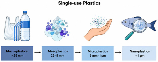
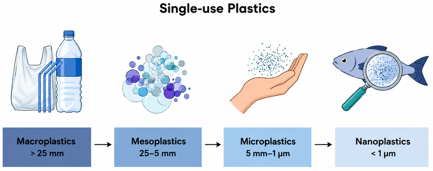
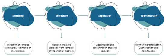
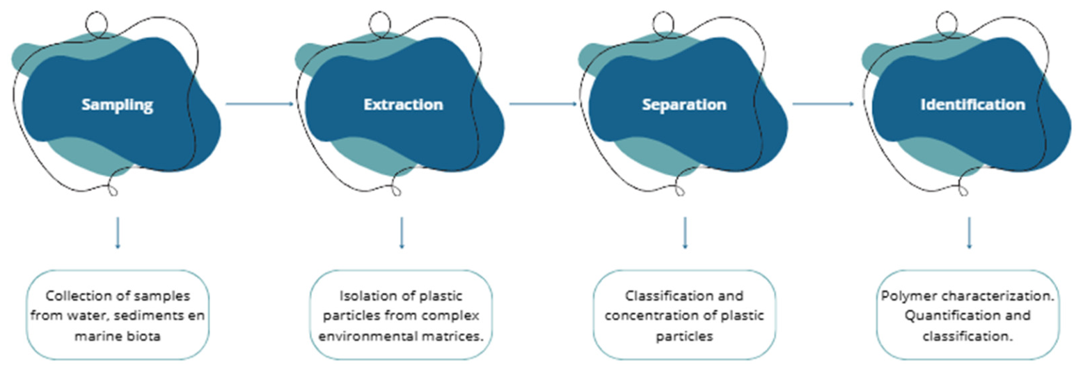
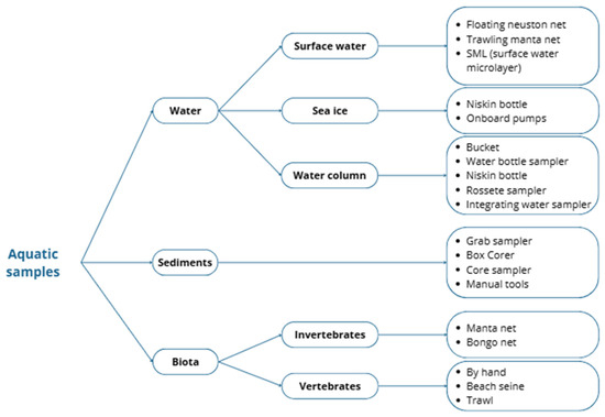
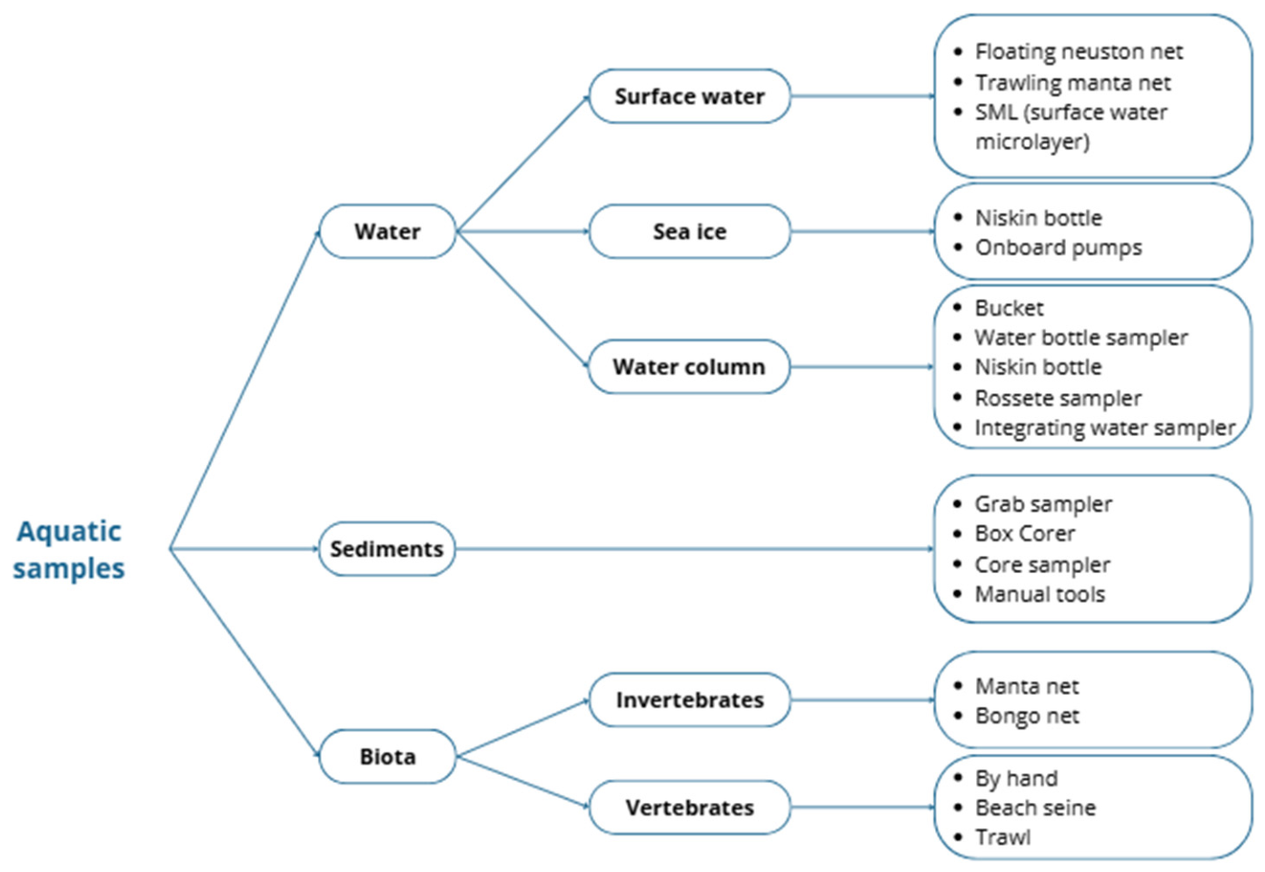
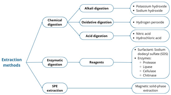
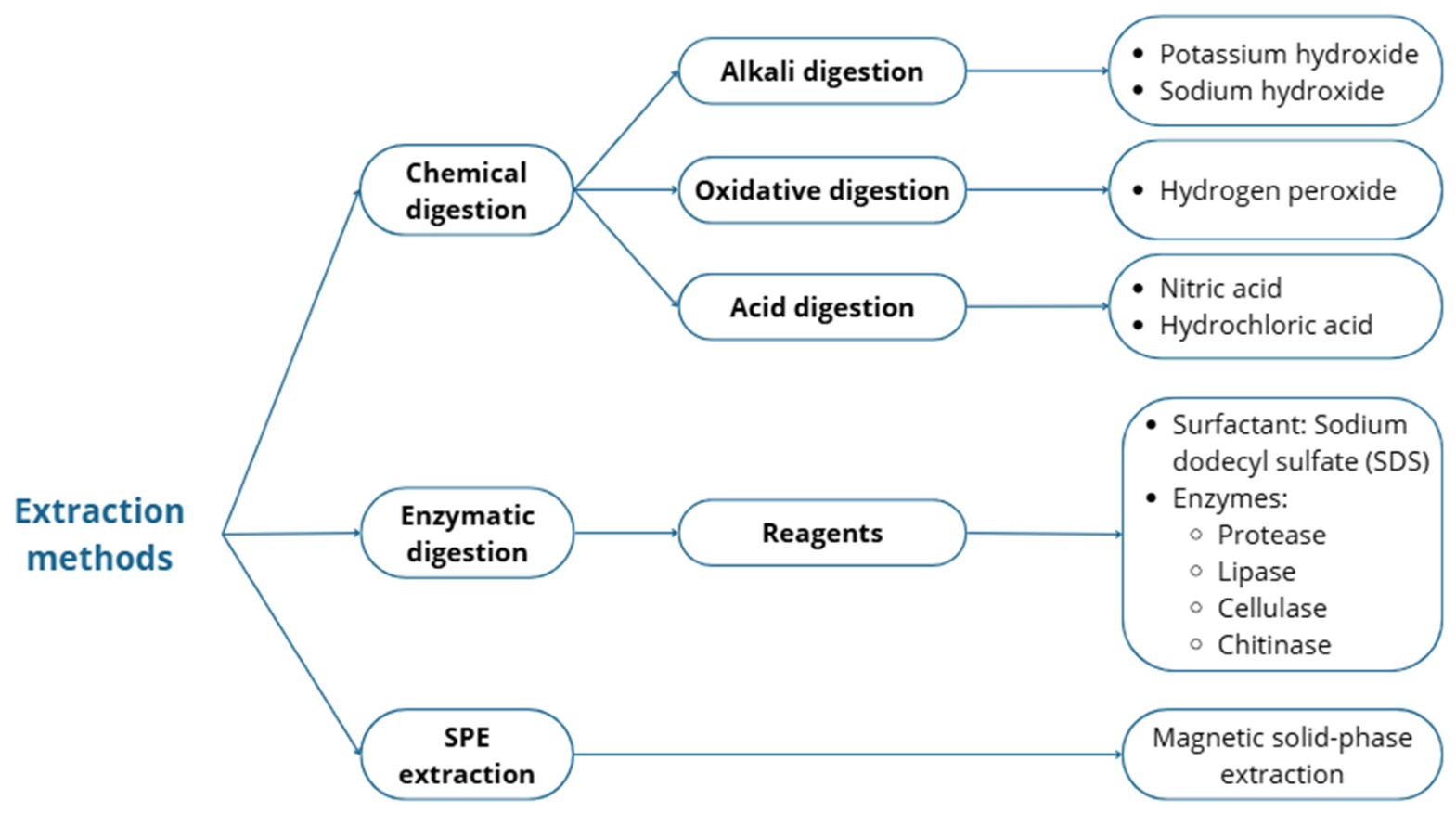
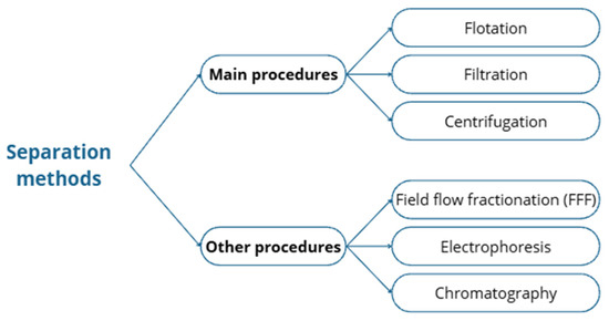
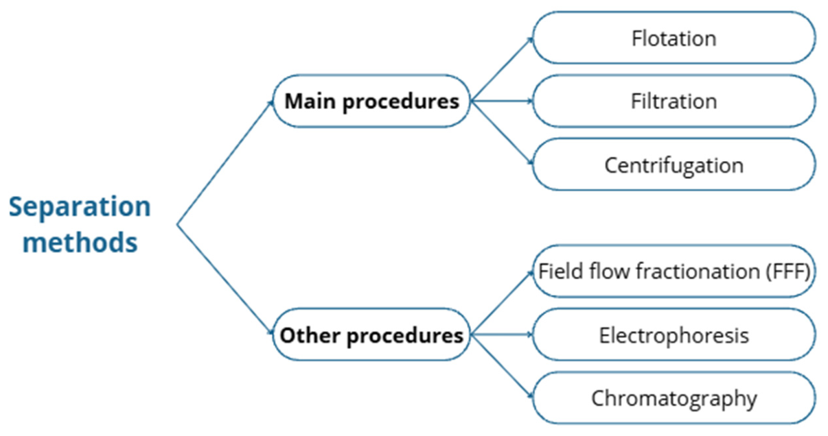
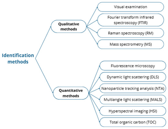
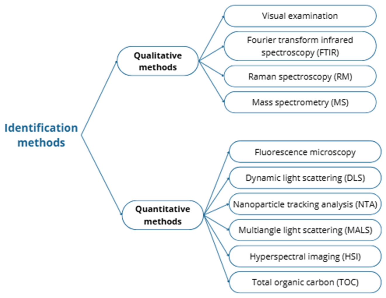

Disclaimer/Publisher’s Note: The statements, opinions and data contained in all publications are solely those of the individual author(s) and contributor(s) and not of MDPI and/or the editor(s). MDPI and/or the editor(s) disclaim responsibility for any injury to people or property resulting from any ideas, methods, instructions or products referred to in the content. |
© 2026 by the authors. Licensee MDPI, Basel, Switzerland. This article is an open access article distributed under the terms and conditions of the Creative Commons Attribution (CC BY) license.