


Abstract
Many filter-feeding invertebrates consume microplastics (MP) under laboratory conditions, but little is known about newly settled, field-collected juveniles. To address this information gap, we collected 3439 juvenile invertebrates in the Indian River Lagoon (IRL), FL, USA. Previous studies suggest that the IRL is a MP hotspot. A total of 70% of IRL adult oysters (Crassostrea virginica) contained MP (mean: 2.3 MP/individual), and MP number and MP length were positively correlated with animal size. We predicted that juvenile C. virginica and other sessile invertebrates would contain MP with a positive correlation to animal size. Five species were examined; 51% were C. virginica (mean shell length ± SD: 6.3 ± 4.7 mm). Overall, 117 (3.4%) animals contained potential MP (fibers: 90.7%). Of these particles that matched FTIR databases with a score of 70% or greater, 51% were plastic and 49% were anthropogenically modified particles. No correlations to animal size were found for particle presence (logistic regressions: p ≥ 0.20 for all species) or particle length (linear regressions: p ≥ 0.23 for all species). Thus, even though found in a MP hotspot, our extrapolated results suggest few juveniles (<1%) contained MP. This information is important for understanding the relationship between MP and the life histories of filter-feeding animals, especially for species considered biological indicators of MP.
1. Introduction
Oysters are filter-feeding invertebrates that have a two-stage life cycle, which begins with the release of gametes into the water column which grows into a dispersal, feeding larval form, followed by radical metamorphosis into a sessile, benthic filter-feeding animal [1]. During metamorphosis, parts of the larva body are resorbed and the animal cements itself to a hard surface [1]. Larval feeding on particles in the size range of 2–30 microns starts 1–2 days post-fertilization and lasts for 2–4 weeks until metamorphosis occurs [2]. The newly metamorphosed individual lives off lipid reserves for ~6 days until adult feeding structures are functional [3]. Laboratory investigations of plastic pollution have focused on all life stages of oysters (e.g., gametes, larvae, adults) to quantify any negative impacts on survival, growth rates, and reproductive outputs [4]. Although no acute toxicity values have been reported to date for any oyster species [5], documented negative plastic particle impacts include physical damage (e.g., gut blockage, perforation of the digestive tract, lysosomal stability), altered feeding behaviors, reduced energy allocation, reduced shell growth rates, chemical toxicity, and increased oxygen consumption and respiratory rates [4,6,7,8]). Many of these published studies focused on responses to polystyrene beads or high-density polyethylene (HDPE) particles in concentrations ranging from 0 (controls) to higher than environmentally relevant values with the commercially and ecologically important Pacific oyster Crassostrea gigas [5,9,10]. For example, Sussarellu et al. [9] reported in 2016 that when reproductive-age adults of C. gigas were exposed to polystyrene, results included significantly smaller and fewer oocytes, reduced sperm velocity, and reduced oyster survival. Exposure to polystyrene beads reduced survival and development in some, but not all, in additional studies with larvae of C. gigas [7,10,11,12]. Bringer et al. [13] fed HDPE particles to C. gigas larvae and found that these particles induced malformations, arrested development, and resulted in abnormal swimming behaviors as well as significantly slower spat growth post-settlement. Similar responses were reported in trials with juvenile and adult C. gigas with greater negative impacts, higher concentrations, and larger MP [4,7]. When larvae of the eastern oyster Crassostrea virginica were exposed to HDPE particles, Bhatt et al. [14] reported no impact on survival at 10 mg/L concentrations, but they did find delayed larval development.
The number of MP studies conducted on non-commercial sessile, filter-feeding species was historically more limited in the literature but is now rapidly increasing for both laboratory and field studies [15,16,17]. For example, the cosmopolitan barnacle Amphibalanus amphitrite ingested and egested polystyrene with no observed negative impacts on larval development at environmentally relevant concentrations in the laboratory [15]. Gut retention time, however, was increased with smaller microbeads [18]. Wang et al. [19] exposed the same species to MP particles with and without biofilms and found that the presence of the biofilm did not affect ingestion, while Xu et al. 2023 [20] found no difference in ingestion abundances for microspheres versus microfibers. Lo et al. 2018 [16] fed polystyrene beads to larvae of the slippersnail Crepidula onyx and found that at environmentally relevant concentrations, larval and juvenile C. onyx were not negatively affected. At higher concentrations of plastic, however, slower growth over 65 days, settlement at smaller sizes, and a negative legacy impact on post-larval juvenile individuals were reported. While these studies provide important data, few provide information on recently metamorphosed sessile invertebrates (shell length: ≤1 cm) that were not reared in a laboratory nor fed MP. Our research goal was to address this information gap in a Florida estuary, the Indian River Lagoon (IRL). This estuary was previously described as a microplastic (MP) hotspot [21], thus suggesting that invertebrate larvae and juveniles in these waters would likely encounter MP.
The IRL is a 251 km long subtropical estuary located along the east coast of central Florida, USA, and has been described as a MP hotspot based on studies of surface waters [8] and atmospheric deposition above and immediately surrounding the lagoon [22]. Water sampling documented 1.5 MP/L lagoon-wide with 1224 MP/m2/day entering the system via atmospheric sources. Extrapolated, both sources of data yielded 1.1–1.4 trillion MP in the IRL [22]. These high levels of plastics in the water have translated into high densities of plastics in adult IRL organisms. Previously studied adult IRL organisms with high MP loads include crabs, coastal birds, and oysters [21,23,24]. Craig et al. [25] determined that 70% of adult IRL oysters contained MP (mean: 2.3 MP/individual). Some MP were excreted in feces (1 MP/h), while 1 MP was removed every 2 h in pseudofeces (particles captured and bound in mucus, but never entered the digestive tract). Craig et al. [25] also reported that MP size was positively correlated to retention in oyster soft tissues; the larger the MP, the more likely that the MP were retained in C. virginica with egestion efficiency decreasing by 0.8% per 1 g increase in tissue weight. The average lengths of MP (±S.E.) in feces and pseudofeces were 1.5 ± 0.1 and 1.7 ± 0.2 mm, respectively, while the average length of MP collected from soft tissues of the same oysters was larger at 2.5 ± 0.2 mm.
Our research focused on understanding the abundances of MP in recently settled C. virginica and co-occurring sessile invertebrates under field conditions. MP data are very limited on sessile invertebrates in the early post-metamorphosis life-history stage for individuals not reared in the laboratory with exposure to high concentrations of a single polymer of virgin plastics of specific dimensions. Based on the results for adult IRL oysters [10], we asked the following: (1) Do juvenile oysters contain MP? (2) Are there increases in MP numbers with increases in oyster size? (3) Are there increases in the mean length of MP with increases in oyster size? As 49% of the collected recruits in this study were species other than C. virginica, we asked the same questions for all collected sessile invertebrates that were co-located on the oyster reefs. Moreover, we also report the results for anthropogenic particles (AP), defined here as human-modified natural materials, such as microfibrillated cellulose [9].
2. Materials and Methods
2.1. Sample Collection Location
All collections occurred in Mosquito Lagoon (ML), the northernmost region of the IRL system. Mosquito Lagoon is a shallow, microtidal, high-salinity (30+ ppt) estuary with restricted water movement due to a single ocean inlet [26]. Reefs of C. virginica in ML are intertidal. Boat wakes, boat strikes, hurricanes, sea level rise, algal blooms, mangrove encroachment and MP are some of the stressors that C. virginica encounter in this system [27].
2.2. Field Collections
Recruitment substrates were deployed for 96 days starting in September 2022. On the edge of each of ten oyster reefs (28°56′28.36″ N, 80°52′10.67″ W), two mats made from 23 × 46 cm non-plastic BESE-elements (Biodegradable EcoSystem Engineering Elements, Culemborg, The Netherlands) produced from the biopolymer Solanyl were deployed on bare sediment [28]. Solanyl is biodegradable, primarily composed of poly(lactic) acid from reclaimed starch, and considered to be non-toxic in the environment [29,30]. Eighteen recycled, adult oyster shells were attached to each mat with stainless-steel cable ties. Mats were held in place with cement weights and stainless-steel cable ties.
Collected mats were individually wrapped in aluminum foil to prevent contamination. All mats were kept wrapped in foil at room temperature in University of Central Florida Biology laboratory (20 °C) until processing.
2.3. Laboratory and Data Processing
To minimize contamination, standard MP protocols were followed throughout processing [22]. All researchers in the field and laboratory wore 100% natural fiber clothing. All Petri dishes, filtration units, ceramic pestles, metal probes and forceps were first triple-rinsed with deionized (DI) water filtered to 0.45 µm. Additionally, while microscopy was underway, five “blank” Petri dishes were placed in a half-circle around each microscope, with new blanks used for each individual [22]. Each blank dish contained a 0.45 µm filter to which 2 mL of filtered water was added to dampen the filter and promote atmospheric particle capture. Blank dishes were open to the environment throughout the duration of microscopy. Once an individual animal was completed, all particles in blank dishes were recorded and a contamination rate per sample was calculated [25].
For sample processing, all mats, including the external surfaces of all individual animals, were first triple-rinsed with 0.45 µm filtered, deionized water to remove any external particles. Next, all sessile animals were identified to the species level, and the shell length of each settled individual was recorded with calipers. Individuals were then removed from the settlement substrate using stainless-steel forceps and probes. All individuals were placed into separate dishes. Additional gentle crushing with a ceramic pestle was used, if necessary, to expose all of the animal’s soft tissue. While chemical digestion is required with larger organisms (e.g., adult oysters) to enable scientists to accurately view all soft tissue under a microscope, this step was not needed with these small individuals (mean length: <1 cm), as the small amount of tissue present was liquid and transparent after crushing [25].
Filtered DI water (0.45 µm) was added to each dish containing a single animal, and all contents were then filtered through gridded (3 mm2) 0.45 µm filter paper using a GAST vacuum pump (Gast Manufacturing, Inc., Benton Harbor, MI, USA). Each dish was rinsed and filtered three additional times, using a total of ~100 mL of filtered water per animal. Filters were then placed in washed, labeled Petri dishes for microscopy. All samples were examined under a Leica EZ4 dissecting microscope (Leica Microsystems, Wetzlar, Germany) at 20–40X magnification to allow for observation of particles as small as 4–10 µm. Due to the small size of some particles, it was not possible to always test for the breakage of particles to separate natural materials from potential MP. Data were collected for all potential MP as particle type (fiber, fragment, film, foam, bead, pellet) and particle length (mm).
Attenuated Total Reflection Fourier Transform Infrared Spectroscopy (ATR-FTIR) was performed on all potential MP particles to identify polymers. Polymer composition analysis was performed using a Shimadzu IR Spirit-T spectrometer (Shimadzu, Kyoto, Japan). Particles were scanned in the 400 to 4000 cm−1 range. Spectra were matched with a qualitative approach, in which the acquired spectra were compared against reference spectra from the KnowItAll FTIR Spectral Database Collection™.
Each spectrum was baseline-corrected and smoothed using a Savitzky–Golay filter with an average of three prior to library matching. Only responses that had a 70% or higher match with the standards database were deemed reliable and categorized as plastic. The matching score was derived from the Euclidean distance between the sample and the reference spectra, representing a nonlinear metric, as variations in environmental or instrument conditions can influence the similarity value. Additionally, the library was designed to match to many types of cellulose/modified cellulose. Modified cellulose particles are reported here as anthropogenic cellulose and include materials such as rayon. Any remaining particles were reported as natural materials (e.g., cellulose).
All statistical analyses were conducted in R version 4.4.1 [31]. Each species was analyzed separately. Organism length and particle length were log transformed to account for the significant positive skew in the data. Models for particle presence/absence were logistic regressions (glm() function with family = “binomial” from the stats package version 4.4.1). Models for MP length used the linear model lm() function from the R statistical package. Confidence intervals for logistic regressions and linear models were determined using the confint() function [31]. Confidence intervals for logistic regressions are reported as odds ratios. Logistic regression model fits were assessed using the Hosmer–Lemeshow test (hoslem.test() function in the ResourceSelection package version 0.3–6)]. Linear model assumptions were assessed visually using diagnostic plots, and no major violations were observed.
3. Results
A total of 3439 sessile invertebrates representing five species settled within 96 days on the deployed oyster shell substrate (Table 1). Overall, 117 (3.4%) of the 3439 individuals contained 118 potential MP. Of these 118 particles, 107 were fibers (90.7%), 9 were fragments (7.6%), and 2 were films (1.7%). No foams or beads/pellets were found. A total of 100% of the particles were fibers for the Atlantic ribbed mussel Geukensia demissa, while the ivory barnacle Amphibalanus eburneus and the Atlantic jingle shell Anomia simplex contained both fibers and fragments. Fibers, fragments, and films were found in Crassostrea virginica and Crepidula fornicata (Table 1). Overall particle length ranged from 0.1 to 12 mm, with mean lengths for C. virginica at 1.5 mm (n = 58 particles) and C. fornicata at 1.7 mm (n = 47 particles) (Table 1).
Table 1.
Animal and particle metrics. Particle lengths were only calculated when particles were detected (no zeros). SD = standard deviation.
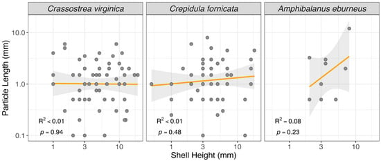
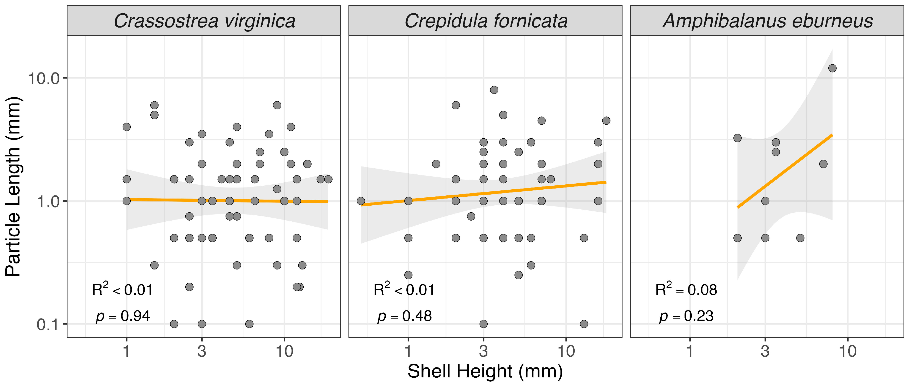
We found no significant relationships between organism size and anthropogenic particle presence or size for the three species with ≥5 individuals containing particles (Table 2 and Table 3). We observed no relationship between the presence/absence of particles and shell height (logistic regression, p-values ≥ 0.05; Table 2). All Hosmer–Lemeshow (HL) test p-values for our logistic regression models were not significant (p values > 0.05), suggesting our models appropriately fit the data. We also observed no relationship between shell length and the particle size in these three species (linear model, p-value ≥ 0.05; Table 3, Figure 1).
Table 2.
Results of logistic regression analysis of particle detection (presence/absence of particles) versus organism shell length. Beta 0 and Beta 1 values are reported as odds ratios. CI = confidence intervals.
Table 3.
Results of linear regression analysis of shell length versus particle length. CI = confidence interval.
Figure 1.
Relationship between shell height and particle length for each C. virginica, C. fornicata, and A. eburneus. Note log10 scaling of both axes. Orange lines are the log-log linear line of best fit, and the gray shaded areas show the 95% confidence interval for each line. R2 and p-values are also reported for each species.
All particles from all individuals were examined using ATR-FTIR. Due to the small size of many particles and their states of degradation, many of these particles produced scores that were below the 70% matching threshold. This resulted in usable results from 45 of the 118 particles (38.1% of the total). The breakdown of totals of MP versus AP by species is included in Table 1. For each species, the percentage of MP to AP was similar. A total of 51.1% of these confirmed particles were plastic polymers (PET: 17 particles, 37.8%; nylon: 3, 6.7%; HDPE: 1, 2.2%; acrylonitrile film: 1, 2.2%; modified rubber: 1, 2.2%). A total of 48.9% of particles were anthropogenic cellulose fibers (microfibrillated cellulose: 20, 44.4%, Tencel: 1, 2.2%). The only other anthropogenic compound observed was one calcium stearate particle. Contamination rates associated with atmospheric particles in blanks were very low (0.009/sample) and thus not included in the FTIR data analyses [9]. In the contamination blanks, there were seven fibers that produced FTIR matches at 70% or higher; included was one PET fiber and six microfibrillated cellulose fibers.
4. Discussion
Overall, 117 of 3439 (3.4%) sessile, filter-feeding, field-collected animals from the Indian River Lagoon, a shallow estuary on the east coast of central Florida, contained potential MP (fibers: 90.7%). Of these particles that matched FTIR databases with a score of 70% or greater (38.1%), 51.1% were plastic and 48.9% were anthropogenically modified. However, particles at the lower end of the size range observed in this study are typically identified using μ-FTIR rather than ATR-FTIR. The use of ATR-FTIR for smaller particles could add uncertainty to our results. Additionally, we did not sample surface waters at oyster reefs during the study, which limits our ability to discern if the particles observed were ingested or were present in larvae prior to settlement. No correlations to animal size were found for particle presence (logistic regressions: p ≥ 0.20 for all species) nor particle length (linear regressions: p ≥ 0.23 for all species). Thus, even though found in a MP hotspot, our extrapolated results suggest few juveniles (<1%) that were less than 1 cm in shell length contained MP. No published articles found during our literature review focused on newly metamorphosed, wild-caught individuals, and only a few laboratory-based studies tracked these sessile filter-feeders post-metamorphosis [13,16]. Chiani et al. (2025) [32] did, however, report from field-collected animals that smaller individuals of the mussel Mytilaster lineatus and barnacle Amphibalanus improvisus had significantly more MP than larger individuals. Although the animal size ranges were not large (mussel: 1.0–1.4 cm; barnacle: 0.66–1.07 cm), the authors suggest that future studies on MP biomonitoring include size-related variables. Moreover, they thus suggested that small individuals have greater potential as biological indicators.
Good biological indicators of ecosystem health should focus on cosmopolitan species that are common, easy to identify, have limited mobility, feed at lower trophic levels, and resist ecological change [33,34]. Sessile, filter-feeding invertebrates have been suggested as potentially being good bioindicators of MP pollution in many studies (bivalves: [34,35,36]; mussels: [17]; barnacles: [37,38]; gastropods including Crepidula: [39]). As mentioned in the previous paragraph, it is important to consider the size of the potential biological indicator, as our results document that very few (3.4%) of the 3439 study individuals contained any visible particles and <1% contained MP. It is likely that nanoparticles would have been found in these animals as well. However, the tools needed to quantify nanoparticles are less available, making nanoplastic metrics less useful for a biological indicator.
Ward et al. [40,41] suggested that bivalves (oysters, mussels, clams) are poor biological indicators due to their selective feeding capabilities. Although both filter-feeders, bivalves and barnacles feed in different ways that may impact their ingestion of MP. Bivalves actively feed by bringing water into their body, sorting and selecting particles on their gills, followed by additional sorting on their labial palps before the particles enter their mouth [40,41]. Sorting is based on many particle criteria including size, shape, density, chemical, and nutritional properties [40,41]. Unselected particles are removed as pseudofeces. The selected particles are then transported to the bivalve’s mouth for ingestion. MP have been reported in both pseudofeces and feces in oysters [25]. Alternatively, barnacles are considered passive, mechanical feeders with no, or very limited, pre-ingestive particle sorting [19]. Barnacles create water movement by rhythmic movement of their cirri [19]. The cirri have fine setae on them that collect all particles and transfer them to the barnacle’s mouth with limited sorting or selection [19]. Another non-selective feeder is the slippersnail Crepidula sp.; this genus uses a mucus net to capture particles and then consumes this mucus [42]. Sponges, tunicates, and suspension-feeding polychaetes are also non-selective filter-feeding animals in estuarine and marine waters [43]. Khan and Rountos (2025) [43] evaluated 144 publications (133 species) and compared MP metrics in selective versus non-selective filter-feeders. They found similar percentages of fibers and fragments, as well as the same dominant polymers (PE, PET, and PP). However, higher concentrations of MP were found in the non-selective feeding species [43]. While C. virginica takes ~6 days until filter-feeding commences post-metamorphosis, this duration is not known for most sessile invertebrates [3]. Nor is it known how selective the “selective” filter feeders are when the animal sizes are <1 cm. Our reported data suggest similar numbers of particles per individual regardless of whether the species was a selective or non-selective feeder.
Unlike our results with newly settled C. virginica from the IRL, Craig et al. [25] found positive correlations with adult IRL oysters for both particle counts and particle lengths. This difference is not a sample size issue as a large number of C. virginica were examined in both studies; 70.0% (981 of 1402) of adults contained MP, versus our 3.3% (58 of 1741) of juvenile C. virginica. The type of particle was similar in both Craig et al. [25] and our study. Ninety-five percent of particles were fibers for Craig et al. [25] and 91.4% of C. virginica for our study. However, the mean number of particles per individual in our study for juvenile C. virginica was two orders of magnitude smaller (0.04) versus 2.3 particles for Craig et al. [25] in adult oysters from the same area. Moreover, the mean length of particles was 1 mm shorter (1.5 mm) in our juvenile C. virginica, while it was 2.5 mm in the adult IRL oysters [25]. We initially predicted that individuals ≤ 96 days old would follow similar patterns to adult C. virginica, but this was not the case. To better understand the capability of each species of these size ranges to consume MP, it would have been necessary to culture each species in a laboratory, and that was out of scope for this observational field study.
Because the days since settlement was only known to be <96 days, our focus was on animal size (e.g., shell length) rather than age. Shell length and wet weight are widely adopted indicators of bivalve growth [7,44]. As our invertebrates all averaged less than one centimeter in mean length (≤6.3 mm) and were tightly cemented to the substrate, as is common in newly settled animals, we focused on shell length as our proxy to enable comparisons with global populations of each genus and species. Using shell length additionally makes sense with these sessile animals collected in the fall in warm Florida waters, as growth rates in the field are dependent on temperature and food availability rather than the number of days since settlement [45].
Although much more research is needed, concerns are rising over the wide-ranging negative impacts of plastic particles on humans (e.g., microplastic syndrome) [46]. Humans unintentionally consuming plastic is an important source of pollution [47,48]. Shellfish, mussels and, in some locations, barnacles, are consumed whole, so whatever was contained in the invertebrate moves to the human consumer. Ribeiro et al. 2020 [49] calculated that ~0.7 mg of plastic are consumed by humans eating just 10 adult oysters. Recent studies suggest that humans consume up to 11,000 MP particles annually by consuming shellfish [50]. This type of bioaccumulation via predation represents an important vector for trophic transfer of MP from sessile, filter-feeding invertebrates to humans and to many other animals in food webs. Common predators of oysters and mussels in estuaries include crabs, coastal birds, fish, and small vertebrates such as raccoons [17,24,51]. Additionally, egestion of MP by bivalves onto the sediment surface can provide MP that are ingested by nearby deposit-feeding organisms such as worms [25,52]. MP loads in shellfish may increase if grown in aquaculture settings, especially for those industries reliant on plastic tanks, cages, ropes, or bags [53]. Bringer et al. 2020 [13] found slower initial growth of C. gigas larvae when grown in tanks spiked with aged aquaculture plastics. Moreover, liquid algal feed for shellfish that is packaged in plastic bottles may increase MP concentrations in tanks [L. Nguyen, pers. comm]. A future research area should focus on understanding the “tipping point” at which field-collected filter-feeding invertebrates go from being mostly MP-free to being a valuable bioindicator and potential source of microplastic syndrome in humans.
In conclusion, we found very small numbers of small plastic particles (23 MP, 0.7% of total animals) and 22 anthropogenically modified particles (0.6% of total animals) in filter-feeding invertebrates that averaged less than 1 cm in length in this shallow, subtropical Florida estuary. Extrapolated to all individuals from this study, we suggest that 1.3% of the total invertebrates contained some sort of human-produced particles (MP + AP). Further research is needed to understand and distinguish if any observed particles were larval legacy particles retained post-metamorphosis or if they were captured post-settlement via filter-feeding. Moreover, additional research is needed to better understand juvenile capture and egestion rates of various sized fibers, fragments, and films under realistic conditions. Only with this information will we begin to understand the impact of plastics on newly recruited animals that are ecologically and economically important in Florida, throughout the temperate and subtropical western Atlantic estuaries, and beyond.
Author Contributions
Conceptualization, L.J.W.: Methodology, L.J.W., L.B., P.E.S., F.J. and L.Z.; Formal Analysis, L.J.W., J.S.S. and L.B.; Investigation, L.J.W., L.B., P.E.S., F.J. and L.Z.; Resources, L.J.W. and L.Z.; Writing—Original Draft Preparation, L.J.W.; Writing—Review and Editing, L.B., P.E.S., J.S.S., F.J. and L.Z.; Funding Acquisition, L.J.W. All authors have read and agreed to the published version of the manuscript.
Funding
This study received financial support from the National Science Foundation (#2149866), and the University of Central Florida.
Institutional Review Board Statement
Institutional Review Board Statement: Ethics approval was not required as the study involved only invertebrate organisms.
Data Availability Statement
Data will be uploaded to University of Central Florida STARS Digital Repository at: https://stars.library.ucf.edu/ (accessed on 6 May 2026).
Acknowledgments
The authors thank the many students who assisted with this project, and W. Kumler for statistical assistance.
Conflicts of Interest
The authors declare no conflicts of interest.
Abbreviations
The following abbreviations are used in this manuscript:
| MP | Microplastics |
| IRL | Indian River Lagoon |
| FTIR | Fourier Transform Infrared Spectroscopy |
| ML | Mosquito Lagoon |
| ATR-FTIR | Attenuated Total Reflection Fourier Transform Infrared Spectroscopy |
| AP | Anthropogenic Particle (non-plastic, human-modified) |
References
- Kennedy, V.S. Biology of larvae and spat. In The Eastern Oyster Crassostrea virginica; Kennedy, V.S., Newell, R.I.E., Eble, A.F., Eds.; Maryland Sea Grant College: College Park, ML, USA, 1996; pp. 371–421. [Google Scholar]
- Newell, R.I.E.; Langdon, C.J. Mechanisms and physiology of larval and adult feeding. In The Eastern Oyster Crassostrea virginica; Kennedy, V.S., Newell, R.I.E., Eble, A.F., Eds.; Maryland Sea Grant College: College Park, ML, USA, 1996; pp. 185–229. [Google Scholar]
- Hickman, R.W.; Gruffydd, L.D. The histology of the larva of Ostrea edulis during metamorphosis. In Fourth European Marine Biology Symposium, Cambridge University, Cambridge, United Kingdom, 1971; Crisp, D.J., Ed.; Cambridge University Press: Cambridge, UK; pp. 281–294.
- Thomas, M.; Jon, B.; Craig, S.; Edward, R.; Ruth, H.; John, B.; Dick, V.A.; Heather, L.A.; Matthew, S. The world is your oyster: Low-dose, long-term microplastic exposure of juvenile oysters. Heliyon 2020, 6, e03103. [Google Scholar] [CrossRef]
- Kim, H.J.; Rhee, J.S. Molecular and cellular effects of nano- and microplastics on oysters: A review. Mol. Cell. Toxicol. 2026. [Google Scholar] [CrossRef]
- Galloway, T.S.; Lewis, C.N. Marine microplastics spell big problems for future generations. Proc. Natl. Acad. Sci. USA 2016, 113, 2331–2333. [Google Scholar] [CrossRef] [PubMed]
- Seong, T.; Makino, M.; Yagi, M.; Nakatani, H.; Kyozuka, Y.; Rhee, J.S.; Satuito, G.; Kim, H.J. Physiological impacts of microplastics on the Pacific oyster Crassostrea gigas: The role of particle size and concentration. Mol. Cell. Toxicol. 2025, 22, 277–287. [Google Scholar] [CrossRef]
- Martín, J.; Santos, J.L.; Aparicio, I.; Alonso, E. Microplastics and associated emerging contaminants in the environment: Analysis, sorption mechanisms and effects of co-exposure. Trends Environ. Anal. Chem. 2022, 35, e00170. [Google Scholar] [CrossRef]
- Sussarellu, R.; Suquet, M.; Thomas, Y.; Lambert, C.; Fabioux, C.; Pernet, M.E.J.; Le Goic, N.; Quillien, V.; Mingant, C.; Epelboin, Y.; et al. Oyster reproduction is affected by exposure to polystyrene microplastics. Proc. Natl. Acad. Sci. USA 2016, 113, 2430–2435. [Google Scholar] [CrossRef]
- Seong, T.; Onizuka, D.; Satuito, G.; Kim, H.J. Impact of nano- and micro-sized polystyrene beads on larval survival and growth of the Pacific oyster Crassostrea gigas. J. Hazard. Mater. 2024, 469, 133952. [Google Scholar] [CrossRef]
- Cole, M.; Galloway, T.S. Ingestion of nanoplastics and microplastics by Pacific oyster larvae. Environ. Sci. Technol. 2015, 49, 14625–14632. [Google Scholar] [CrossRef]
- Tallec, K.; Huvet, A.; Di Poi, C.; Gonzalez-Fernandez, C.; Lambert, C.; Peton, B.; Le Goic, N.; Berchel, M.; Soudant, P.; Paul-Pont, I. Nanoplastics impaired oyster free living stages, gametes and embryos. Environ. Pollut. 2018, 242, 1226–1235. [Google Scholar] [CrossRef]
- Bringer, A.; Thomas, H.; Prunier, G.; Dubillot, N.; Churlaud, C.; Clerandeau, C.; Le Bihanic, F. High density polyethylene (HDPE) microplastics impair development and swimming activity of Pacific oyster D-larvae, Crassostrea gigas, depending on particle size. Environ. Pollut. 2020, 260, 113978. [Google Scholar] [CrossRef]
- Bhatt, S.; Fan, C.; Liu, M.; Wolfe-Bryant, B. Effect of high-density polyethylene microplastics on the survival and development of eastern oyster (Crassostrea virginica) larvae. Int. J. Environ. Res. Public Health 2023, 20, 6142. [Google Scholar] [CrossRef] [PubMed]
- Yu, S.P.; Chan, B. Effects of polystyrene microplastics on larval development, settlement, and metamorphosis of the intertidal barnacle Amphibalanus amphitrite. Ecotoxicol. Environ. Saf. 2020, 194, 110362. [Google Scholar] [CrossRef] [PubMed]
- Lo, H.; Lo, A.; Chan, K. Negative effects of microplastic exposure on growth and development of Crepidula onyx. Environ. Pollut. 2018, 223, 588–595. [Google Scholar] [CrossRef]
- Ashley, J.; Pilat, A.; Ohlweiler, A.; Ogden, C.; Bradley, O.; Modi, P.; Talbot, S.; Smith, C.; O’Pella, J.; Oxbay, G. Microplastics in Atlantic ribbed mussels (Geukensia demissa) from the Delaware Inland Bays, USA. Microplastics 2024, 3, 147–164. [Google Scholar] [CrossRef]
- Yu, S.; Nakaoka, M.; Chan, B.K.K. The gut retention time of microplastics in barnacle naupliar larvae from different climatic zones and marine habitats. Environ. Pollut. 2021, 268, 115865. [Google Scholar] [CrossRef]
- Wang, Y.; Wong, A.C.W.; Chiu, J.M.Y.; Cheung, S.G. Non-selective feeding on microplastics in the acorn barnacle Amphibalanus amphitrite: The implications in assessing barnacles as global microplastics bioindicators. Front. Mar. Sci. 2025, 12, 1646294. [Google Scholar] [CrossRef]
- Xu, X.Y.; Fang, J.K.-H.; Wong, C.-Y.; Cheung, S.G. Experimental accumulation of microplastics in acorn barnacle Amphibalanus amphitrite and its use in estimating microplastic concentration in coastal waters. Front. Mar. Sci. 2023, 9, 1081329. [Google Scholar] [CrossRef]
- Walters, L.; Craig, C.; Dark, E.; Wayles, J.; Encomio, V.; Coldren, G.; Sailor-Tynes, T.; Fox, D.; Zhai, L. Quantifying spatial and temporal trends of microplastic pollution in surface water and in the eastern oyster Crassostrea virginica for a dynamic Florida estuary. Environments 2022, 9, 131. [Google Scholar] [CrossRef]
- Walters, L.; Serrate, M.; Blanchard, T.; Sacks, P.; Joshua, F.; Zhai, L. Raining plastics: Quantification of atmospheric deposition of plastic and anthropogenic particles in an estuary of national significance with the assistance of citizen scientists. Environments 2025, 12, 424. [Google Scholar] [CrossRef]
- Waite, H.R.; Donnelly, M.J.; Walters, L.J. Quantity and types of microplastics in the organic tissues of the eastern oyster Crassostrea virginica and the Atlantic mud crab Panopeus herbstii from a Florida estuary. Mar. Pollut. Bull. 2018, 129, 179–185. [Google Scholar] [CrossRef]
- Carlin, J.; Donnelly, M.; Walters, L. Microplastic accumulation in the gastrointestinal tracts in birds of prey in central Florida, USA. Environ. Pollut. 2021, 264, 114633. [Google Scholar] [CrossRef]
- Craig, C.A.; Fox, D.W.; Zhai, L.; Walters, L. In-situ microplastic egestion efficiency of the eastern oyster Crassostrea virginica. Mar. Pollut. Bull. 2022, 178, 113653. [Google Scholar] [CrossRef]
- Phlips, E.; Badylak, S.; Lasi, M.; Chamberlain, R.; Green, W.; Hall, L.; Hart, J.; Lockwood, J.; Miller, J.; Morris, L.; et al. From red tides to green and brown tides: Bloom dynamics in a restricted subtropical lagoon under shifting climatic conditions. Estuaries Coasts 2015, 38, 886–904. [Google Scholar] [CrossRef]
- Benson, G.; Donnelly, M.; Sacks, P.; Walters, L. Documenting loss and fragmentation of intertidal oyster (Crassostrea virginica) reefs in a subtropical estuary. Environments 2023, 10, 133. [Google Scholar] [CrossRef]
- Nitsch, C.; Walters, L.; Sacks, J.; Sacks, P.; Chambers, L. Biodegradable material for oyster reef restoration: First-year performance and biogeochemical considerations in a coastal lagoon. Sustainability 2021, 13, 7415. [Google Scholar] [CrossRef]
- Parres, F.; Peydro, M.A.; Juarez, D.; Arrieta, M.P.; Aldas, M. Study of the properties of a biodegradable polymer filled with different wood flour particles. Polymers 2020, 12, 2974. [Google Scholar] [CrossRef]
- Biodegradable EcoSystem Engineering Products. Available online: https://www.bese-products.com/biodegradable-ecosystem-engineering-products-bese/ (accessed on 23 April 2026).
- Posit Team. RStudio: Integrated Development Environment for R. Posit Software, PBC, Boston, MA. 2025. Available online: http://www.posit.co/ (accessed on 26 December 2025).
- Chiani, M.M.; Rasta, M.; Taleshi, M.S.; Elmi, F. The role of organisms’ size in microplastic pollution monitoring: Insights from Mytilaster lineatus and Amphibalanus improvisus. Mar. Environ. Res. 2025, 204, 106863. [Google Scholar] [CrossRef]
- Hilty, J.; Merenlender, A. Faunal indicator taxa selection for monitoring ecosystem health. Biol. Conserv. 2000, 92, 185–197. [Google Scholar] [CrossRef]
- Kargar, M.; Hamidian, A.H.; Basaran, B. A review on microplastics in bivalves: Analysis, quantification, and effects. Environ. Monit. Assess. 2026, 198, 70. [Google Scholar] [CrossRef]
- Cho, Y.; Shim, W.J.; Jang, M.; Han, G.M.; Hong, S.H. Nationwide monitoring of microplastics in bivalves from the coastal environment of Korea. Environ. Pollut. 2021, 270, 116175. [Google Scholar] [CrossRef]
- Li, H.X.; Ma, L.S.; Lin, L.; Ni, Z.X.; Xu, X.R.; Shi, H.H.; Yan, Y.; Zheng, G.M.; Rittschof, D. Microplastics in oysters Saccostrea cucullata along the Pearl River estuary, China. Environ. Pollut. 2018, 236, 619–625. [Google Scholar] [CrossRef]
- Raufanda, M.S.; Aunurohim, A.; Prabowo, R.E. Barnacle analysis as a microplastic pollution bioindicator on the East Coast of Surabaya. PeerJ 2024, 12, e17548. [Google Scholar] [CrossRef]
- Ribero, V.V.; Castro, I.B. Barnacles as emerging sentinels of microplastic contamination: A global synthesis and analytical framing. Mar. Pollut. Bull. 2026, 226, 119425. [Google Scholar] [CrossRef] [PubMed]
- Curren, E.; Yu, D.C.Y.; Leong, S.C.Y. From the seafloor to the surface: A global review of gastropods as bioindicators of marine microplastics. Water Air Soil Pollut. 2024, 235, 45. [Google Scholar] [CrossRef]
- Ward, J.E.; Levinton, J.; Shumway, S.E.; Cucci, T. Site of particle selection in a bivalve molluscs. Nature 1997, 390, 31–132. [Google Scholar] [CrossRef]
- Ward, J.E.; Levinton, J.; Shumway, S.E.; Cucci, T. Particle sorting in bivalves: In vivo determination of the pallial organs of selection. Mar. Biol. 1998, 131, 283–292. [Google Scholar] [CrossRef]
- Beninger, G.P.; Decottignies, P.; Guiheneui, F.; Barillé, L.; Rincé, Y. Comparison of particle processing by two introduced suspension feeders: Selection in Crepidula fornicata and Crassostrea gigas. Mar. Ecol. Prog. Ser. 2007, 334, 165–177. [Google Scholar] [CrossRef]
- Khan, A.; Rountos, K.J. Evaluation of microplastics in marine selective and non-selective suspension-feeding in benthic invertebrates. Mar. Environ. Res. 2025, 209, 107205. [Google Scholar] [CrossRef]
- Carriker, M.R. The shell and ligament. In The Eastern Oyster Crassostrea virginica; Kennedy, V.S., Newell, R.I.E., Eble, A.F., Eds.; Maryland Sea Grant College: College Park, ML, USA, 1996; pp. 75–168. [Google Scholar]
- Carlton, J.T.; Mann, R. Transfers and world-wide introductions. In The Eastern Oyster Crassostrea virginica; Kennedy, V.S., Newell, R.I.E., Eble, A.F., Eds.; Maryland Sea Grant College: College Park, ML, USA, 1996; pp. 691–706. [Google Scholar]
- Cornelli, U.; Casella, C.; Belcaro, G.; Cesarone, M.R.; Marucci, S.; Rondanelli, M.; Recchia, M.; Zanoni, G. Definition of emerging microplastic syndrome based on clinical and epidemiological evidence: A narrative review. Microplastics 2025, 4, 93. [Google Scholar] [CrossRef]
- Khatoon, N.; Ali, S.; Huang, J.; Yu, Z.; Liu, H. Microplastics in essential food commodities: A systematic review of identification, detection, contamination, and health implications (2017–2024). Food Rev. Int. 2025, 42, 1151–1185. [Google Scholar] [CrossRef]
- Baby, A.; Revathy, V.S. Review on microplastic pollution in marine ecosystems: Sources, distribution, ecological impacts, and future directions. Proc. Zool. Soc. 2025, 78, 186–199. [Google Scholar] [CrossRef]
- Ribeiro, F.; Okoffo, E.D.; O’Brien, J.W.; Fraissinet-Tachet, S.; O’Brien, S.; Gallen, M.; Samanipour, S.; Kaserzon, S.; Mueller, J.F.; Galloway, T.; et al. Quantitative analysis of selected plastics in high-commercial-value Australian seafood by pyrolysis gas chromatography mass spectrometry. Environ. Sci. Technol. 2020, 715, 136924. [Google Scholar] [CrossRef]
- Ciesielski, M.; Hanke, M.; Jurgens, L.J.; Kamalanathan, M.; Mortuza, A.; Gahn, M.B.; Hala, D.; Kaiser, K.; Quigg, A. A comparison of methods to quantify nano- and/or microplastic (NMPs) deposition in wild-caught eastern oysters (Crassostrea virginica) growing in a heavily urbanized, subtropical estuary (Galveston Bay, USA). J. Mar. Sci. Eng. 2025, 13, 2065. [Google Scholar] [CrossRef]
- Blanchard, T.L.; Sacks, P.E.; Donnelly, M.J.; Walters, L.J. Monitoring faunal responses to biodegradable oyster reef restoration materials with camera traps in a subtropical estuary. Restor. Ecol. 2025, 33, e70186. [Google Scholar] [CrossRef]
- Fernández, B.; Albentosa, M. Insights into the uptake, elimination and accumulation of microplastics in mussels. Environ. Pollut. 2019, 249, 321–329. [Google Scholar] [CrossRef] [PubMed]
- Hossain, S.; Shukri, Z.N.A.; Iber, B.T.; Yatim, N.I.; Mana, H.; Rahman, T.; Islam, Z.; Minhaz, T.M.; Khatoon, H.; Waiho, K.; et al. Microplastic pollution in shellfish aquaculture: Occurrence, impact, and possible remedies. Water Air Soil Pollut. 2024, 235, 240. [Google Scholar] [CrossRef]

Disclaimer/Publisher’s Note: The statements, opinions and data contained in all publications are solely those of the individual author(s) and contributor(s) and not of MDPI and/or the editor(s). MDPI and/or the editor(s) disclaim responsibility for any injury to people or property resulting from any ideas, methods, instructions or products referred to in the content. |
© 2026 by the authors. Licensee MDPI, Basel, Switzerland. This article is an open access article distributed under the terms and conditions of the Creative Commons Attribution (CC BY) license.