


Abstract
There are currently no published methods for the controlled introduction of microplastic particles into the European rabbit (Oryctolagus cuniculus) as an animal model. The aim of this pilot study was to establish a novel rabbit-based experimental model for assessing the impact of microplastic particles by evaluating the physiological and biochemical responses to an eight-day oral administration of polystyrene latex (1 and 5 mg/kg/b.w./day), providing a foundation for future studies. This study was also aimed at evaluating the possibility of using Raman spectroscopy and Fourier-transform infrared spectroscopy to analyze the distribution of microplastics in rabbit samples. We observed a dose-dependent decrease in water and food consumption in the high-dose (5 mg/kg) study group. In addition, a decrease in alanine aminotransferase and total calcium levels, along with an increase in phosphorus levels, was detected. The rabbit’s stomach was the only organ where polystyrene microparticles were identified, with the colon, kidneys, ovaries, and uterus not showing any evidence of polystyrene presence. The selected doses of microplastics did not lead to pronounced toxic effects in rabbits and may be used on larger animal samples. Physiological and biochemical data obtained indicate predominantly negative metabolic shifts associated with the intake of microplastics, which warrants further study.
1. Introduction
It has been 165 years since the chemist Alexander Parkes invented plastic materials [1], which contributed immensely to the development of mankind. However, it is now becoming clear that plastic has a serious negative impact on the environment and human health. There has been a significant increase in plastic production and, as a result, the accumulation of plastic waste. By 2050, it is projected that more than 25 billion metric tons of plastic will have been produced, with an average of more than ten thousand metric tons of plastic waste polluting the environment [2]. Microplastics, defined as plastic fragments and particles with a diameter of less than 5 mm [3], have become a ubiquitous environmental contaminant. According to the latest data, an average individual consumes from 74 to 121 thousand microplastic particles per year [4]. The particles can enter the human body through the respiratory tract due to the use of various products for both household and professional purposes, such as medical masks made of synthetic polymer materials [5,6]. Pollution of the environment with plastics, followed by pollution of aquatic ecosystems and, as a consequence, the consumption of microplastics by terrestrial freshwater and marine animals, can lead to the entry of synthetic polymer microparticles into the human body through food consumed [7,8], as well as bottled and tap water drunk [3]. It is estimated that people consume an average of 5 g of plastic per week through their standard diet and drinks [9].
Research aimed at assessing the impact of microplastics on the human body has only begun recently. The first publication on microplastics in human biological samples appeared only in 2019 [3]. Later, it was demonstrated that microplastics were found in the blood and could spread throughout the body [10]. It was noted that plastic particles were found in 15 human tissues and organs, such as the spleen, liver, colon, lungs, placenta, breast milk, etc. [3,11]. Meanwhile, methods for extracting microplastics from human organs vary significantly. The most reliable identification methods are infrared spectroscopy and Raman spectroscopy, which provide high accuracy and reliability of results [11,12].
In addition, microplastics are carbon-based polymer structures into which a number of chemical additives are integrated to impart specific characteristics to the polymers. Many of these additives are highly toxic. These include carcinogenic compounds, neurotoxins, and endocrine disruptors such as phthalates, bisphenols, per- and polyfluoroalkyl substances, as well as brominated and organophosphate flame-retardants [13]. The above chemicals, being an integral part of polymeric materials, make a significant contribution to the negative impact of plastics on human health and the environment. Furthermore, numerous studies predominantly describe the adverse effects of polystyrene (PS), a polymer of significant environmental concern due to its extensive industrial application and persistence [14]. Recent systematic analysis confirms that PS microplastics elicit both neurotoxic and genotoxic effects in different models, manifesting as behavioral disruptions, neurochemical imbalance, and elevated markers of DNA damage [12].
However, at present, a limited number of studies describe the effect of synthetic polymer microparticles on human health. Assessing the effect of PS particles in various animal models is an urgent task of experimental pharmacology, toxicology, and medicine. The predominant model organisms for studying the effects of microplastics are rodents (Mus, Rattus), but there are also studies conducted on the fish Danio rerio [9,12,15]. However, such models have a number of limitations that can be overcome by conducting experiments on rabbits (Oryctolagus). One of the limitations is the amount of biological material obtained, which is critical with respect to the sensitivity threshold of methods used for the isolation and/or detection and analysis of microplastic particles [16]. The daily food consumption of Oryctolagus is almost 5% of its body weight (b.w.), while water consumption is about 10% [17], which allows for the repeated collection of large volumes of biological material (blood, urine, feces) in quantities sufficient for analysis. In addition, rodents and fish, Danio rerio, may not fully reflect the physiological and metabolic processes characteristic of humans, which limits the possibility of extrapolating the results to the human body. Rabbits are considered more sensitive to pharmaceuticals compared to the mouse-like rodents often used in experimental studies [18]. It has been shown that the introduction of a number of pharmacological drugs over the course of seven days is capable of causing a dose-dependent effect in the body of a rabbit [19,20]. At the same time, the large size of Oryctolagus allows for the introduction of the necessary volumes and/or doses of a drug into its body. Thus, the rabbit is a convenient animal model for pharmacokinetic studies of the absorption and excretion of various substances.
Research on the specific effects of microplastics on rabbits is limited to a few isolated studies. One original article published in 1993 described intraintestinal administration of a latex suspension by surgery [21], which could result in death of the animal, while a later 2024 paper used a method of allowing animals free access to food containing microplastics [22]. However, the feeding design of the experiment carried the risk of uneven consumption of microplastic particles by individual animals, which could lead to significant variability in the doses received. Meanwhile, oral intake is the main route of microplastic penetration into the human body [3], which makes the use of animal models with oral administration (per os) of microparticles highly relevant. Despite this, to date, there are no published methods in the literature for strictly controlled oral administration of microplastics to the European rabbit (Oryctolagus cuniculus) as an animal model.
Therefore, the aim of this pilot study was to develop a baseline model of microplastic ingestion in the European rabbit (Oryctolagus cuniculus) by assessing effects thereof on key biochemical and physiological parameters and analyzing the animal organs for microplastics following controlled oral administration of PS latex.
2. Materials and Methods
2.1. Experimental Design
Forasmuch as oral consumption of polymer particles is the main route of microplastic entry into the human body [3], this experiment was based on the per os entry of polymer microparticles into the body of experimental animals. To create conditions for minimal contact of animals with elements of the environment containing synthetic polymers, the rabbits were kept in a metal cage with a metal feeder and a glass drinking bowl during the experiment. The laboratory staff and researchers were wearing leather shoes, cotton coats, and cotton gloves throughout the experiment.
So far as microplastics are defined as plastic fragments and particles with a diameter of less than 5 mm [3] and PS is one of the most common polymeric microplastics in the environment [14], we used the PS latex suspension (N#K-5 series; Institute of Macromolecular Compounds, St. Petersburg, Russia). A latex concentration was 1 mg/mL, and a particle diameter of 5 μm.
The effect of microplastics was studied on sexually mature female European rabbits (Oryctolagus cuniculus) with an average weight of 5.1 ± 0.6 kg and a body temperature of 38.6 ± 0.5 °C. Due to their unique reproductive trait of induced ovulation, female rabbits remain anestrous without a male, resulting in a stable hormonal baseline [23], which makes them a consistent animal model for short-term investigations.
This pilot study included an analysis of two different doses of PS administration: Group 1 (n = 3) and Group 2 (n = 3) were administered a PS latex suspension at a rate of 5 mg/kg b.w. and 1 mg/kg b.w. once a day, respectively, using a glass syringe and a metal cannula. The experimental duration was set at eight days to capture early physiological and biochemical changes.
2.2. Biochemical Blood Analysis
Biochemical blood analysis was performed on the first and last days of the experiment using a UniCel Dxl 600 automatic immunochemical analyzer (Beckman Coulter, Indianapolis, IN, USA). The following biochemical parameters were determined using standard biochemical kits: alanine aminotransferase (ALT) (Beckman Coulter), aspartate aminotransferase (AST) (Beckman Coulter), urea (Beckman Coulter), creatinine (Beckman Coulter), phosphorus (Beckman Coulter), amylase (Beckman Coulter), and total calcium (Calcium Arsenazo, Dialab, Wiener Neudorf, Austria), being the key indicators of liver and kidney function, pancreatic health, and mineral metabolism [24]. Blood was collected into a glass test tube by accessing the ear vein with a metal scalpel.
2.3. Physiological Monitoring
A comprehensive assessment of the animals’ general health and well-being was conducted daily through a set of non-invasive physiological measurements. Throughout the experiment, we recorded the rabbits’ weight and rectal temperature, and visually examined a visible sclera for signs of redness (injected sclera) and mucous membranes in the mouth (oral mucosa). From the second day of PS administration, the mass of food eaten and the volume of water drunk per day were recorded as well. These parameters are established to be sensitive indicators of systemic toxicity and stress in animal models [25]. The weight of the rabbits and the mass of the food consumed were measured using steel scales. The volume of water drunk was determined using steel scales as the difference between the weight of the water with the drinker and the weight of the drinker. Rectal temperature was measured with a glass mercury thermometer. The visible mucous membrane in the mouth and the sclera were assessed visually.
2.4. Microplastic Identification
Internal organs (stomach, liver, small intestine, colon, kidneys, ovaries, and uterus) were removed post mortem using metal instruments. All collected biomaterial was placed in a glass Petri dish or glass containers and stored in a freezer (−80 °C) until further analysis.
After thawing, all biological samples were treated with H2O2 to completely decompose the organic matrix. To do this, 0.5–1.5 g of each sample was placed in a 100 mL single-neck round-bottom flask equipped with a heater and reflux condenser, followed by adding 50 mL of a 30% aqueous H2O2 solution to the flask. The suspension was left unstirred at room temperature for 24 h. The flask was then heated to boiling and was simmered for 50 h. After cooling down to room temperature, the resulting suspension was filtered through a polytetrafluoroethylene 0.22 µm membrane filter (Haining Delv New Material Technology Co., Ltd., Haining, China). The filter precipitate was washed successively with 5% aqueous K2CO3 and HCl solutions (3 mL each) and distilled water (10 mL, 3 times). The membrane containing the precipitate was removed from the membrane filter and air-dried at room temperature for 48 h. The dried membrane containing the precipitate was placed in a glass-weighing bottle with a tightly sealed lid.
To prevent plastic contamination at the sample preparation stage we used the following preventative measures: (a) all working surfaces were thoroughly cleaned with 70% ethanol prior to each procedure; (b) personnel wore cotton laboratory coats throughout the experiment; (c) all liquids (deionized water, hydrogen peroxide, and organic solvents) were filtered through a 1.6 µm pore-size membrane filter (Whatman GF/A, Cytiva, Maidstone, UK) before use; (d) all instruments (scissors, forceps, scalpels) and glassware (flasks, beakers, stir bars) were thoroughly washed, rinsed with deionized water, and given a final rinse with 1.6 µm-filtered deionized water.
Following the above, detection and identification of PS microparticles in all the filtrates were performed with the help of Fourier-transform infrared (FTIR) microscopy. The method was performed on Micran3 FTIR Microscope (Simex, Novosibirsk, Russia) equipped with 4× and 15× objectives. The instrument was purged with N2 1 h before and during the entire experiment to suppress CO2 and H2O signals from the atmosphere. FTIR spectra were recorded in the spectral range from 4500 to 600 cm−1 with the wavenumber accuracy of 2 cm−1. Filters bearing samples were mounted directly on the microscope stage using stainless steel tweezers. The final spectra were obtained by averaging 200 single spectra. Spectral identification was performed by the comparison of experimental spectra with those of the PS standard.
The independent validation of the detected PS microparticles was done by Raman spectroscopy, as far as this method successfully complements FTIR and increases the overall reliability of the spectroscopic analysis [26,27,28]. Raman spectra were recorded in the spectral range from 4000 to 100 cm−1 with the wavenumber accuracy of 1 cm−1 using the SENTERRA Raman spectrometer (Bruker Optik GmbH, Ettlingen, Germany). Filters bearing samples were mounted directly on the microscope stage using stainless steel tweezers. Exposition times were 4 min. Manual baseline correction was performed on OMNIC Specta 2.0 software (Thermo Nicolet, Waltham, MA, USA). Spectral identification was performed by the comparison of experimental spectra with those of the PS standard.
2.5. Statistical Analysis and Study Limitations
This pilot study was conducted on a small sample of animals (n = 3 per group) over a short administration period, which imposes limitations on the statistical power attainable in analyses. Consistent with the ARRIVE 2.0 guidelines for feasibility studies, a concurrent unexposed control group was not included; instead, within-subject baseline comparisons were employed [29]. To analyze trends in the studied parameters in the absence of statistical differences between the study groups (p > 0.05), all obtained values were pooled into an expanded study group (n = 6 per group). Consequently, the statistical pilot study outputs reported herein should be regarded as preliminary indicators of effect direction.
Statistical analyses were performed using Statistica 10 and SPC for Excel 2013. For independent samples, we used the Mann–Whitney U-test or Student’s t-test. For dependent samples, the Wilcoxon signed-rank W-test or the paired Student’s t-test was applied. The choice of statistical method was based on an assessment of the data distribution, the normality of which was tested using the Shapiro–Wilk test. Data are presented as mean (M) and standard deviation (SD) or as medians (Me) and interquartile range (IQR, 25–75%). Differences were considered statistically significant at p < 0.05.
3. Results
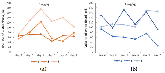
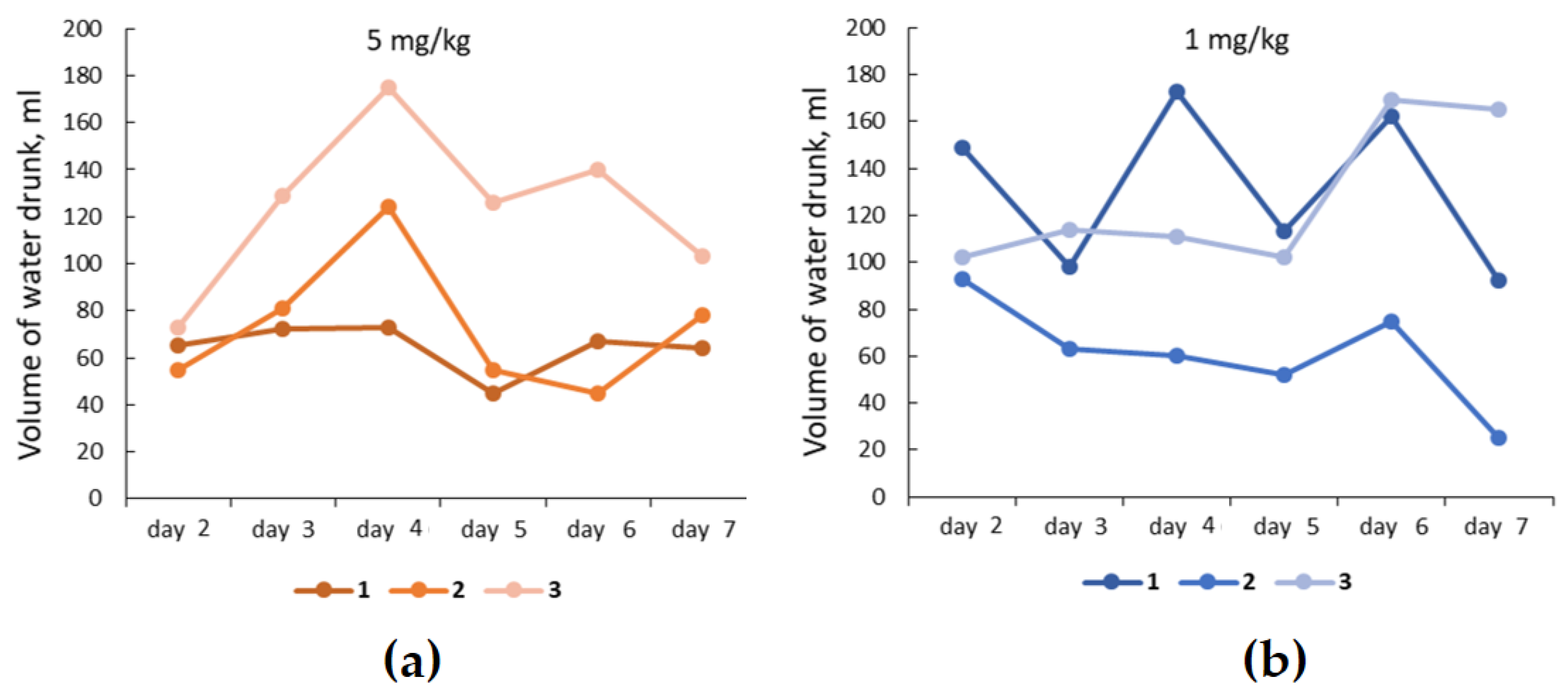
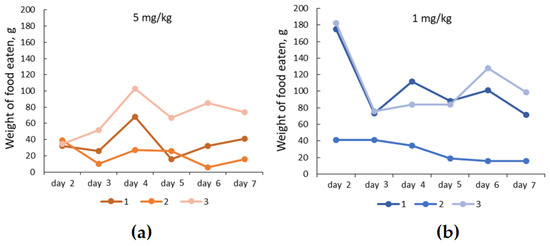
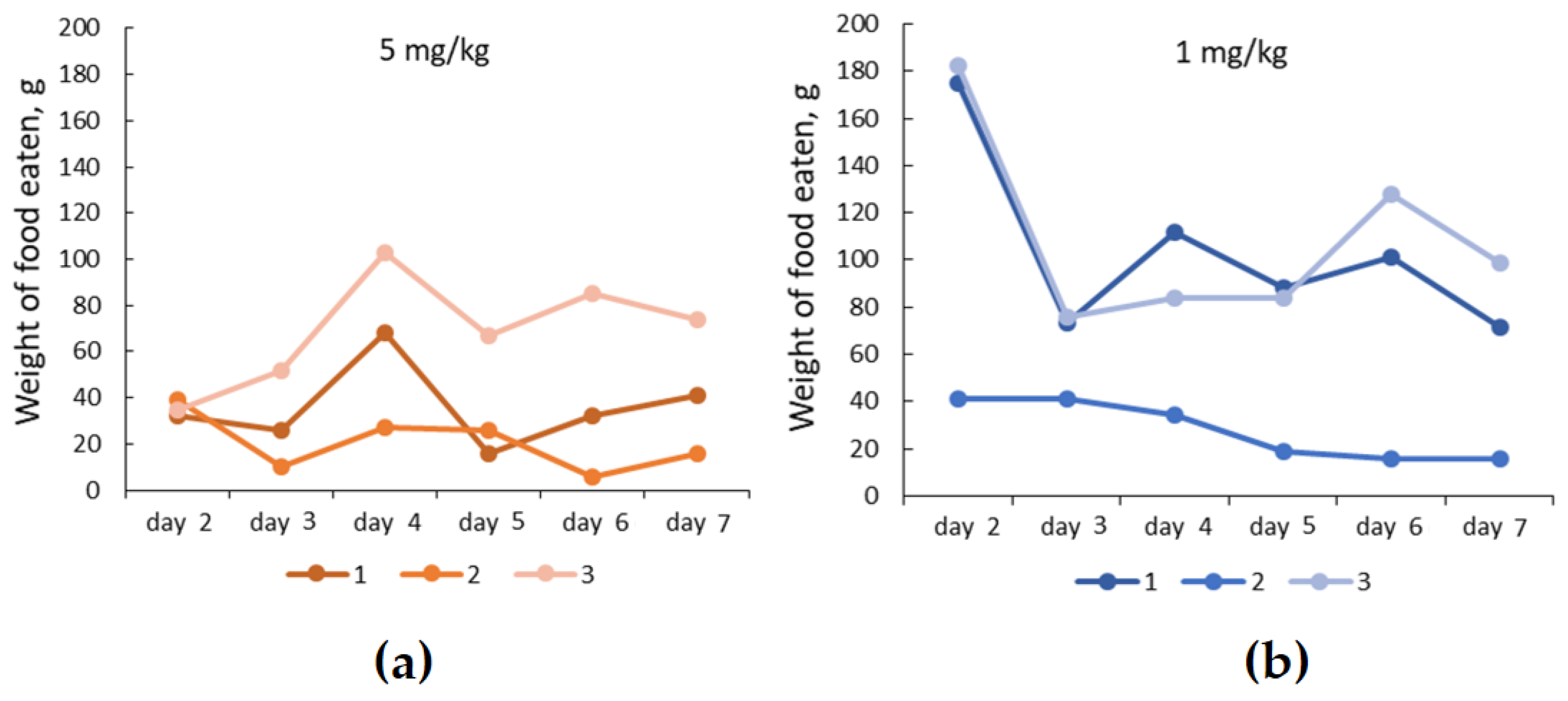
We found that the mean of the medians of daily water consumption during the experiment in the study group receiving 1 mg/kg b.w. was higher (111 ± 26 mL) than that in the group of animals receiving a higher dose of latex particles (78 ± 24 mL) (p = 0.045) (Figure 1). Conversely, the mean daily food consumption, calculated across all individual intake values pooled over the experimental period, was significantly lower in the high-dose group (42 ± 13 g) than in the low-dose group (80 ± 27 g) (p = 0.020) (Figure 2). Figure 1 and Figure 2 depict the daily trajectories of water and food intake, respectively, for each individual animal (#1, #2, and #3) over the experimental period at both administered doses of PS latex particles (5 and 1 mg/kg b.w./day).
Figure 1.
The volume of water drunk by each individual animal (#1, #2, #3) from the second day of the experiment, with the introduction of PS microparticles in dosages of 5 mg/kg b.w. (a) and 1 mg/kg b.w. (b) per day.
Figure 2.
The mass of food consumed by each individual animal (#1, #2, #3) from the second day of the experiment, with the introduction of PS microparticles in dosages of 5 mg/kg b.w. (a) and 1 mg/kg b.w. (b) per day.
The average weight of all of the animals did not change over the course of the experiment (p > 0.05), with initial and final weights measuring 5.1 ± 0.6 kg and 5.0 ± 0.6 kg, respectively.
To assess the general physical condition of the animals, rectal temperature was measured. When comparing the daily temperature of the animals between the study groups, we found no differences (p > 0.05). On the last day of the experiment, the average temperature of all rabbits was 39.1 ± 0.4 °C (p = 0.173). The visually assessed sclera was not characterized by redness (vasodilation) during the experiment in any of the study groups. The visible mucous membrane in the mouth did not change throughout the experiment, either.
Analysis of AST, urea, creatinine, and amylase levels showed no differences between the study groups (Table 1). Meanwhile, all obtained values were pooled into the expanded study group, with no changes observed (p > 0.05) in the same blood parameters (Table 2).
Table 1.
Blood parameters in the study groups. Data are presented as M ± SD; * p < 0.05.
Table 2.
Blood parameters in the expanded study group. Data are presented as Me (IQR, 25–75%); * p < 0.05.
However, in Group 1 (5 mg/kg b.w. per day), a decrease in ALT levels was observed with microplastic exposure (p = 0.034) (Table 1), with no differences in ALT levels found between the study groups on the first and last days of the experiment (p > 0.05). In addition, in the expanded study group, we observed a decrease in ALT levels by 27% by the end of the experiment (p = 0.028) (Table 2). Analysis of serum phosphorus and total calcium concentrations only showed differences in the expanded study group between the first and eighth days of the experiment. Thus, by the end of the experiment, the phosphorus level increased by 7% (p = 0.028), while the total calcium level decreased by 4% (p = 0.046).
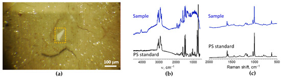
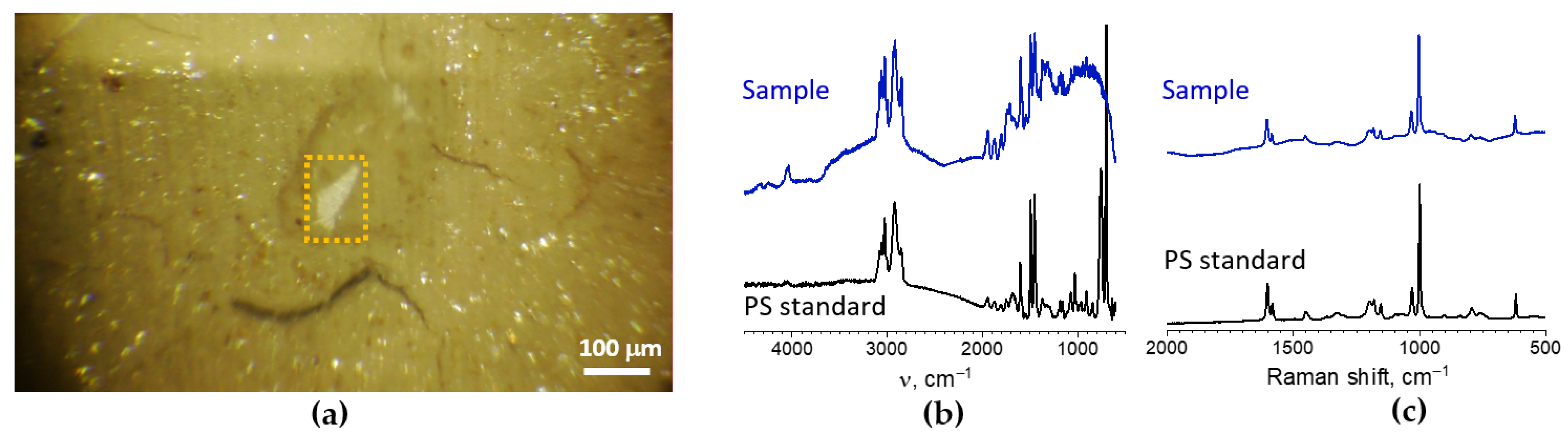
Subsequently, the sample prepared from the rabbit’s stomach was the only one where PS microparticles were identified (the samples prepared from liver, small intestine, colon, kidneys, ovaries, and uterus did not show any presence of PS). In the filtrates, all the detected PS microparticles appeared as irregularly shaped white formations with sizes ranging from ca. 40 μm to ca. 200 μm (Figure 3a). It is worth noting that the investigation of PS latex treated in ‘blank’ experiments, where PS latexes were treated without tissue samples, gave almost the same particle sizes. FTIR spectra of these particles showed a high degree of similarity with that of the PS standard, demonstrating all the main spectral bands of PS (Figure 3b). The method demonstrated coincidence of broad peaks of aromatic and aliphatic C–H bond stretching at 3110–2990 and 2960–2840 cm−1, respectively, and narrow peaks of C=C bond stretching vibrations inside the aromatic ring at 1600, 1490, and 1450 m−1.
Figure 3.
(a) Optical microphotograph of a typical PS microparticle found in the stomach sample (highlighted in a yellow box). Scale bar: 100 μm. (b) Overlaid FTIR spectra measured from PS microparticle and from the PS standard. (c) Overlaid Raman spectra measured from PS microparticle and from the PS standard.
Presence of PS in the detected particles was further corroborated by Raman spectra that appeared to be virtually identical to those of the PS standard (Figure 3c). The method demonstrated coincidence of the most pronounced narrow peaks at 1604 cm−1 (C=C stretching vibration of the aromatic ring), 1033 cm−1 (in-plane bending of the C–H bonds on the aromatic ring), and 1003 cm−1 (“breathing” mode of the aromatic ring).
Differences in the shape and size of PS microparticles found in the stomach sample indicate that filters contained the aggregates of particles formed during the sample preparation stage rather than the original single particles.
4. Discussion
The European rabbit (Oryctolagus cuniculus) is widely used as an animal model for studying infectious and non-infectious conditions, including the immune response, reproductive function, and intestinal pathologies [30,31,32]. Often, when compared to rodents, a small number of rabbits (on average 6–7 animals) is used in experimental studies [33,34,35], whereas in test groups for pharmacokinetic studies, the number of rabbits can be reduced to three [36,37] in order to determine humanized dosing regimens for a used drug. The size of these animals, their anatomy and metabolism are supposed to contribute to more successful modeling of the impact of microplastics on a living organism.
However, despite all the advantages of using rabbits as experimental subjects, there is currently no data in the open literature on the method of controlled oral administration of synthetic polymer particles to such model organisms. Moreover, methods for isolating microplastics in tissues have been described primarily for human and rodent samples [11,15]. Therefore, on the one hand, this pilot study was designed with a small sample size (n = 3 per study group) for testing the methodology for isolating microplastics from rabbit organs and assessing the feasibility of this animal model before committing to a larger-scale study. On the other hand, the eight-day exposure timing was chosen as an initial timeframe to detect early biochemical and physiological responses without expecting profound chronic or pathological changes, which typically require longer exposure times [19,20]. The primary limitation of this pilot study is the small sample size, which indicates that while the observed trends are plausible, they require validation in a larger cohort in future studies.
Recent literature demonstrates that microplastics are transported throughout the human body via the bloodstream and are found in almost all human biological organs and tissues [3,11]. Studies in rodents have shown that microplastics can also accumulate in a number of organs, disrupt the intestinal barrier, reduce mucus secretion, and cause metabolic changes and neurotoxicity [3,9,15]. However, studies of the effects of microplastics on rodents often lack information on changes in physiological parameters, which further highlights the relevance of this pilot study. The observed differences in food and water consumption of rabbits in this study may be likely accounted for by the influence of microplastic particles administered at a dose of 5 mg/kg b.w.
Microplastics can alter the intestinal microbiota composition in animals, leading to dysbiosis, as demonstrated in other models [38,39], which is likely to affect feeding behavior. Despite these, it should be noted that no changes in the weight of the animals were found by us in this pilot study, which may be due to the relatively short period of administration of microplastic particles. Likewise, in another rabbit model with free access to food containing microplastics (polyvinyl chloride), no changes in weight were observed either after one week or even after four weeks of the experiment [22]. The average temperature of the animals during this experiment is consistent with the literature and corresponds to normal values for rabbits of this breed [40].
The oral mucosa, as an easily accessible indicator and critical, non-invasive diagnostic mirror for gastrointestinal health, is of considerable importance [41]. This is particularly relevant for orally administered compounds, for which it can reveal potential local irritant effects. External parameters such as injected sclera and visible mucous membrane in the mouth did not change visually in our study, which together emphasize the absence of acute effects of exposure to the administered doses of microplastics.
At the biochemical level, several changes were observed. ALT and AST are common liver enzymes in many mammals. In rabbits, AST is widely distributed in various tissues (including skeletal muscle, cardiac muscle, and liver), while ALT is considered less specific to the liver and, like AST, has a relatively short half-life of approximately five hours [24]. It is likely that the changes in ALT levels we observed in the extended study group reflect a physiological response to microplastic particles, with a significant effect observed at a dose of 5 mg/kg b.w. Notably, feed restriction in rabbit models has been shown to result in significantly reduced serum ALT levels compared to ad libitum feeding controls [42]. Thus, this result may be related to the changes in eating behavior observed in the higher dose group. However, the absence of concomitant changes in AST levels suggests the absence of acute toxic injury to highly vascularized organs such as the liver.
We found no changes in the blood levels of nitrogen metabolites (urea, creatinine), which are excreted mainly by the kidneys, and the digestive enzyme amylase, which in rabbits, unlike other biological species, is a pancreatic enzyme [24]. The absence of changes in these parameters at different doses of PS administration, as in the expanded study group, may indicate no critical damage to the kidneys and pancreas with daily consumption of the selected doses of microplastics.
Phosphorus, in its turn, is involved in many enzymatic systems in rabbits, and the kidneys are the main organ that maintains mineral balance [24]. Blood phosphate levels should always be considered together with calcium levels, which is important for assessing calcium-phosphorus metabolism [43]. It is noted that calcium metabolism in rabbits differs from that in other animals, as far as this biological species is characterized by constant tooth growth. Rabbits absorb calcium in proportion to its concentration in the intestine, and the kidneys remove its excess [24,44]. Thus, by the end of the experiment in the expanded study group, we found a decrease in calcium levels and an increase in phosphorus levels in the blood serum. However, the magnitude of these shifts is relatively small and may fall within the range of normal physiological fluctuation. On the other hand, these alterations may be due to the effect of microplastics, for example, on the absorption of these macronutrients in the intestine. In a recent investigation, young rats orally administered microplastics for 28 days exhibited altered blood calcium and phosphorus metabolism, which was associated with impaired bone development and growth plate endochondral ossification disorder [45]. In many mammals, absorption of phosphorus and calcium occurs in the small intestine [46,47]. However, we recorded no accumulation of microplastic particles in the small intestine, which requires further study.
It is known from the literature that microplastic particles can be found in the stomach, small intestine, colon, and liver within two to four days after administration, but the particles administered in those studies were of smaller dimensions [48,49]. In our per os model, microplastic particles were indeed detected in the stomach of rabbits after eight days of administration. The fact that PS latex particles were detected in gastric tissue confirms that the selected analytical methods (FTIR spectroscopy and Raman spectroscopy) have sufficient sensitivity and specificity for working with the biomatrix of this animal species. The observed discrepancy in sizes of PS particles detected in the treated stomach (40 to 200 μm) compared to the starting latex particles (5 μm) is mainly due to the sample preparation protocol, since the same change in sizes was observed in ‘blank’ experiments where PS latexes were treated without tissue samples.
Previous studies in rodent models have demonstrated that 5 µm PS microparticles can accumulate in the liver, kidneys, and gastrointestinal tract following oral administration for 28 consecutive days and can even reach the brain following twice-weekly oral gavage over 4 weeks [9,50]. By contrast, in the present study, there was no evidence of microplastic accumulation in the liver, kidneys, colon, small intestine, or reproductive organs of rabbits, which may be associated with both the duration of administration of the PS latex suspension, the size and material of the particles. It has been shown that the absorption and translocation of particles depend on their size and that particles larger than 1 µm are absorbed to a lesser extent than smaller particles [15,51]. Evidence from in vitro indicates that polyethylene particles demonstrate markedly greater translocation efficiency relative to PS particles of identical dimensions [52]. Furthermore, some authors emphasized in their comprehensive review that the genotoxic and neurotoxic potential of plastic particles is intimately linked to intracellular uptake, which is strongly governed by particle dimensions [12].
Thus, the European rabbit (Oryctolagus cuniculus) model of microplastic effects established in our pilot study may be considered effective, as the administration of PS microparticles did not induce acute or severe pathological toxicity in the animals’ liver and kidneys. The early metabolic shifts detected on eight day likely reflect the sensitivity of the rabbit model to PS microplastic exposure. In addition, the methods for detecting PS latex particles are adequate for this model. The data obtained by us may possibly be accounted for by biochemical and physiological mechanisms described in the literature on other animal models. However, given the limitations of the present pilot study, including the small sample size and the short exposure duration, the observed metabolic changes should be regarded as preliminary. Future investigations should incorporate longer exposure periods and extended post-exposure observation windows to capture chronic and delayed effects of microplastic particles, including potential bioaccumulation, progressive organ damage, and long-term metabolic dysregulation.
5. Conclusions
Despite limitations, the conducted pilot study demonstrated that the European rabbit (Oryctolagus cuniculus) represents an effective and promising model organism for investigating the effects of microplastics under controlled oral exposure. Meanwhile, the administration of a PS latex suspension at the studied doses did not lead to the development of an acute toxic effect. Moreover, the chosen methodology (combining FTIR microscopy and Raman spectroscopy) for studying the accumulation of PS latex particles is suitable for their determination in rabbit tissues. In turn, eight-day oral administration of the suspension resulted in the occurrence of a number of metabolic and physiological changes, but they were not critical and are consistent with the literature. Thus, this animal model holds considerable promise to conduct more in-depth and prolonged studies of the effects of PS microparticles on a living organism.
Author Contributions
Conceptualization, A.B. and A.K.; Methodology, A.B., M.G. and P.C.; Formal analysis, A.M. and P.C.; Investigation, A.B., M.K., M.G. and P.C.; Writing—original draft, M.K. and A.M.; Writing—review & editing, A.M. and A.K.; Visualization, A.M. and P.C.; Project administration, A.K. All authors have read and agreed to the published version of the manuscript.
Funding
This study was supported by the Ministry of Science and Higher Education of the Russian Federation (state contract no. 075-15-2025-016 from 28.02.2025 MegaGrant).
Institutional Review Board Statement
The study was conducted in accordance with the Declaration of Helsinki and approved by the Ethics Committee of the D.O. Ott Institute of Obstetrics, Gynecology, and Reproductive Medicine, Approval No. 137, date of approval 21 October 2024.
Data Availability Statement
Data is contained within the article.
Acknowledgments
FTIR microscopy and Raman spectroscopy were carried out using equipment of the Center for Optical and Laser Materials Research in the Research Park of St. Petersburg State University, Russia.
Conflicts of Interest
The authors declare no conflicts of interest.
Abbreviations
The following abbreviations are used in this manuscript:
| ALT | Alanine aminotransferase |
| AST | Aspartate aminotransferase |
| FTIR | Fourier transform infrared microscopy |
| PS | Polystyrene |
References
- Rasmussen, S.C. From Parkesine to Celluloid: The Birth of Organic Plastics. Angew. Chem. Int. Ed. 2021, 60, 8012–8016. [Google Scholar] [CrossRef] [PubMed]
- Geyer, R.; Jambeck, J.R.; Law, K.L. Production, use, and fate of all plastics ever made. Sci. Adv. 2017, 3, e1700782. [Google Scholar] [CrossRef]
- Kutralam-Muniasamy, G.; Shruti, V.C.; Pérez-Guevara, F.; Roy, P.D. Microplastic diagnostics in humans: “The 3Ps” Progress, problems, and prospects. Sci. Total Environ. 2023, 856, 159164. [Google Scholar] [CrossRef]
- Cox, K.D.; Covernton, G.A.; Davies, H.L.; Dower, J.F.; Juanes, F.; Dudas, S.E. Human Consumption of Microplastics. Environ. Sci. Technol. 2019, 53, 7068–7074. [Google Scholar] [CrossRef]
- Shukla, S.; Khan, R.; Saxena, A.; Sekar, S. Microplastics from face masks: A potential hazard post Covid-19 pandemic. Chemosphere 2022, 302, 134805. [Google Scholar] [CrossRef]
- Eberhard, T.; Casillas, G.; Zarus, G.M.; Barr, D.B. Systematic review of microplastics and nanoplastics in indoor and outdoor air: Identifying a framework and data needs for quantifying human inhalation exposures. J. Expo. Sci. Environ. Epidemiol. 2024, 34, 185–196. [Google Scholar] [CrossRef]
- Li, J.; Green, C.; Reynolds, A.; Shi, H.; Rotchell, J.M. Microplastics in mussels sampled from coastal waters and supermarkets in the United Kingdom. Environ. Pollut. 2018, 241, 35–44. [Google Scholar] [CrossRef]
- Wong, J.K.H.; Lee, K.K.; Tang, K.H.D.; Yap, P.-S. Microplastics in the freshwater and terrestrial environments: Prevalence, fates, impacts and sustainable solutions. Sci. Total Environ. 2020, 719, 137512. [Google Scholar] [CrossRef]
- Garcia, M.M.; Romero, A.S.; Merkley, S.D.; Meyer-Hagen, J.L.; Forbes, C.; Hayek, E.E.; Sciezka, D.P.; Templeton, R.; Gonzalez-Estrella, J.; Jin, Y.; et al. In Vivo Tissue Distribution of Polystyrene or Mixed Polymer Microspheres and Metabolomic Analysis after Oral Exposure in Mice. Environ. Health Perspect. 2024, 132, 47005. [Google Scholar] [CrossRef] [PubMed]
- Leslie, H.A.; van Velzen, M.J.M.; Brandsma, S.H.; Vethaak, A.D.; Garcia-Vallejo, J.J.; Lamoree, M.H. Discovery and quantification of plastic particle pollution in human blood. Environ. Int. 2022, 163, 107199. [Google Scholar] [CrossRef]
- Roslan, N.S.; Lee, Y.Y.; Ibrahim, Y.S.; Tuan Anuar, S.; Yusof, K.M.K.K.; Lai, L.A.; Brentnall, T. Detection of microplastics in human tissues and organs: A scoping review. J. Glob. Health 2024, 14, 04179. [Google Scholar] [CrossRef]
- Casella, C.; Ballaz, S.J. Genotoxic and neurotoxic potential of intracellular nanoplastics: A review. J. Appl. Toxicol. 2024, 44, 1657–1678. [Google Scholar] [CrossRef]
- Landrigan, P.J.; Raps, H.; Cropper, M.; Bald, C.; Brunner, M.; Canonizado, E.M.; Charles, D.; Chiles, T.C.; Donohue, M.J.; Enck, J.; et al. The Minderoo-Monaco Commission on Plastics and Human Health. Ann. Glob. Health 2023, 89, 23. [Google Scholar] [CrossRef]
- Siddiqui, S.A.; Singh, S.; Bahmid, N.A.; Shyu, D.J.H.; Domínguez, R.; Lorenzo, J.M.; Pereira, J.A.M.; Câmara, J.S. Polystyrene microplastic particles in the food chain: Characteristics and toxicity—A review. Sci. Total Environ. 2023, 892, 164531. [Google Scholar] [CrossRef] [PubMed]
- Banerjee, A.; Shelver, W.L. Micro- and Nanoplastic-Mediated Pathophysiological Changes in Rodents, Rabbits, and Chickens: A Review. J. Food Prot. 2021, 84, 1480–1495. [Google Scholar] [CrossRef]
- Wu, D.; Lim, B.X.H.; Seah, I.; Xie, S.; Jaeger, J.E.; Symons, R.K.; Heffernan, A.L.; Curren, E.E.M.; Leong, S.C.Y.; Riau, A.K.; et al. Impact of Microplastics on the Ocular Surface. Int. J. Mol. Sci. 2023, 24, 3928. [Google Scholar] [CrossRef]
- Yanni, A.E. The laboratory rabbit: An animal model of atherosclerosis research. Lab. Anim. 2004, 38, 246–256. [Google Scholar] [CrossRef] [PubMed]
- Theunissen, P.T.; Beken, S.; Beyer, B.; Breslin, W.J.; Cappon, G.D.; Chen, C.L.; Chmielewski, G.; de Schaepdrijver, L.; Enright, B.; Foreman, J.E.; et al. Comparing rat and rabbit embryo-fetal developmental toxicity data for 379 pharmaceuticals: On systemic dose and developmental effects. Crit. Rev. Toxicol. 2017, 47, 402–414. [Google Scholar] [CrossRef]
- Sokolowski, K.; Turner, P.V.; Lewis, E.; Wange, R.L.; Fortin, M.C. Exploring rabbit as a nonrodent species for general toxicology studies. Toxicol. Sci. 2024, 199, 29–39. [Google Scholar] [CrossRef]
- Fennessy, P.A.; Godwin, S.; Head, G.A.; Campbell, J.H.; Campbell, G.R. Short-term and long-term cardiovascular actions of different doses of perindopril in the rabbit. Pharmacol. Res. 1996, 34, 135–141. [Google Scholar] [CrossRef]
- Jepson, M.A.; Simmons, N.L.; Savidge, T.C.; James, P.S.; Hirst, B.H. Selective binding and transcytosis of latex microspheres by rabbit intestinal M cells. Cell Tissue Res. 1993, 271, 399–405. [Google Scholar] [CrossRef]
- Papp, P.P.; Hoffmann, O.I.; Libisch, B.; Keresztény, T.; Gerőcs, A.; Posta, K.; Hiripi, L.; Hegyi, A.; Gócza, E.; Szőke, Z.; et al. Effects of Polyvinyl Chloride (PVC) Microplastic Particles on Gut Microbiota Composition and Health Status in Rabbit Livestock. Int. J. Mol. Sci. 2024, 25, 12646. [Google Scholar] [CrossRef]
- Zhang, B.; Wei, Y.; Hu, S.; Qiu, Y.; Tang, X.; Wang, S.; Sun, X. Study on the Mechanism of Induced Ovulation in Rabbits. J. Anim. Sci. 2025, 103, skaf241. [Google Scholar] [CrossRef]
- Melillo, A. Rabbit Clinical Pathology. J. Exot. Pet Med. 2007, 16, 135–145. [Google Scholar] [CrossRef]
- Tangthong, J.; Ayimbila, F.; Nakphaichit, M.; Keawsompong, S. Evaluation of Acute and Subchronic Oral Toxicity of Copra Meal Hydrolysate: A Novel Candidate for Prebiotic in Sprague Dawley Rats. J. Toxicol. 2025, 2025, 7235371. [Google Scholar] [CrossRef]
- Käppler, A.; Fischer, D.; Oberbeckmann, S.; Schernewski, G.; Labrenz, M.; Eichhorn, K.-J.; Voit, B. Analysis of Environmental Microplastics by Vibrational Microspectroscopy: FTIR, Raman or Both? Anal. Bioanal. Chem. 2016, 408, 8377–8391. [Google Scholar] [CrossRef]
- Cabernard, L.; Roscher, L.; Lorenz, C.; Gerdts, G.; Primpke, S. Comparison of Raman and Fourier Transform Infrared Spectroscopy for the Quantification of Microplastics in the Aquatic Environment. Environ. Sci. Technol. 2018, 52, 13279–13288. [Google Scholar] [CrossRef]
- Zorin, I.M.; Chelushkin, P.S.; Senchukova, A.S.; Fetin, P.A.; Kenny, J.M.; Lyulin, S.V. Study on Primary Microplastics in Cosmetics: Their Isolation, Spectral and Thermal Analysis. Anal. Methods 2025, 17, 4599–4606. [Google Scholar] [CrossRef]
- Percie du Sert, N.; Ahluwalia, A.; Alam, S.; Avey, M.T.; Baker, M.; Browne, W.J.; Clark, A.; Cuthill, I.C.; Dirnagl, U.; Emerson, M.; et al. Reporting animal research: Explanation and elaboration for the ARRIVE guidelines 2.0. PLoS Biol. 2020, 18, e3000411. [Google Scholar] [CrossRef]
- Esteves, P.J.; Abrantes, J.; Baldauf, H.-M.; BenMohamed, L.; Chen, Y.; Christensen, N.; González-Gallego, J.; Giacani, L.; Hu, J.; Kaplan, G.; et al. The wide utility of rabbits as models of human diseases. Exp. Mol. Med. 2018, 50, 1–10. [Google Scholar] [CrossRef]
- Soares, J.; Pinheiro, A.; Esteves, P.J. The rabbit as an animal model to study innate immunity genes: Is it better than mice? Front. Immunol. 2022, 13, 981815. [Google Scholar] [CrossRef]
- Fischer, B.; Chavatte-Palmer, P.; Viebahn, C.; Navarrete Santos, A.; Duranthon, V. Rabbit as a reproductive model for human health. Reproduction 2012, 144, 1–10. [Google Scholar] [CrossRef]
- Jiménez-García, A.; Balongo-García, R.; Alconero, F.F.; Araji, O.A.; Martínez, G.J.; Haba, M.G.; Morales, L.C.; Beviá, J.M.; Martínez, J.C. Intestinal wall damage in simple ileus in rabbits: Immune-modulator role of somatostatin. Hepatogastroenterology 2004, 51, 1030–1036. [Google Scholar]
- Tian, J.; Hu, S.; Sun, Y.; Ban, X.; Yu, H.; Dong, N.; Wu, J.; Yu, B. A novel model of atherosclerosis in rabbits using injury to arterial walls induced by ferric chloride as evaluated by optical coherence tomography as well as intravascular ultrasound and histology. J. Biomed. Biotechnol. 2012, 2012, 121867. [Google Scholar] [CrossRef]
- Li, T.; Tang, J.; Li, C.; Liu, G.; Li, Y.; Guo, S.; Fang, Q.; Li, J.; Qi, X.; Liu, X.; et al. Evaluating the efficacy and safety of polyglycolic acid-loading mitomycin nanoparticles in inhibiting the scar proliferation after glaucoma filtering surgery. Ann. Med. 2025, 57, 2436458. [Google Scholar] [CrossRef]
- Kumar, P.V.; Maki, M.A.A.; Wei, Y.S.; Tatt, L.M.; Elumalai, M.; Cheah, S.C.; Raghavan, B.; Majeed, A. Rabbit as an Animal Model for Pharmacokinetics Studies of Enteric Capsule Contains Recombinant Human Keratinocyte Growth Factor Loaded Chitosan Nanoparticles. Curr. Clin. Pharmacol. 2019, 14, 132–140. [Google Scholar] [CrossRef]
- Gras, E.; Vu, T.T.T.; Nguyen, N.T.Q.; Tran, V.G.; Mao, Y.; Tran, N.D.; Mai, N.H.; Dong, O.X.; Jung, D.H.; Iorio, N.; et al. Development and validation of a rabbit model of Pseudomonas aeruginosa non-ventilated pneumonia for preclinical drug development. Front. Cell. Infect. Microbiol. 2023, 13, 1297281. [Google Scholar] [CrossRef]
- Jin, Y.; Xia, J.; Pan, Z.; Yang, J.; Wang, W.; Fu, Z. Polystyrene microplastics induce microbiota dysbiosis and inflammation in the gut of adult zebrafish. Environ. Pollut. 2018, 235, 322–329. [Google Scholar] [CrossRef]
- Jin, Y.; Lu, L.; Tu, W.; Luo, T.; Fu, Z. Impacts of polystyrene microplastic on the gut barrier, microbiota and metabolism of mice. Sci. Total Environ. 2019, 649, 308–317. [Google Scholar] [CrossRef]
- Pericin, C.; Grieve, A.P. Seasonal variation of temperatures in rabbits. Lab. Anim. 1984, 18, 230–236. [Google Scholar] [CrossRef]
- Jajam, M.; Bozzolo, P.; Niklander, S. Oral manifestations of gastrointestinal disorders. J. Clin. Exp. Dent. 2017, 9, e1242–e1248. [Google Scholar] [CrossRef]
- Chen, D.; Sun, S.; Chen, Y.; Wang, J.; Sang, L.; Gao, C.; Xie, X. Effects of feeding methods on growth and slaughter performance, blood biochemical indices, and intestinal morphology in Minxinan black rabbits (Oryctolagus cuniculus). Trop. Anim. Health Prod. 2023, 55, 51. [Google Scholar] [CrossRef]
- Taylor, J.G.; Bushinsky, D.A. Calcium and phosphorus homeostasis. Blood Purif. 2009, 27, 387–394. [Google Scholar] [CrossRef]
- Varga, M. Rabbit Basic Science. In Textbook of Rabbit Medicine, 2nd ed.; Butterworth-Heinemann: Edinburgh, UK, 2014; pp. 3–108. [Google Scholar] [CrossRef]
- Zhang, Q.; Lang, Y.; Tang, X.; Cheng, W.; Cheng, Z.; Rizwan, M.; Xie, L.; Liu, Y.; Xu, H.; Liu, Y. Polystyrene microplastic-induced endoplasmic reticulum stress contributes to growth plate endochondral ossification disorder in young rat. Environ. Toxicol. 2024, 39, 3314–3329. [Google Scholar] [CrossRef]
- Liesegang, A.; Burger, B.; de Vries de Heekelingen, T.; Schroeter-Vogt, C.; Hatt, J.; Kowalewski, M.P.; Clauss, M. Rabbits (Oryctolagus cuniculus) increase caecal calcium absorption at increasing dietary calcium levels. J. Anim. Physiol. Anim. Nutr. 2024, 108, 185–193. [Google Scholar] [CrossRef]
- Borowitz, S.M.; Granrud, G.S. Ontogeny of intestinal phosphate absorption in rabbits. Am. J. Physiol. 1992, 262, G847–G853. [Google Scholar] [CrossRef]
- Sanders, E.; Ashworth, C.T. A study of particulate intestinal absorption and hepatocellular uptake: Use of polystyrene latex particles. Exp. Cell Res. 1961, 22, 137–145. [Google Scholar] [CrossRef]
- Jani, P.; Halbert, G.W.; Langridge, J.; Florence, A.T. The Uptake and Translocation of Latex Nanospheres and Microspheres after Oral Administration to Rats. J. Pharm. Pharmacol. 1989, 41, 809–812. [Google Scholar] [CrossRef]
- Deng, Y.; Zhang, Y.; Lemos, B.; Ren, H. Tissue accumulation of microplastics in mice and biomarker responses suggest widespread health risks of exposure. Sci. Rep. 2017, 7, 46687. [Google Scholar] [CrossRef]
- Fröhlich, E. Local and systemic effects of microplastic particles through cell damage, release of chemicals and drugs, dysbiosis, and interference with the absorption of nutrients. J. Toxicol. Environ. Health B Crit. Rev. 2024, 27, 315–344. [Google Scholar] [CrossRef]
- Stock, V.; Laurisch, C.; Franke, J.; Dönmez, M.H.; Voss, L.; Böhmert, L.; Braeuning, A.; Sieg, H. Uptake and cellular effects of PE, PP, PET and PVC microplastic particles. Toxicol. Vitr. 2021, 70, 105021. [Google Scholar] [CrossRef]

Disclaimer/Publisher’s Note: The statements, opinions and data contained in all publications are solely those of the individual author(s) and contributor(s) and not of MDPI and/or the editor(s). MDPI and/or the editor(s) disclaim responsibility for any injury to people or property resulting from any ideas, methods, instructions or products referred to in the content. |
© 2026 by the authors. Licensee MDPI, Basel, Switzerland. This article is an open access article distributed under the terms and conditions of the Creative Commons Attribution (CC BY) license.