


Abstract
Sunlight-driven UV weathering is a major transformation pathway of environmental microplastics, promoting surface oxidation, molecular degradation, embrittlement, and progressive fragmentation toward smaller size fractions. However, comparisons across studies remain difficult because weathering is often described using descriptors that probe different aspects of degradation without being clearly distinguished. Surface-sensitive oxidation metrics, such as carbonyl or oxidation indices (CI/OI), are frequently emphasized, whereas fragmentation and embrittlement are more directly governed by bulk molecular-weight loss, mechanical weakening, and particle-size evolution. This review examines UV weathering of common polymers through a coupled chemico-mechanical perspective relevant to the micro-to-nano transition. We distinguish surface chemical descriptors, bulk molecular and mechanical descriptors, and fragmentation-related metrics, and critically assess the analytical methods used to measure them, including FTIR, Raman spectroscopy, GPC/SEC, thermal methods, mechanical testing, and particle-size analyses. We argue that no single metric is sufficient to describe weathering progression, and that meaningful interpretation requires joint reporting of oxidation state, Mn/Mw changes, mechanical deterioration where available, and particle-size distribution as a function of cumulative or spectrum-weighted UV dose. We further propose a minimal QA/QC reporting framework including UV metadata, temperature, oxygen availability, blanks, replicates, recovery tests, and matrix-specific detection limits. By separating what different methods actually probe and linking them to fragmentation mechanisms, this review provides a more operational basis for interpreting UV-aged microplastics in environmental sampling and biomonitoring.
1. Introduction
Microplastics (MPs; ~1 µm–5 mm) are widely reported in air, surface and groundwater, soils, sediments, and biota, underscoring their relevance for exposure assessment and biomonitoring. Human biomonitoring studies have also detected polymer particles in blood and placenta, confirming internal exposure pathways and the need for standardized detection in biological matrices [1,2]. However, environmental microplastics are rarely pristine. Once released, they undergo weathering processes that modify their size, chemistry, surface properties, and analytical detectability. Among these processes, sunlight-driven UVA–B weathering is particularly important because it changes not only the surface chemistry of plastic particles, but also the form in which they are measured and interpreted in environmental and biological samples.
UV photo-oxidation introduces carbonyl and other oxygen-containing functionalities, generates peroxy and hydroperoxy intermediates, and shifts the balance between chain scission and crosslinking depending on polymer structure, formulation, and exposure conditions [3,4,5]. These reactions increase surface polarity, roughness, and brittleness, thereby promoting crack formation, fragmentation into smaller size fractions, and the release of oxidized low-molecular-weight products and dissolved organic carbon (DOC) [3,4,5,6]. Thus, UV exposure does not simply increase particle number; it progressively changes the physicochemical identity of microplastics and affects their environmental fate, analytical behaviour, and biological interpretation.
This transformation is especially important from a measurement perspective. As microplastics undergo UV aging, they may shift toward submicron and nanoscale fractions, where chemical confirmation becomes substantially more difficult [3,6]. At the same time, weathering alters the very descriptors commonly used to infer degradation state, including carbonyl/oxidation indices, molecular weight, mechanical integrity, and particle-size distribution. These descriptors, however, do not probe the same aspect of degradation. Carbonyl and oxidation indices obtained by vibrational spectroscopy primarily reflect surface or near-surface chemical change, whereas fragmentation and embrittlement are more directly linked to bulk molecular-weight loss, mechanical weakening, and the development of crack-prone structures. Consequently, UV-aged particles cannot be adequately interpreted through surface oxidation markers alone.
Detection of the smallest fractions remains a major analytical bottleneck. Routine polymer-identification workflows are still strongest in the micron range: µFTIR/FPA-FTIR performs most robustly above ~10–20 µm, whereas µRaman can extend into the low-micron range under optimized conditions [7,8,9,10]. Below ~1 µm, confident chemical confirmation usually requires lower-throughput approaches such as O-PTIR, AFM-IR, AF4-based fractionation, or combinations of thermal and spectroscopic methods [7,10,11,12,13,14,15,16]. These analytical limitations are compounded by inconsistent reporting of UV exposure conditions. Many studies still describe weathering primarily in terms of exposure time, although spectral distribution, irradiance, cumulative dose, temperature, oxygen availability, and humidity strongly influence degradation kinetics. Differences in blanks, replicates, recovery tests, sample preparation, and validated LOD/LOQ further limit comparability, especially for biological tissues and other complex matrices [17,18,19].
A more comparable interpretation of UV-weathered microplastics requires measurable degradation descriptors to be reported within a harmonized framework that distinguishes surface chemical aging, bulk molecular degradation, mechanical weakening, and fragmentation outcomes. Carbonyl and oxidation indices (CI/OI), changes in molecular weight (Mn/Mw), and shifts in particle-size distribution (PSD) are among the most practical indicators of UV-weathering progression, but they should not be treated as analytically equivalent. CI/OI values are useful markers of oxidation state, Mn/Mw changes provide more direct information on chain scission and loss of bulk molecular integrity, and PSD shifts reflect the physical outcome of fragmentation. Reporting these parameters as a function of cumulative or spectrum-weighted UV dose (MJ m−2), rather than exposure time alone, provides a more transferable basis for comparison across light sources, polymers, and environmental conditions [20].
Recent progress in QA/QC infrastructure makes such harmonization increasingly realistic. Interlaboratory initiatives and methodological guidance emphasize contamination control, matrix-specific recovery tests, validated detection limits, and the use of reference materials where available [17,18,19]. The release of EURM-060, a European Commission Joint Research Centre reference material consisting of PET microplastic particles in water, represents an important step toward improved calibration and comparability in microplastic analysis [21,22,23]. These developments support a shift from descriptive reporting of UV-aged plastics toward a measurement-oriented framework in which weathering state, analytical limitations, and QA/QC performance are considered together.
This review therefore focuses on sunlight-driven UVA–B weathering of microplastics and its implications for analytical comparability and biomonitoring. We summarize polymer-specific degradation fingerprints relevant to UV-aged particles, distinguish surface-sensitive oxidation metrics from bulk molecular and fragmentation-related descriptors, examine dose-normalized reporting, and outline a minimal QA/QC framework for complex matrices. By separating what different analytical methods actually probe, this review provides a more operational basis for interpreting UV-weathered microplastics in environmental sampling and biomonitoring studies.
2. UV-Induced Oxidation Mechanisms and Polymer-Specific Weathering Fingerprints
Solar UV weathering of plastics begins with photon absorption by chromophoric sites already present in the material or formed during prior aging, including aromatic groups, residual carbonyls, pigments, catalysts, additives, and structural defects. Excited singlet or triplet states can undergo homolytic bond cleavage or transfer energy to oxygen, generating primary macroradicals on the polymer backbone. Even polymers with weak intrinsic absorption in the solar range, such as PE and PP, may become photoresponsive once trace chromophores are introduced during processing or early thermo-oxidation. This initiation step is critical because it triggers a self-sustaining oxidation cycle that can continue beyond the initial photon event [24,25,26].
Once formed, macroradicals are rapidly trapped by molecular oxygen to produce peroxy radicals (POO•), which abstract hydrogen from neighboring chains and generate hydroperoxides (POOH) while regenerating new radicals. Hydroperoxides are key intermediates in UV aging because they are thermally and photochemically unstable and decompose into highly reactive alkoxy (PO•) and hydroxyl (HO•) radicals. These species promote β-scission of the polymer backbone, leading to shorter chains and the formation of oxygenated products such as ketones, aldehydes, and carboxylic acids. At the same time, radical recombination can produce crosslinks, so the balance between chain scission and crosslinking ultimately determines whether the material becomes brittle and fragmentation-prone or develops a more resistant partially crosslinked structure. As summarized in Figure 1, this sequence links radical initiation, oxygen uptake, hydroperoxide formation, and chain breakdown to the chemical signatures commonly used to track weathering [24,25,26,27].
Figure 1.
Schematic illustration of UV-induced oxidation and fragmentation of microplastics.
From an analytical perspective, the most immediate outcome of this process is the accumulation of oxygen-containing functional groups, particularly carbonyl species that are readily detected by vibrational spectroscopy. Growth of the C=O stretching region around ~1710–1730 cm−1 is therefore widely used to calculate carbonyl or oxidation indices (CI/OI), while concurrent reductions in Mn/Mw reflect progressive chain cleavage [24,25,26,27,28]. However, these descriptors do not probe the same aspect of degradation. CI/OI primarily describes oxidation at the surface or near-surface region, whereas Mn/Mw changes more directly reflect loss of bulk molecular integrity. This distinction is important because fragmentation is not driven by oxidation alone, but by the coupled evolution of chemical aging, molecular-weight loss, and mechanical weakening.
These chemical changes nevertheless contribute directly to physical deterioration. Oxidized polymers become more polar, rougher, and more brittle, and increasingly develop microcracks, pits, and other stress-concentrating defects. Once sufficient molecular degradation and structural weakening have occurred, even relatively mild environmental forces such as wave action, wind, or sediment abrasion can drive fragmentation into smaller particles. UV weathering should therefore be understood not only as a chemical process, but as the trigger for a coupled chemico-mechanical loop that accelerates the shift in particle populations toward the micro- and nano-size range [24,25,26,27,28].
The rate and selectivity of these reactions depend strongly on environmental conditions. UVA dominates at the Earth’s surface and sustains long-term photochemical aging, whereas UVB, although less abundant, carries higher photon energy and can be particularly effective at bond activation where it penetrates. Temperature promotes radical reactions and hydroperoxide decomposition, oxygen availability controls propagation efficiency, and moisture can either facilitate oxidation by increasing mobility in amorphous regions or dampen specific radical pathways. In real environmental settings, additional factors further modulate aging. Biofilms, mineral particles, soot, and metal-rich coatings may shield the surface, introduce photosensitizers, or catalyze radical formation, while dissolved organic matter, nitrate, and Fe(III) species in aquatic systems can generate secondary reactive oxygen species such as •OH and 1O2 that further attack the polymer surface [24,25,26,27].
Additives and fillers are also decisive in controlling early-stage stability. Commercial plastics often contain antioxidants and UV stabilizers that quench excited states or scavenge radicals, thereby extending the induction period. However, once these protective components are consumed, leached, or photolyzed, oxidation can accelerate sharply. Inorganic fillers may either retard or enhance degradation depending on their chemistry and dispersion; for example, properly treated rutile TiO2 may provide protection, whereas anatase or poorly stabilized oxides can act as photosensitizers and promote ROS generation. Because these formulation effects can substantially alter apparent degradation kinetics, meaningful comparison among studies requires dose-normalized reporting of oxidation, molecular-weight loss, and particle-size changes together with the relevant exposure metadata, including spectrum, irradiance, temperature, humidity, and oxygen availability [24,25,26,27].
From the perspective of this review, the key implication is that no single analytical descriptor is sufficient to describe UV-weathering progression. Surface-sensitive oxidation metrics are highly informative for early chemical aging, but they do not by themselves quantify embrittlement or fragmentation readiness. A more robust interpretation of the micro-to-nano transition therefore requires joint consideration of surface oxidation, bulk molecular degradation, and physical fragmentation outcomes. This distinction provides the basis for the following sections.
Polymer-Specific Analytical Fingerprints of UV Weathering
Common environmental microplastics are dominated by a limited group of high-production polymers, particularly polyethylene (PE), polypropylene (PP), polystyrene (PS), polyethylene terephthalate (PET), and polyvinyl chloride (PVC). As illustrated in Figure 2, these polymers are commonly represented in the environment by familiar debris items such as plastic bags, bottles, caps, foams, and PVC-based materials. Although all of them undergo sunlight-driven aging, their degradation pathways, analytical signatures, and fragmentation behavior are not equivalent. This matters directly for microplastic analysis, because UV weathering does not generate a single generic “aged plastic” state, but a set of polymer-specific fingerprints that influence how degradation is detected, compared, and interpreted across studies.
Figure 2.
Representative examples of common environmental plastic debris and their corresponding polymer types (PE, PP, PS, PET, and PVC).
From a measurement perspective, the most relevant outputs of UV weathering include changes in oxidation state, molecular mass, and particle-size distribution. These changes may be expressed through carbonyl/oxidation indices (CI/OI), Mn/Mw decrease, the appearance of characteristic FTIR or Raman bands, and visible or morphological changes such as yellowing, chalking, cracking, recrystallization, or dehydrochlorination. However, the balance between these outcomes depends strongly on polymer chemistry. Some polymers are dominated by chain scission, others show a greater contribution of crosslinking or gelation, and some develop highly diagnostic optical signatures before major carbonyl accumulation occurs. For this reason, UV-weathered microplastics should be interpreted polymer by polymer rather than through a single generalized degradation scheme [24,25,26,27].
PE is one of the most abundant sources of secondary microplastics in environmental compartments. Because its methylene backbone has little intrinsic absorbance in the solar range, UV aging is typically initiated by trace chromophores introduced during processing or early thermo-oxidation. Once initiated, PE follows the classical radical oxidation sequence, with chain scission generally prevailing over crosslinking. As a result, PE commonly shows progressive Mn/Mw decrease, growth of oxygenated end groups, and increasing embrittlement under prolonged UVA exposure [24,25,26,27]. Analytically, PE weathering is most often tracked by FTIR growth of the carbonyl band near ~1715 cm−1, accompanied by OH broadening and the appearance of vinyl or trans-vinylene bands associated with scission. Raman, GPC/SEC, and XPS provide complementary evidence for backbone modification, molecular-mass loss, and surface oxidation [26,27,28,29,30,31,32,33,34]. Because oxidation preferentially starts in amorphous regions, PE may also show an apparent increase in crystallinity as shorter chains recrystallize, which further promotes brittle fracture and fragmentation. For PE-rich debris, CI is therefore best interpreted together with Mn/Mw and particle-size changes when evaluating progression from weathering toward fragmentation [24,25,26,27,32,33,34,35,36].
PP undergoes faster UV oxidation than PE because hydrogen abstraction is facilitated at tertiary carbon sites along the backbone. This makes radical initiation more efficient and often accelerates early photo-oxidation under environmental UV conditions [24,25]. Like PE, PP follows the auto-oxidative radical cycle, but its degradation often reflects a stronger competition between chain scission and radical recombination, which can lead to partial gelation or crosslinking effects at advanced stages [24,25,26,27,28].
The most common analytical markers of PP weathering are growth of carbonyl bands in FTIR, the appearance of unsaturated C=C modes, and changes in Raman bands linked to CH3 groups and backbone order [26,27]. In practice, PP often exhibits a two-stage behavior: relatively rapid chemical oxidation at early exposure, followed by a later and more abrupt loss of mechanical integrity once hydroperoxide accumulation and structural destabilization reach a critical level. Accordingly, oxidation metrics in PP should not be treated as stand-alone proxies for fragmentation, but should be interpreted together with molecular-weight and mechanical evidence where available [24,25,34,37,38,39,40,41,42].
PS differs from the polyolefins because its aromatic rings act as efficient chromophores and photosensitizers. As a result, PS can undergo relatively rapid surface-dominated photo-oxidation under UVA exposure, with benzylic radical formation, yellowing, and crack development occurring at comparatively moderate doses [43,44,45]. In addition to fragmentation, PS may also contribute to dissolved transformation products and DOC, making it particularly relevant in discussions of both particle formation and chemical transformation [46].
Its degradation is analytically distinctive. FTIR detects carbonyl growth and oxygenated aromatic products, while Raman is especially useful for monitoring aromatic ring modes and conjugated structures formed during aging [43,44,47,48,49]. PS therefore offers a relatively clear spectroscopic route for identifying weathering progression, but it also tends to embrittle earlier than many other common polymers. For this reason, PS-rich samples are especially relevant when linking surface oxidation markers with crack formation, brittleness, and the release of smaller oxidized fragments [46,47,48,49,50].
PET shows a different UV-weathering behavior because of its aromatic–ester backbone. Photolysis at ester-linked carbonyl sites, including Norrish-type reactions, promotes chain cleavage, oxidation, and decline in intrinsic viscosity or molecular mass [51,52,53]. Compared with polyolefins, PET often exhibits analytically recognizable oxidation without immediately entering the same rapid fragmentation regime, because surface-confined oxidation and secondary crystallization can delay visible mechanical collapse [53,54,55,56].
In spectroscopic terms, PET weathering is reflected in changes in the carbonyl and C–O–C regions, OH growth, and XPS-detectable increases in surface oxygen functionalities [51,52,53,54]. With continued exposure, shorter chains may recrystallize, stiffening the matrix and preconditioning it for later crack propagation [53,56]. For environmental microplastic analysis, PET is therefore best interpreted using combined oxidation and molecular-mass or viscosity-related descriptors, rather than oxidation markers alone [51,52,53,54,55,56].
PVC follows yet another pathway, with dehydrochlorination preceding extensive oxidation. Under solar exposure, elimination of HCl generates conjugated polyene sequences that absorb increasingly strongly in the UV–visible range, producing the characteristic yellow-to-brown discoloration of weathered PVC [24,25,26,27,48,50,54,56]. This early-stage chemistry means that PVC can develop strong optical and spectroscopic weathering signals even before carbonyl accumulation becomes dominant.
Its most diagnostic markers include growth of C=C-related bands, polyene-associated UV–Vis shifts, and resonance-enhanced Raman signatures associated with conjugation length. Secondary oxidation later introduces OH and carbonyl features, while HCl evolution can serve as a kinetic indicator of degradation [48,52,53,57]. From a measurement perspective, PVC is especially important because its weathering history depends strongly on formulation, stabilizers, pigments, and fillers. As a result, apparently similar PVC particles may show very different degradation behavior depending on additive depletion and prior exposure [49,54,57]. The main polymer classes differ in their UV-reactive structural motifs and degradation pathways, as summarized schematically in Figure 3 and comparatively in Table 1.
Figure 3.
Polymer repeat units and main UV-reactive/degradation-prone sites relevant to weathering.
Table 1.
Polymer-specific UV-weathering fingerprints and descriptor classes relevant to microplastic analysis [24,25,26,27,28,29,30,31,32,33,34,35,36,37,38,39,40,41,42,43,44,45,46,47,48,49,50,51,52,53,54,55,56,57].
Overall, these polymers do not weather in analytically equivalent ways. PE and PP are dominated by oxidation and scission chemistry characteristic of polyolefins, PS shows rapid aromatic photo-oxidation and earlier embrittlement, PET is shaped by ester-linked chain cleavage and secondary crystallization, and PVC is distinguished by early dehydrochlorination and polyene formation. These contrasts are not merely mechanistic; they directly affect which degradation markers are most informative, how weathering progression should be compared, and how fragmentation propensity is inferred from analytical data. To make these differences operational, Table 1 summarizes the dominant UV-weathering pathways, key analytical markers, and typical degradation trends for PE, PP, PS, PET, and PVC, with emphasis on their relevance for microplastic studies [24,25,26,27,28,29,30,31,32,33,34,35,36,37,38,39,40,41,42,43,44,45,46,47,48,49,50,51,52,53,54,55,56,57].
Table 1 emphasizes that polymer-specific UV-weathering fingerprints should not be interpreted through surface oxidation markers alone. For each polymer, fragmentation inference requires integration of surface-sensitive descriptors with bulk molecular or mechanical evidence and particle-level metrics such as PSD, particle number, crack formation, or morphology.
3. Micro-to-Nano Transformation as a Chemico-Mechanical Process
The transformation of microplastics (MPs) into progressively smaller particles, including submicron and nanoscale fractions, is one of the most important consequences of environmental weathering. This transition is not a single-step process, but the cumulative result of surface oxidation, loss of molecular integrity, and mechanical fragmentation. Its significance lies not only in particle-size reduction, but also in associated changes in oxidation state, molecular mass, surface reactivity, and analytical detectability [15,24,25].
As described in the previous section, UV-induced oxidation introduces oxygen-containing functional groups and promotes polymer-chain scission. However, for understanding the micro-to-nano transition, it is essential to distinguish surface oxidation markers from descriptors of bulk degradation. CI/OI primarily reflects surface or near-surface oxidation, whereas Mn/Mw changes more directly indicate loss of bulk molecular integrity. This distinction is important because fragmentation is not governed by oxidation alone, but by the coupled development of chemical and mechanical degradation.
As aging progresses, polymers lose ductility and develop microcracks, voids, and other stress concentrators. Under these conditions, even moderate environmental forces such as wave action, sediment abrasion, or freeze–thaw cycling can break the weakened material into progressively smaller fragments [24,58,59,60,61,62,63]. This establishes a chemico-mechanical feedback loop: newly generated fragments expose fresh surface area, which promotes further oxidation and continued size reduction. Over time, particle-size distributions shift toward the submicron and nano range [63,64,65,66,67].
This transition is also analytically significant. As particle size decreases, chemical confirmation becomes more difficult, while weathering simultaneously alters the descriptors used to interpret degradation state, including oxidation indices, molar-mass trends, and particle-size distributions. In parallel, prolonged UV exposure may generate dissolved transformation products and DOC, so the transition should not be viewed only as a decrease in size, but as a broader redistribution of polymer mass across particulate and dissolved fractions [4,40,46].
From the perspective of biomonitoring and interstudy comparability, the central challenge is therefore to relate the micro-to-nano transition to measurable descriptors without conflating surface chemical aging with bulk fragmentation readiness. In the following sections, we distinguish between surface-sensitive oxidation metrics, bulk molecular-weight and structural descriptors, and physical fragmentation outcomes, and discuss their polymer dependence and the environmental factors that control their interpretation [20,24,59,63,67].
3.1. Quantitative Descriptors: CI/OI, Mn/Mw, Mechanical Weakening, and PSD
A quantitative description of the micro-to-nano transition should link chemical aging, loss of molecular integrity, and progressive particle-size reduction. In practice, the most useful descriptors are carbonyl/oxidation indices (CI/OI), changes in molar mass (Mn/Mw), and shifts in particle-size distribution (PSD), especially when these parameters are related to spectrum-weighted UV dose rather than exposure time alone [20,24,25,67].
Among these descriptors, CI is one of the most widely used markers of photo-oxidation because it reflects the accumulation of oxygenated functionalities during UV aging. In contrast, decreases in Mn and Mw more directly indicate chain scission and loss of bulk molecular integrity. PSD evolution, in turn, reflects the physical outcome of fragmentation. These descriptors should therefore not be treated as analytically equivalent. Rather, the transition toward accelerated fragmentation is best interpreted as a coupled process in which surface oxidation, molar-mass loss, mechanical weakening, and size reduction evolve together [24,25,59,67].
As aging progresses, polymers lose ductility and develop microcracks, voids, and other stress concentrators. Where available, these trends should be interpreted together with direct mechanical descriptors such as tensile strength or elongation at break, because fragmentation is governed not by oxidation alone but by the combined development of chemical and mechanical deterioration [24,25,59,67].
Kinetic approaches are useful for describing this progression, but they should be interpreted with caution. Arrhenius-type relationships are often used to describe temperature-dependent acceleration of photo-oxidation, and reported activation energies generally fall within ranges consistent with radical-mediated oxidation and hydroperoxide decomposition [24,25,67]. However, the apparent rate of degradation depends not only on temperature, but also on irradiance, spectral distribution, oxygen availability, humidity, polymer formulation, and the presence of additional stressors. Accordingly, quantitative thresholds reported in the literature should be treated as indicative rather than universal.
A similar caution applies to fragmentation thresholds expressed through CI. In some studies, values around 0.15–0.20 for polyolefins have been reported together with substantial loss of tensile performance and the onset of more rapid fragmentation, but these values vary with polymer type, thickness, additives, and exposure conditions [53,60]. Thus, CI is best used as a comparative weathering indicator, not as a fixed universal criterion that automatically predicts fragmentation in all environmental settings. Accordingly, oxidation markers derived from FTIR or Raman spectroscopy should be interpreted as indicators of chemical weathering progression, not as stand-alone predictors of fragmentation.
PSD evolution provides a complementary descriptor of the transition. Weathering commonly shifts PSDs toward smaller median sizes, while the number of particles increases disproportionately relative to the original mass. Log-normal descriptions are often sufficient for empirical comparison, whereas population-balance approaches are useful when fragmentation and aggregation are considered simultaneously [63,66]. In both cases, PSD data become most informative when interpreted together with oxidation and molar-mass trends.
Overall, the most robust quantitative approach is to combine CI/OI, Mn/Mw, and PSD changes and to report them as a function of spectrum-weighted UV dose, together with essential metadata such as temperature, relative humidity, and oxygen availability [20,61,67]. In this form, quantitative descriptors do not define a universal fragmentation law, but they do provide a practical basis for comparing weathering progression across polymers, exposure conditions, and analytical workflows.
3.2. Polymer-Dependent Transition Tendencies and Indicative Timescales
The micro-to-nano transition does not proceed at the same rate, or through the same dominant descriptors, for all polymers. Differences in backbone chemistry, chromophore content, stabilizer package, crystallinity, and formulation lead to substantial variability in UV sensitivity, oxidation patterns, molecular-weight loss, and fragmentation behavior. For this reason, polymer-specific comparisons are more informative when expressed as relative transition tendencies rather than as fixed universal timescales [24,25,26,27,53,68].
Among the common environmental polymers, PP and PS are generally considered more UV-sensitive than PE under comparable exposure conditions, although the mechanisms differ. In PP, radical initiation is facilitated by tertiary C–H bonds, which tends to accelerate oxidation and embrittlement relative to PE. In PS, aromatic chromophores promote efficient light absorption and rapid surface oxidation, which can lead to yellowing, crack formation, and early fragmentation at comparatively modest doses [24,25,26,27,37,38,39,40,41,68]. PET also undergoes substantial UV-induced degradation, but through ester-linked chain cleavage and oxidation of its aromatic–ester backbone, while secondary crystallization may influence the timing of visible mechanical failure [42,43,44,45,46,47,51,52,53,54,55,56]. PVC follows a distinct pathway in which dehydrochlorination and polyene formation dominate early weathering, making optical and spectroscopic changes particularly important, while fragmentation remains strongly dependent on formulation and stabilizer depletion [53].
Because of these differences, the literature supports only indicative—not universal—timescales for transition toward smaller and more bioavailable fractions. Thin films, foils, and fibers generally weather and fragment more rapidly than bulkier items, while outdoor timescales vary strongly with climate, UV dose, humidity, oxygen supply, and mechanical forcing. Accordingly, statements such as “months” or “years” should be interpreted as exposure-dependent ranges rather than fixed polymer constants.
From an analytical perspective, the most useful conclusion is therefore not that one polymer always reaches the nano-fraction after a specific number of months, but that different polymers require different primary descriptors of transition. For PE and PP, CI/OI are useful indicators of oxidation progression, but they are most informative when interpreted together with Mn/Mw loss, crystallinity changes, and PSD evolution. For PS, aromatic and oxidation-related spectroscopic changes may provide particularly sensitive evidence of early weathering, but these surface-dominated signals should not be taken alone as proof of bulk fragmentation readiness. For PET, oxidation metrics are most informative when paired with viscosity- or molar-mass-related indicators, whereas for PVC, polyene growth, discoloration, and HCl-related degradation signatures may be more informative than CI alone [24,25,26,27,42,43,44,45,46,47,48,49,50,51,52,53,54,55,56,57].
These polymer-dependent tendencies matter because they shape both analytical expectations and exposure interpretation. Polymers that oxidize and embrittle rapidly may contribute disproportionately to small-fragment and nanoplastic inventories, even if they are not the most abundant by mass. Conversely, polymers with slower or more surface-confined oxidation may remain detectable as larger particles for longer periods while still undergoing substantial chemical transformation. For this reason, the transition from microplastics to nanoplastics should be interpreted polymer by polymer and through a combination of surface-sensitive oxidation metrics, bulk molecular-weight or structural descriptors, and fragmentation-related particle-size changes, rather than through any single weathering marker. To summarize these polymer-dependent differences in UV sensitivity, dominant descriptor classes, and indicative fragmentation tendencies, Table 2 provides a comparative overview for the most common environmental polymers.
Table 2.
Polymer-dependent UV sensitivity, dominant descriptor classes, and indicative fragmentation tendencies [24,25,26,27,32,33,34,35,36,37,38,39,40,41,42,43,44,45,46,47,48,49,50,51,52,53,54,55,56,57,68].
3.3. Biofilm as a Modifier of UV Weathering and Fragmentation
Biofilm formation can substantially modify the micro-to-nano transition of environmental plastics. As microorganisms colonize the particle surface and produce extracellular polymeric substances (EPS), they create a dynamic interfacial layer that alters light exposure, oxygen transfer, moisture retention, and local chemical conditions. In this way, biofilm does not represent an independent degradation pathway, but an important modifier of UV-driven weathering and subsequent fragmentation [69,70,71,72].
In the early stages, biofilm may slow photo-oxidation by partially shielding the polymer surface from incident radiation and limiting oxygen diffusion to the interface. The EPS matrix can scatter and absorb part of the incoming light, while the attached biomass may reduce access of oxygen to reactive sites, thereby prolonging the induction period of oxidation [62,73,74]. This effect is likely to be most pronounced in nutrient-rich aquatic environments, where surface colonization proceeds rapidly and particles may become extensively covered.
As the biofilm matures, however, its role may shift. Microbial communities can promote oxidation indirectly through the production of extracellular enzymes, siderophores, pro-oxidative metabolites, and localized chemical gradients. These processes may enhance carbonyl formation, increase surface roughness, and create chemically heterogeneous surface “hot spots” that facilitate crack initiation and propagation [62,69,73,74]. Biofilm also retains water and modifies the local redox environment, which may further influence oxidation kinetics and shorten the time required for a weathered surface to become mechanically fragile.
Biofilm can additionally affect the physical behavior of small fragments once they are generated. By altering roughness, stickiness, and surface charge, it may promote aggregation or attachment to other particles, thereby changing sedimentation, transport, and residence time [72,75]. These effects are especially relevant when interpreting particle-size distributions in natural samples, because the observed size spectrum may reflect not only fragmentation itself, but also post-fragmentation aggregation and surface modification.
Overall, biofilm should be regarded as an important environmental modifier of UV weathering rather than as a separate process. Its net effect may be either retarding or accelerating, depending on biofilm maturity, environmental conditions, and polymer type. For this reason, studies of the micro-to-nano transition should consider biofilm status explicitly when comparing oxidation metrics, fragmentation tendencies, and PSD changes under environmentally realistic conditions [62,69,72,73,74,75]. Biofilm effects highlight that UV weathering and fragmentation are strongly environment-dependent processes. The following section therefore considers the broader environmental factors that control the rate of the micro-to-nano transition.
3.4. Environmental Factors Controlling Transition Rates
The rate of the micro-to-nano transition is strongly controlled by environmental conditions. Although UV-driven oxidation provides the initial chemical preconditioning, the extent and pace of fragmentation depend on the combined effects of light regime, temperature, oxygen availability, humidity, water chemistry, and mechanical forcing. Transition rates reported in the literature should therefore be interpreted as condition-dependent outcomes rather than fixed material constants [20,59,61,63,70,76].
Solar irradiance is a primary driver because both UV intensity and spectral composition determine the effective photochemical dose received by the polymer surface. Regions with higher annual UV flux, stronger seasonal peaks, or reduced atmospheric attenuation expose plastics to larger spectrum-weighted UV doses over the same nominal exposure period [20,59,63]. For this reason, exposure time alone is insufficient for comparing degradation across studies; dose-normalized descriptors provide a more transferable basis for linking laboratory and field observations.
Temperature further modifies transition rates by accelerating radical reactions, hydroperoxide decomposition, and the associated loss of molecular and mechanical integrity. Oxygen availability controls the efficiency of oxidative propagation, while relative humidity and water exposure influence oxidation kinetics, particularly in amorphous domains and at weathered interfaces [61,67]. Thus, two samples exposed to similar nominal UV conditions may degrade differently if they experience different thermal, oxygenation, or moisture regimes.
In aquatic environments, the effective role of UV radiation also depends on optical conditions such as water depth, transparency, dissolved organic matter (DOM), and turbidity. Clear waters allow deeper penetration of photochemically active radiation, whereas DOM-rich or turbid systems attenuate UV and may shift the relative importance toward mechanical abrasion, biofilm-mediated modification, or sediment interactions [59,63]. Hydrodynamic forcing, wave action, sediment abrasion, and repeated wetting–drying cycles can then accelerate size reduction once oxidation has weakened the polymer structure.
On land and in coastal zones, additional physical stressors such as sediment grinding, freeze–thaw cycles, wind-driven abrasion, and storm-related disturbance may further promote fragmentation. These processes do not replace UV weathering, but amplify its consequences by acting on already oxidized and embrittled materials. The micro-to-nano transition should therefore be understood as an environmentally controlled chemico-mechanical process, rather than as a purely photochemical one.
From an analytical and QA/QC perspective, this means that environmental metadata are essential for interpreting transition rates. At minimum, studies should report spectrum-weighted UV dose, irradiance spectrum, temperature, relative humidity or water exposure, oxygen availability, and the main sources of mechanical forcing relevant to the exposure scenario [20,61]. In aquatic systems, DOM, turbidity, and optical attenuation should also be documented where possible. Such reporting is necessary to compare fragmentation trends meaningfully across polymers, habitats, and experimental designs.
3.5. Implications of the Micro-to-Nano Transition for Biomonitoring
The micro-to-nano transition has important implications for biomonitoring because decreasing particle size is accompanied by changes in mobility, surface reactivity, bioavailability, and analytical detectability. Nanoplastics and submicron fragments are not simply smaller versions of larger microplastics; their higher surface-area-to-volume ratio, colloidal behavior, and often more oxidized surfaces can alter their interactions with natural organic matter, pollutants, biofilms, and biological membranes [63,65,77].
From an exposure-assessment perspective, the formation of smaller and more oxidized particles may increase the probability of uptake by organisms and may affect particle retention, translocation, and trophic transfer. Weathered nanoplastic fractions can also interact with metals, persistent organic pollutants, pharmaceuticals, or microbial components, potentially modifying contaminant transport and mixture exposure [63,65,78]. However, the extent of these effects depends strongly on polymer type, particle size, surface oxidation state, aggregation behavior, environmental matrix, and biological model. Therefore, nanoplastic hazard should not be inferred from particle size alone.
For the purpose of this review, the key point is not to provide a complete toxicological assessment, but to emphasize that UV-induced fragmentation changes the fraction of plastic particles that is most difficult to detect and most relevant for biomonitoring interpretation. As particles approach the submicron and nanoscale range, conventional µFTIR and Raman workflows become less reliable, while particle number, surface area, and oxidation state may become more informative than mass concentration alone. This creates a direct link between weathering metrics, analytical limitations, and biological interpretation.
Accordingly, biomonitoring studies should not only report polymer identity and particle abundance, but should also document weathering state and size distribution wherever possible. A combined framework including oxidation descriptors, molecular or mechanical degradation indicators, and particle-size metrics would improve the interpretation of whether detected particles represent relatively pristine debris, weathered microplastics, or fragments approaching the nanoplastic range. Such reporting is essential for connecting environmental sampling with realistic exposure assessment and for avoiding underestimation of the smaller, more analytically challenging fractions.
3.6. Harmonized Reporting of the Micro-to-Nano Transition: Current Needs and Practical Priorities
The comparison of micro-to-nano transition studies remains limited by the lack of harmonized reporting. Many published experiments report oxidation indices, molar-mass loss, or particle-size changes, but these data are often not directly comparable because exposure conditions and analytical metadata are described inconsistently. As a result, fragmentation trends observed under laboratory irradiation, outdoor weathering, and environmentally realistic mixed-stressor conditions are difficult to align across studies [20,61,62].
Among the available exposure descriptors, spectrum-weighted UV dose provides a practical basis for interstudy comparison because it accounts for differences in spectral distribution more effectively than exposure time alone [20,61]. However, cumulative dose is not sufficient by itself. The same total UV dose may be delivered under different irradiance intensities or exposure rates, and this can influence oxidation kinetics, hydroperoxide decomposition, crack formation, and embrittlement. High-intensity irradiation may accelerate surface oxidation and mechanical deterioration compared with lower-intensity exposure at the same cumulative dose, especially when oxygen transport, temperature, or radical recombination become rate-limiting. Therefore, UV intensity should be reported together with cumulative spectrum-weighted dose.
UV exposure metadata should therefore include the wavelength range, irradiance spectrum, average and/or peak irradiance intensity, exposure rate, exposure duration, photoperiod or irradiation schedule, and cumulative spectrum-weighted dose. Where possible, irradiance should be reported separately for UVA and UVB regions, using SI units such as W m−2 for irradiance and MJ m−2 for cumulative dose. Reporting only exposure time, or only cumulative dose without intensity, limits the comparability of laboratory and field-weathering studies.
UV exposure parameters are informative only when interpreted together with degradation and fragmentation descriptors, particularly CI/OI, Mn/Mw loss, mechanical weakening where available, and PSD shifts. Reporting these parameters in parallel is more useful than presenting any single metric in isolation, because the transition to smaller fractions reflects coupled surface oxidation, bulk molecular degradation, mechanical deterioration, and particle fragmentation rather than one independent process.
Comparability is further limited when environmental boundary conditions are not reported in sufficient detail. Temperature, relative humidity, oxygen availability, and relevant mechanical stress strongly influence oxidation kinetics, embrittlement, and subsequent fragmentation. In aquatic systems, optical attenuation, dissolved organic matter (DOM), and turbidity may additionally determine the effective photochemical conditions experienced by the particles [20,59,61,63]. Without such metadata, apparent differences between studies may reflect exposure design rather than genuine polymer behaviour.
Recent initiatives, including interlaboratory harmonization efforts and the development of reference materials such as EURM-060, represent important progress toward more consistent microplastic analysis [17,21,23]. Nevertheless, important gaps remain. Available reference materials are still limited in polymer diversity and are particularly scarce in the submicron range, where the analytical challenges are greatest. In addition, many studies still examine UV aging in isolation, whereas environmental fragmentation often reflects the combined action of light, mechanical stress, and surface colonization [79,80].
At present, the most realistic near-term goal is not a single universal fragmentation threshold, but improved consistency in how transition studies are reported and interpreted. For this reason, studies addressing the micro-to-nano transition should, at minimum, document UV spectrum, wavelength range, irradiance intensity or exposure rate, exposure duration, photoperiod or irradiation schedule, cumulative spectrum-weighted UV dose, and key environmental metadata. A practical minimum reporting set should include: (i) UV spectrum/wavelength range, UVA/UVB irradiance intensity, exposure rate, exposure duration, photoperiod, and cumulative spectrum-weighted dose; (ii) temperature, relative humidity or water exposure, and oxygen availability; (iii) at least one oxidation-related descriptor such as CI/OI; (iv) one molecular-integrity descriptor such as GPC/SEC-derived Mn/Mw where feasible; (v) one mechanical or structural descriptor, such as tensile strength, elongation at break, modulus, or crystallinity change where feasible; and (vi) one fragmentation descriptor such as PSD shift, particle-count change, or crack/morphology evidence. Where relevant, additional environmental modifiers such as DOM/turbidity, abrasion regime, optical attenuation, or biofilm status should also be reported.
In this form, harmonization is best understood as a tool for improving comparability rather than as a final regulatory endpoint. More consistent reporting would make it easier to interpret polymer-dependent transition tendencies, compare laboratory and field studies, and identify which datasets are robust enough to inform biomonitoring and exposure assessment. For the micro-to-nano transition in particular, better harmonization is essential not because it eliminates variability, but because it makes that variability interpretable [17,20,21,23].
4. Detection and Characterization of the Micro-to-Nano Plastic Transition
Understanding the transition of microplastics into submicron and nanoscale plastic fractions requires methods that probe different, non-equivalent levels of degradation. Surface-sensitive techniques such as FTIR and Raman spectroscopy provide information on oxidation state and functional-group formation, but they do not directly quantify bulk molecular degradation or fragmentation readiness. In contrast, GPC/SEC, intrinsic viscosity, and mechanical testing provide more direct information on molecular-weight loss, chain scission, embrittlement, and loss of material integrity, while particle-size and imaging methods document whether fragmentation has actually occurred.
Therefore, the micro-to-nano transition cannot be characterized by a single analytical method or descriptor. A robust workflow should combine: (i) surface chemical descriptors, such as CI/OI; (ii) bulk molecular or mechanical descriptors, such as Mn/Mw, elongation at break, modulus, or crystallinity changes; and (iii) fragmentation descriptors, such as PSD shifts, particle-count increase, crack formation, or nanoscale particle release. This distinction is essential because surface oxidation may precede, accompany, or sometimes exceed bulk degradation, depending on polymer type, formulation, morphology, and exposure conditions [20,59,63,79].
A critical comparison of analytical platforms also requires separating spatial resolution, chemical specificity, throughput, and matrix applicability. Conventional µFTIR and FPA-FTIR remain powerful for relatively high-throughput polymer identification in the micron range, but their practical performance declines for particles below approximately 10–20 µm because of diffraction limits, weak absorbance, and matrix interference. Raman microscopy can reach smaller particles under optimized conditions, but fluorescence from weathered polymers, pigments, biofilms, or organic residues may limit spectral quality. O-PTIR and AFM-IR partly overcome the spatial-resolution bottleneck by enabling submicron or nanoscale chemical mapping, and O-PTIR combined with Raman has been shown to improve microplastic identification in environmental, nutritional, and biological matrices [80]. However, these methods remain lower-throughput, more expensive, and less standardized than conventional µFTIR/Raman workflows. Therefore, they are best interpreted as high-resolution correlative tools rather than direct replacements for routine screening methods.
4.1. Surface Chemical Descriptors: FTIR and Raman Spectroscopy
Vibrational spectroscopy is central for identifying polymer type and assessing chemical weathering, particularly through the detection of oxygen-containing functional groups formed during photo-oxidation. µFTIR, including ATR-FTIR and FPA-FTIR imaging, is widely used to monitor carbonyl, hydroxyl, ester, and unsaturated groups, and to calculate carbonyl or oxidation indices (CI/OI) as indicators of surface or near-surface oxidation [81,82,83]. These descriptors are especially useful for comparing oxidative aging within a given polymer system and exposure design.
However, CI/OI should not be interpreted as direct proxies for fragmentation. In some polyolefin studies, increasing CI has been reported together with molecular-weight loss and reduced mechanical performance, but this relationship is not universal. The connection between oxidation state and fragmentation depends on polymer type, thickness, crystallinity, additive package, prior aging, and exposure conditions. Therefore, CI/OI values are best treated as early chemical-aging descriptors that require interpretation together with bulk molecular-weight data, mechanical descriptors, and particle-size metrics.
Raman spectroscopy provides complementary information, particularly for polymers with aromatic or conjugated structures such as PS and PET. It offers higher spatial resolution than conventional µFTIR and can be useful for detecting changes in aromatic ring modes, backbone order, and conjugated oxidation products. Raman spectroscopy is also less affected by water than FTIR, which can be advantageous for hydrated or biofilm-associated samples. Nevertheless, fluorescence from organic matter, pigments, additives, or biofilm components can obscure Raman signals and limit its applicability in complex environmental matrices [83,84,85,86,87].
Combining µFTIR and Raman mapping on the same particles can improve confidence in polymer identification and surface-aging assessment, especially when chemical changes are linked with visible cracks, pits, or surface roughening. However, such multimodal spectroscopic workflows still mainly describe chemical and near-surface structural changes. They should therefore be combined with molecular-weight, mechanical, and particle-size analyses when the aim is to evaluate the micro-to-nano transition rather than chemical oxidation alone.
Polymer-specific fingerprints should also be interpreted mechanistically rather than only descriptively. For example, PP is generally more UV-sensitive than PE because hydrogen abstraction is facilitated at tertiary carbon sites, which promotes faster radical formation, carbonyl growth, and mechanical deterioration. In PS, aromatic rings act as chromophores and photosensitizers, making Raman and UV–Vis-sensitive aromatic/conjugated bands particularly informative. In PET, ester-linked chain cleavage and changes in carbonyl/C–O–C regions should be interpreted together with intrinsic viscosity or molecular-weight data, whereas PVC weathering is often dominated initially by dehydrochlorination and polyene formation. Thus, the most informative spectroscopic marker depends on polymer chemistry and should not be interpreted independently of bulk or particle-level descriptors.
4.2. Bulk Molecular-Weight and Mechanical Descriptors
Because fragmentation and embrittlement are governed by the loss of bulk molecular integrity, molecular-weight and mechanical descriptors are essential complements to surface-sensitive spectroscopic methods. Gel permeation chromatography or size-exclusion chromatography (GPC/SEC) provides direct information on changes in number-average and weight-average molecular weight (Mn and Mw), as well as dispersity. These parameters are particularly important for identifying chain scission during UV aging and for distinguishing surface oxidation from deeper molecular degradation [88,89]. This distinction is essential because carbonyl or oxidation indices may indicate surface photo-oxidation, but they do not necessarily quantify the extent of bulk chain scission or the mechanical readiness of a material to fragment [90].
SEC coupled with multi-angle light scattering (SEC-MALS) can provide more absolute molar-mass information and reduce dependence on polymer calibration standards. However, GPC/SEC-based methods require polymer dissolution and are therefore limited by solvent compatibility, extraction efficiency, and the presence of crosslinked, gelled, or highly oxidized insoluble fractions. These limitations are especially relevant for aged polyolefins, where oxidation, branching, or crosslinking may produce fractions that are not fully soluble under standard analytical conditions. Thus, GPC/SEC is highly informative for molecular degradation, but its results should be interpreted in relation to polymer solubility, sample recovery, and the possible formation of insoluble residues [88,89,90].
For polyester-based materials such as PET, intrinsic viscosity can serve as a practical descriptor of molecular degradation because viscosity loss reflects chain scission and reduction in average molecular size. Intrinsic-viscosity measurements are therefore particularly useful when evaluating PET aging, where molecular degradation may occur before extensive visible fragmentation. In PET studies, intrinsic viscosity is often interpreted together with DSC, XRD, or SEC data to connect molecular degradation with crystallinity changes, structural reorganization, and loss of mechanical performance [91,92].
Mechanical testing provides the most direct evidence of embrittlement and fragmentation readiness. Tensile strength, elongation at break, Young’s modulus, and dynamic mechanical analysis (DMA) can reveal the loss of ductility and the development of brittle behavior during UV aging. These measurements are especially informative for films, fibers, pellets, and standardized test specimens, where mechanical failure can be related to molecular-weight loss and oxidation state [89,93]. However, they are often difficult to apply to irregular environmental particles because sample geometry, thickness, prior weathering history, and particle heterogeneity strongly affect mechanical response. Nevertheless, where available, mechanical descriptors provide a critical bridge between molecular degradation and physical fragmentation.
Thermal analysis provides supportive structural information. Differential scanning calorimetry (DSC) can track changes in melting temperature, glass transition, enthalpy of melting, cold crystallization, and degree of crystallinity. These parameters may reflect recrystallization of shorter chains, lamellar reorganization, or changes in amorphous-phase mobility during aging. For polyolefins, apparent increases in crystallinity may occur when chain scission produces shorter segments that can reorganize more readily, while in PET, crystallization behavior and thermal transitions can provide useful information on degradation state and recycling potential [91,92]. However, DSC does not directly measure molecular weight, mechanical failure, or particle fragmentation. It should therefore be interpreted as a complementary structural method rather than as a stand-alone indicator of the micro-to-nano transition.
Overall, bulk molecular-weight and mechanical descriptors are necessary for linking chemical aging to fragmentation. CI/OI values can identify surface oxidation, but GPC/SEC-derived Mn/Mw loss, intrinsic-viscosity changes, tensile or DMA data, and DSC-derived structural changes provide stronger evidence of whether the polymer has lost molecular and mechanical integrity. For this reason, studies addressing the micro-to-nano transition should report bulk molecular or mechanical descriptors wherever feasible, particularly when surface oxidation metrics are used to infer fragmentation potential [88,89,90,91,92,93].
4.3. Thermal and Pyrolytic Methods for Polymer Confirmation and Mass-Based Quantification
Thermal and pyrolytic methods provide chemically specific information on polymer identity and polymer mass, particularly in complex environmental and biological matrices where optical methods may be limited by particle size, fluorescence, biofilm coverage, or mineral/organic coatings. Unlike µFTIR and Raman spectroscopy, these methods do not rely on optical imaging of individual particles. Instead, they thermally decompose or desorb polymer-related compounds and identify characteristic molecular markers by gas chromatography–mass spectrometry. For this reason, pyrolysis–gas chromatography–mass spectrometry (Py-GC/MS) and thermal extraction/desorption–gas chromatography–mass spectrometry (TED-GC/MS) are valuable validation tools for confirming polymer composition and supporting mass-based quantification [24,25,26,27,63].
Py-GC/MS involves controlled thermal decomposition of polymer material, producing characteristic degradation products such as monomers, oligomers, and polymer-specific hydrocarbon series. These compounds are separated chromatographically and identified by their mass spectra. For example, polystyrene can be recognized through styrene-related fragments, whereas polyethylene produces characteristic aliphatic hydrocarbon patterns [94]. This makes Py-GC/MS particularly useful when particles are strongly weathered, embedded in sediments or tissues, or coated by organic matter, where FTIR or Raman spectra may be difficult to interpret [95].
TED-GC/MS is conceptually related but focuses on the thermal extraction or desorption of volatile and semi-volatile compounds released from the sample before or during thermal decomposition. It can therefore provide information on polymer markers, additives, oligomers, and weathering-related low-molecular-weight products [96]. This is useful for complex matrices such as sediments, soils, wastewater, and biota, especially when the goal is polymer confirmation or mass-based comparison rather than particle-by-particle identification.
However, thermal and pyrolytic methods should not be interpreted as direct descriptors of fragmentation. They are destructive, generally provide bulk or fraction-level chemical information, and do not directly preserve particle number, morphology, crack structure, or particle-size distribution. They also do not measure molecular-weight loss in the same way as GPC/SEC. Their sensitivity at the nanoplastic scale depends on the total polymer mass recovered from the sample, which may be very low for environmentally realistic nanoplastic fractions.
Size-resolved workflows can partly overcome this limitation when thermal analysis is combined with prior fractionation. For example, asymmetric flow field-flow fractionation (AF4), filtration, density separation, or other size-class separation approaches can be coupled with Py-GC/MS or TED-GC/MS to relate polymer identity and mass to specific particle-size fractions [97]. In such workflows, thermal methods contribute chemical specificity, while the size information comes from the preceding separation step.
Overall, Py-GC/MS and TED-GC/MS are best regarded as complementary confirmation and mass-based quantification tools within a multimodal workflow. They strengthen polymer identification in complex matrices and help validate optical or spectroscopic assignments, but they should be combined with particle-size, morphological, molecular-weight, and mechanical descriptors when the aim is to interpret the micro-to-nano transition.
A major analytical bottleneck lies in connecting high-confidence polymer confirmation with size-resolved particle information. Py-GC/MS and TED-GC/MS provide chemically specific, mass-based polymer identification and are valuable for complex matrices, but they are destructive and do not preserve particle number, morphology, or PSD. In contrast, AF4–MALS can separate submicron and nanoscale fractions and provide size-distribution information, but chemical specificity requires coupling to complementary detectors or subsequent polymer-confirmation methods. Recent AF4–MALS–Py-GC/MS workflows illustrate the value of combining size fractionation with polymer-specific mass analysis, but they also highlight persistent challenges, including low recovered polymer mass, polymer-dependent quantification limits, matrix effects, and incomplete recovery [98].
4.4. Particle-Size, Number, and Morphological Descriptors of Fragmentation
Particle-size and morphological methods provide the most direct evidence that fragmentation has occurred. While surface oxidation and molecular-weight loss indicate chemical and molecular degradation, the physical outcome of the micro-to-nano transition must be assessed through changes in particle-size distribution (PSD), particle number, morphology, and crack development. These descriptors are therefore essential for distinguishing weathered but still intact particles from materials that have actually fragmented into smaller size fractions [99,100].
Size-resolved techniques such as asymmetric flow field-flow fractionation coupled with multi-angle light scattering (AF4–MALS) can separate and characterize submicron and nanoscale fractions, providing information on hydrodynamic size and size distribution [101,102]. AF4-based workflows are particularly useful because they can be combined with additional detectors, such as UV–Vis, Raman microscopy, Py-GC/MS, or other chemical-identification methods, allowing size information to be linked with polymer identity or mass-based signals [101,102,103]. However, AF4 results are strongly influenced by particle–membrane interactions, aggregation, recovery efficiency, carrier composition, and calibration strategy. Therefore, AF4-derived PSDs should be interpreted together with recovery tests, reference particles, and orthogonal confirmation methods [102,103].
Nanoparticle tracking analysis (NTA) provides particle-by-particle information on hydrodynamic size and number concentration in liquid suspensions and can be useful for detecting increases in submicron particle populations during degradation [104,105]. In contrast, dynamic light scattering (DLS) provides rapid ensemble-based hydrodynamic sizing, but its intensity-weighted signal is strongly biased toward larger particles and is less reliable for highly polydisperse environmental samples [103,105]. Both NTA and DLS are useful for monitoring particle-size changes in controlled suspensions, but neither method provides polymer-specific chemical identification. In complex environmental matrices, organic colloids, mineral particles, and biofilm-derived material may therefore be counted together with plastic particles unless chemical or separation-based validation is included [103,105].
Microscopy-based methods provide complementary morphological evidence of fragmentation. Optical microscopy, scanning electron microscopy (SEM), transmission electron microscopy (TEM), atomic force microscopy (AFM), and profilometry can reveal cracking, pitting, surface erosion, roughness development, and the release of smaller fragments from weathered surfaces [100,103]. Electron microscopy offers high spatial resolution, while AFM can provide topographic and nanomechanical information at the surface. However, these approaches are usually limited by small analyzed areas, labor-intensive sample preparation, and the difficulty of obtaining statistically representative particle counts. They are therefore most informative when combined with PSD measurements and chemical confirmation.
For micro-to-nano transition studies, particle-size and morphology data should be interpreted together with chemical and molecular descriptors. A shift toward smaller PSD fractions or an increase in particle number provides stronger evidence of fragmentation when accompanied by surface oxidation, Mn/Mw decrease, mechanical weakening, or visible crack formation. Conversely, oxidation without measurable PSD change should be interpreted as chemical aging rather than confirmed fragmentation. In this sense, PSD, particle-count, and morphology data provide the particle-level evidence needed to connect surface oxidation and bulk degradation with actual micro-to-nano transformation.
4.5. Nanoscale and Correlative Chemical Mapping
Nanoscale and correlative chemical-mapping methods can provide information that is not accessible by conventional µFTIR or Raman spectroscopy. Techniques such as optical photothermal infrared spectroscopy (O-PTIR), atomic force microscopy–infrared spectroscopy (AFM-IR), nano-FTIR, and tip-enhanced Raman spectroscopy (TERS) can probe localized chemical heterogeneity, oxidation gradients, surface domains, and crack-tip regions at submicron or nanoscale resolution [80,106,107]. These methods are particularly valuable for mechanistic studies because UV-weathered polymers often degrade heterogeneously, with oxidation and structural weakening concentrated at defects, amorphous domains, interfaces, or surface cracks.
O-PTIR combines infrared absorption contrast with optical photothermal detection and can provide submicron chemical information from small plastic particles. Recent work has shown that O-PTIR, especially when combined with simultaneous Raman spectroscopy, can improve material identification of microplastic particles in complex environmental, nutritional, and biological matrices [80]. Its main advantage is that it provides IR-based chemical information at spatial scales below conventional FTIR limits. However, O-PTIR is still limited by instrument availability, cost, sample preparation requirements, and relatively small fields of view.
AFM-IR combines AFM-based topographic imaging with infrared chemical information and can reach spatial resolutions on the order of tens of nanometres. This makes it useful for identifying nanoscale plastic particles, mapping surface oxidation, and examining chemically heterogeneous domains on weathered polymer surfaces [103,106,107]. Because AFM-IR can relate chemical signatures to topography, it is especially informative for studying crack tips, surface roughening, and localized degradation features that may precede particle release. However, AFM-IR is generally low-throughput, requires clean and relatively flat substrates, and usually examines small numbers of particles or localized surface regions.
Other near-field approaches, such as nano-FTIR and TERS, can provide even higher spatial resolution in selected applications, but they remain technically demanding and are not yet routine for environmental micro- and nanoplastic analysis [103,107]. These methods are therefore best regarded as advanced correlative tools for mechanistic investigation rather than as stand-alone monitoring methods. They can help explain where and how oxidation develops at the nanoscale, but they do not by themselves provide statistically representative PSDs, particle counts, or bulk molecular-weight information.
For the micro-to-nano transition, nanoscale chemical mapping is most informative when integrated with complementary methods. O-PTIR, AFM-IR, nano-FTIR, or TERS can verify the chemical identity and local oxidation state of selected particles or surface domains, while AF4–MALS, NTA, DLS, microscopy, GPC/SEC, and mechanical testing provide the broader size, number, molecular, and mechanical context. In this multimodal role, nanoscale mapping methods help connect surface chemistry with local fragmentation mechanisms, but they should not be used as sole evidence that a bulk particle population has undergone micro-to-nano transformation.
To clarify the complementary role of different analytical approaches, Table 3 summarizes the main methods used to characterize surface oxidation, bulk molecular and mechanical degradation, polymer confirmation, and particle-level fragmentation during the micro-to-nano plastic transition.
Table 3.
Analytical methods for characterizing surface oxidation, bulk degradation, polymer confirmation, and fragmentation during the micro-to-nano plastic transition.
5. Implications for Biomonitoring and Environmental Interpretation
5.1. Why Weathering State Matters in Biomonitoring
Biomonitoring of micro- and nanoplastics should not be limited to reporting polymer identity and particle abundance. Environmental particles are rarely pristine, and their weathering state can strongly influence analytical recovery, particle mobility, surface reactivity, and biological interpretation. UV-aged particles may differ from virgin materials in oxidation state, molecular integrity, surface roughness, brittleness, aggregation behaviour, and particle-size distribution. Therefore, detected particles should be interpreted not only in terms of polymer type and concentration, but also in relation to their transformation state.
This distinction is particularly important for particles approaching the submicron and nanoscale range. Fragmentation changes the relative importance of mass, particle number, surface area, and oxidation state. In such cases, mass concentration alone may underestimate the environmental and biological relevance of smaller fractions, while particle-number and size-resolved descriptors may provide more meaningful information for exposure interpretation [59,70]. However, these descriptors require careful analytical validation because biological and environmental matrices contain natural colloids, mineral particles, organic debris, and biofilm-derived material that can interfere with micro- and nanoplastic detection.
5.2. Integrating Analytical Descriptors in Biological and Environmental Matrices
Reliable biomonitoring requires multimodal workflows that combine chemical identification, size information, and QA/QC controls. µFTIR and Raman spectroscopy remain useful for polymer identification and assessment of surface oxidation in the micron range, but their performance decreases for submicron particles and complex biological matrices. Fractionation-based methods such as AF4–MALS, together with NTA or DLS where appropriate, can provide information on particle-size distributions and number-based trends, while Py-GC/MS and TED-GC/MS can confirm polymer identity and support mass-based quantification in complex matrices [2,3,96,97,98,99].
The interpretation of weathered particles requires linking these analytical outputs to the descriptor classes discussed above. CI/OI values provide information on surface oxidation, GPC/SEC-derived Mn/Mw or intrinsic-viscosity data indicate molecular degradation where feasible, mechanical or structural descriptors reflect embrittlement or reorganization, and PSD or particle-count changes provide evidence of fragmentation. In biomonitoring studies, these descriptors should be treated as complementary rather than interchangeable. For example, an oxidized particle population without a measurable PSD shift should be interpreted as chemically aged, but not necessarily as fragmented.
Sample preparation is a critical source of uncertainty. Digestion, filtration, density separation, enzymatic treatment, and oxidative cleaning may alter particle size, remove surface coatings, fragment brittle particles, or change the recovery of smaller fractions. These effects are especially important for biological tissues, where matrix removal is necessary but may also introduce artefacts. Therefore, biomonitoring workflows should include procedural blanks, matrix-specific recovery tests, size-dependent recovery estimates, contamination control, and clearly reported LOD/LOQ values [17,18,19].
5.3. Biological Interpretation Without Overextending the Analytical Evidence
The formation of smaller and more oxidized plastic fragments may affect uptake, retention, translocation, and interaction with biological surfaces. Weathered particles can also interact with natural organic matter, metals, organic pollutants, and biofilms, potentially modifying exposure conditions. However, biological effects depend on many factors, including polymer type, size distribution, surface chemistry, aggregation state, concentration, exposure route, and organism or tissue type. For this reason, toxicity or bioaccumulation should not be inferred from particle size or CI/OI values alone.
Bioindicators and biomarkers can provide useful biological context, but they should be integrated carefully with particle-level analytical data. Sentinel organisms such as bivalves, zooplankton, fish, or benthic invertebrates can reflect local exposure conditions, while biomarkers such as oxidative-stress responses, genotoxicity, inflammation-related markers, or reproductive endpoints may indicate biological stress. Nevertheless, these responses are often non-specific and can also be caused by metals, organic pollutants, pathogens, or other environmental stressors. Their interpretation therefore requires parallel confirmation of polymer identity, size distribution, weathering state, and matrix-specific analytical recovery.
For the purpose of this review, the key biomonitoring implication is that micro- and nanoplastic data should be reported in a way that connects particle transformation state with analytical reliability. A practical reporting framework should include: polymer identity; particle-size distribution or size fraction; particle number and/or mass where feasible; oxidation/weathering descriptors; sample matrix and preparation protocol; blanks and contamination control; recovery tests; LOD/LOQ; and relevant environmental metadata such as UV exposure conditions, temperature, oxygen availability, salinity, turbidity, or biofilm status. Such reporting would improve comparability across laboratories and help distinguish true environmental differences from methodological artefacts.
Analytical interpretation becomes particularly challenging when weathered micro- and nanoplastics are investigated in biological barriers or internal tissues such as placental tissue, placental blood, brain-associated tissues, or barrier-forming epithelia. In such matrices, the central analytical question is not only whether polymer signals are detectable, but whether particles are truly internalized, retained in the tissue, present in the associated vasculature, or introduced as contamination or preparation artefacts. Therefore, studies should combine spatially resolved particle localization with polymer-specific chemical confirmation wherever possible. For larger particles, µFTIR or Raman mapping can help localize particles relative to tissue structures, but their applicability decreases for submicron and nanoscale fractions. For smaller fractions and complex biofluids, mass-based or multidimensional methods such as Py-GC/MS, TED-GC/MS, or pyrolysis-GC coupled with additional separation dimensions may improve polymer confirmation, although they generally do not preserve particle localization, morphology, or PSD [108,109].
Lipid-rich matrices, including brain-associated tissues, adipose-rich samples, placenta, blood components, and some organ homogenates, require especially careful preparation and validation. Lipids, proteins, pigments, heme-containing compounds, and endogenous organic fragments may interfere with Raman/FTIR spectra, increase fluorescence background, overlap with thermal degradation markers, or contribute to false positives in mass-based workflows. Recent work using multidimensional pyrolysis approaches in placental blood showed that additional separation dimensions can reduce false-positive polymer detection, highlighting the importance of orthogonal confirmation in biological matrices [109]. Therefore, analytical strategies for these matrices should include matrix-matched blanks, lipid removal or digestion protocols validated for polymer preservation, isotopically or compositionally appropriate reference/spike materials where available, recovery by polymer and size class, and LOD/LOQ reported for the specific biological matrix. In addition, tissue-level claims should distinguish between particle presence in digested bulk tissue, vascular-associated material, and spatially confirmed barrier translocation [108,109].
The Trojan horse effect should also be interpreted cautiously in biomonitoring contexts. Weathered plastic particles may adsorb metals, persistent organic pollutants, pharmaceuticals, or microbial components, but vector behavior depends on polymer type, aging state, surface oxidation, contaminant chemistry, matrix composition, and organism-specific uptake and release processes. Therefore, co-contaminant transport should not be inferred from particle detection alone. Where Trojan horse effects are proposed, studies should ideally confirm both the polymer particle and the associated contaminant load, and should distinguish adsorption during environmental exposure from possible redistribution during extraction or digestion [110].
5.4. Practical Priorities for Future Biomonitoring
Future biomonitoring frameworks should prioritize harmonized, size-resolved, and matrix-validated approaches rather than relying on a single analytical endpoint. The most useful workflows will combine: chemical confirmation of polymer identity, size-resolved particle analysis, weathering-state descriptors, and QA/QC controls appropriate for the matrix being studied. This approach is especially important for the submicron and nanoplastic fractions, where analytical uncertainty remains high and where natural colloids can easily lead to false positives.
Near-term priorities include improved reference materials for different polymers and size ranges, standardized recovery tests for biological matrices, clearer reporting of digestion and extraction effects, and interlaboratory comparison of workflows that combine spectroscopy, fractionation, and thermal analysis. Rather than treating biomonitoring as a purely toxicological or regulatory endpoint, it should be used as an integrated measurement framework linking environmental weathering, analytical detectability, and exposure interpretation.
In this form, biomonitoring becomes directly connected to the central argument of this review: UV-weathered microplastics should be interpreted through a combination of surface oxidation, bulk degradation, and fragmentation descriptors. Only by reporting these descriptors together can studies provide reliable and comparable evidence on the micro-to-nano transition in environmental and biological samples.
6. Harmonized Reporting and QA/QC Priorities for the Micro-to-Nano Transition
The interpretation of UV-weathered micro- and nanoplastics depends strongly on the comparability of sampling, preparation, analytical workflows, and QA/QC procedures across studies. At present, many datasets are internally consistent but difficult to compare externally because they differ in matrix type, sampling strategy, particle-size cut-offs, preparation protocols, analytical endpoints, and reporting formats [17,18,19]. For studies addressing the micro-to-nano transition, harmonization is particularly important because small changes in preparation or detection limits can strongly affect the apparent abundance of submicron and nanoscale fractions.
Harmonization should not impose a single preferred analytical method. Instead, it should require studies to specify which level of degradation is being measured: surface oxidation, bulk molecular degradation, mechanical weakening, polymer confirmation, or particle-level fragmentation. This distinction is essential because CI/OI, Mn/Mw, tensile properties, polymer-specific thermal markers, and PSD shifts provide complementary rather than interchangeable information. Therefore, harmonized reporting should focus on method fitness for purpose, transparent uncertainty, and clear linkage between analytical descriptors and the degradation process being interpreted.
6.1. Sampling and Contamination Control
Sampling represents the first critical step in micro- and nanoplastic analysis. Filter pore size, sampler material, sampled volume or mass, field handling, storage conditions, and contamination-control procedures can all influence the final dataset. These effects become more pronounced for submicron and nanoscale fractions, where particle losses, aggregation, adsorption to sampling materials, airborne contamination, or size-selective retention can substantially alter measured concentrations [17,19,111].
A practical harmonization strategy should therefore include matrix-specific standard operating procedures for water, sediment, soil, air, and biological samples. At minimum, studies should report the sampled matrix, retained volume or mass, pore size or size cut-off, sampler material, storage conditions, and all contamination-control measures. Field blanks, travel blanks, procedural blanks, and laboratory-airborne fallout controls should be included where appropriate. For biomonitoring studies, organism-specific information such as species, tissue or organ analyzed, life stage, depuration or gut content control, and relevant environmental metadata should also be reported. These details are necessary to distinguish true environmental differences from sampling-related artefacts.
In studies of UV-weathered particles, sampling can also introduce fragmentation bias. Weathered microplastics are often more brittle because oxidation and molecular-weight loss reduce ductility and mechanical integrity. As a result, filtration, rinsing, transfer, drying, or resuspension may break already weakened particles and artificially shift the apparent particle-size distribution toward smaller fractions. Filter pore-size selection is therefore not only a retention issue, but also a potential source of artefacts. Coarse filters may underestimate the submicron fraction, whereas very small pore sizes may increase clogging, aggregation, particle retention artefacts, and recovery losses. Pore-size selection should therefore be explicitly reported and, where possible, evaluated using size-fractionated recovery tests.
Mechanical and chemical stresses during extraction may further modify weathered particles. Sonication, vigorous shaking, repeated centrifugation, oxidative digestion, acid treatment, and prolonged enzymatic digestion can remove matrix material, but may also detach surface layers, alter biofilm coatings, promote aggregation or disaggregation, or fragment brittle particles. Consequently, an apparent increase in particle number or a shift toward smaller PSD fractions may partly reflect preparation-induced fragmentation rather than environmental fragmentation alone.
To minimize this bias, studies should report all preparation steps that impose mechanical or chemical stress, including sonication time and power, digestion chemistry, temperature, duration, centrifugation conditions, filtration pressure, and drying or resuspension procedures. Whenever feasible, recovery and fragmentation-control experiments should be performed using weathered reference particles rather than only pristine polymers. Comparing PSD before and after preparation, including procedural controls without matrix, and reporting recovery by size bin can help distinguish true environmental fragmentation from artefacts introduced during sampling and extraction.
This issue is directly connected to the chemico-mechanical loop described in Figure 1: UV-induced oxidation and molecular degradation make particles more brittle, while external mechanical stress can accelerate fragmentation. In analytical workflows, sampling and preparation may therefore act as unintended mechanical stressors. QA/QC protocols should treat preparation-induced fragmentation as a potential source of bias, especially when interpreting increases in particle number or nanoscale fractions.
6.2. Sample Preparation, Recovery, and Reference Materials
Sample preparation remains one of the largest sources of variability in micro- and nanoplastic analysis. Digestion, oxidative cleaning, enzymatic treatment, density separation, filtration, and fractionation are not interchangeable procedures because they can introduce selective particle loss, aggregation, contamination, or fragmentation artefacts [7,11,112]. These effects are especially important for brittle, UV-weathered particles and for the nanoplastic fraction, where recovery is often size-dependent.
Preparation protocols should therefore be validated using matrix-specific recovery tests. Recoveries should ideally be reported by polymer type and size bin rather than as a single overall percentage, because losses are rarely uniform across the full particle-size range. Where possible, mass balance or number balance before and after preparation should be assessed. This is particularly important when oxidative or enzymatic digestion is applied to biological tissues, because the removal of organic matter may also alter surface coatings, aggregation state, or the recovery of the smallest fractions.
Reference materials are essential for traceability and interlaboratory comparability. The development of EURM-060 represents an important step toward harmonized microplastic quantification, but one reference material cannot represent the diversity of polymers, sizes, morphologies, aging states, and environmental matrices encountered in real samples [21,22,23,113]. A broader portfolio of reference materials is needed, including PE, PP, PS, PET, and mixed-polymer particles, with assigned values for size distribution, number concentration, polymer mass, and uncertainty. Weathered or oxidized reference materials would be particularly useful for studies focused on UV aging and the micro-to-nano transition.
6.3. Analytical Comparability and Method Pairing
Analytical comparability is complicated by the fact that available techniques measure different properties. µFTIR and Raman spectroscopy provide polymer identification and oxidation-related information, but remain limited by spatial resolution and matrix interference. GPC/SEC and intrinsic-viscosity measurements provide information on bulk molecular degradation where sufficient soluble material is available. Mechanical testing and DMA can assess embrittlement and loss of ductility, but are difficult to apply to irregular environmental particles. Py-GC/MS and TED-GC/MS provide strong polymer confirmation and mass-based information, but do not preserve particle number, morphology, or PSD. AF4–MALS, NTA, DLS, and microscopy-based methods contribute size, number, and morphological information, but require chemical validation in complex matrices [7,11,17,19,21,22,23].
For this reason, harmonization should encourage paired or tiered workflows rather than reliance on a single method. A defensible workflow may combine size-resolved separation with chemical confirmation, for example AF4-based fractionation followed by polymer-specific detection, or microscopy/spectroscopy supported by Py-GC/MS or TED-GC/MS. Similarly, studies that use CI/OI to infer weathering should, where feasible, include molecular-weight, mechanical, structural, or particle-size descriptors to avoid overinterpreting surface oxidation as direct evidence of fragmentation.
AI-assisted particle recognition and emerging sensor-based workflows may improve throughput, but their application to UV-weathered micro- and nanoplastics requires careful validation. Models trained primarily on pristine reference particles may not generalize well to oxidized, biofilm-coated, mineral-associated, or fragmented particles because weathering changes particle morphology, surface roughness, spectral features, fluorescence background, and aggregation behavior. These changes can increase both false positives, for example by misclassifying natural organic or mineral particles as plastics, and false negatives, for example by failing to recognize highly oxidized particles whose spectra no longer match pristine reference libraries. Therefore, automated workflows should be validated using weathered reference particles, matrix-matched test sets, blinded datasets, and clearly reported performance metrics such as accuracy, precision, recall, false-positive rate, and false-negative rate. AI-assisted classification should be regarded as a screening and prioritization tool unless supported by independent chemical confirmation.
Reporting requirements are as important as method selection. Studies should clearly report calibration strategy, LOD, LOQ, uncertainty, particle-size cut-offs, detection efficiency, and the basis for polymer confirmation. If digital image analysis or AI-assisted particle recognition is used, the training/validation approach should be described, and performance should be tested against reference particles or blinded datasets. Such transparency is necessary for reproducibility and for meaningful comparison between laboratories.
6.4. Minimum Reporting Standard for UV-Weathered Particles
A practical minimum reporting standard is needed to make studies of the micro-to-nano transition comparable. For each dataset, the following information should be reported: sample matrix; sampled volume or mass; sampling material and pore size; contamination-control procedures; blanks; preparation protocol; digestion or oxidation conditions; density or separation media; recovery by polymer type and size bin; analytical method and calibration; LOD/LOQ by size range where possible; uncertainty; polymer-confirmation pathway; and reference material used, if applicable [17,18,19,21,22,23,114,115,116,117,118,119,120,121,122,123].
For UV-weathered particles, additional degradation metadata are essential. These should include the UV spectrum or light source, irradiance, cumulative spectrum-weighted UV dose, exposure duration, temperature, humidity or water exposure, oxygen availability, and relevant environmental modifiers such as salinity, turbidity, DOM, abrasion, or biofilm status [20,61,68]. Analytical descriptors should also be clearly assigned to their relevant degradation level: CI/OI for surface oxidation, Mn/Mw or intrinsic viscosity for bulk molecular degradation, tensile/DMA or DSC data for mechanical or structural change, and PSD, particle number, or morphology for fragmentation.
For biomonitoring applications, the reporting standard should also include biological context. This includes species or sample type, tissue or organ analyzed, depuration or gut-content control where relevant, digestion protocol, matrix-specific recovery, and any biomarker endpoints if included. Biological endpoints should not be interpreted independently of particle-level data. Instead, they should be linked to polymer identity, size distribution, weathering state, and analytical recovery.
Because degradation descriptors are strongly affected by matrix type and particle size, QA/QC reporting should be descriptor-specific rather than limited to a general statement of contamination control. Surface oxidation metrics, bulk molecular descriptors, and particle-size indicators are affected by different sources of bias. Biofilms, mineral particles, natural organic matter, biological residues, and digestion procedures can alter spectral baselines, reduce particle recovery, promote aggregation, or interfere with polymer-specific marker signals. For this reason, studies should report recovery, LOD/LOQ, and validation status by matrix and size bin wherever possible. Table 4 summarizes the main matrix-induced biases and the minimum QA/QC information required for interpreting CI/OI, Mn/Mw, mechanical descriptors, polymer-confirmation data, and PSD-based fragmentation metrics.
Table 4.
Matrix effects and QA/QC validation requirements for degradation metrics used in micro-to-nano transition studies.
This comparison highlights that recovery and LOD/LOQ should not be reported as single global values when the aim is to interpret micro-to-nano transformation. Instead, they should be matrix-specific and, wherever possible, size-bin-specific, because losses and detection limits differ substantially between micron-sized particles, submicron particles, and nanoplastic fractions.
6.5. Implementation Priorities
The most realistic path toward harmonization is staged rather than immediate standardization of a single universal protocol. In the near term, priority should be given to minimum reporting templates, routine use of blanks and recovery tests, wider adoption of available reference materials, and interlaboratory comparisons for key matrices. A second step should include matrix-specific SOPs, improved reporting of uncertainty and LOD/LOQ, and expansion of reference materials beyond a limited number of polymers and size classes. Over the longer term, harmonized datasets may support more reliable exposure assessment and trend analysis across regions, matrices, and monitoring programs [17,18,19,21,22,23,121,123].
The goal of harmonization is not to eliminate variability across polymers, matrices, or environments, but to make that variability interpretable. For the micro-to-nano transition, this means that surface oxidation, bulk molecular degradation, mechanical weakening, polymer confirmation, and particle-size evolution should be reported as complementary descriptors within a transparent QA/QC framework. Such an approach would improve the reliability of biomonitoring studies and provide a stronger basis for comparing UV-weathered microplastics across experimental, environmental, and biological contexts.
The regulatory applicability of QA/QC metrics for micro- and nanoplastics remains uneven across matrices and particle-size ranges. Some elements are close to regulatory or monitoring implementation. For example, harmonized sampling and reporting procedures, contamination control, polymer confirmation, particle-size reporting, and reference-material-based quality control are increasingly aligned with European monitoring efforts. The JRC methodology for microplastics in drinking water, developed in support of the recast Drinking Water Directive, represents an important step toward comparable monitoring data. Similarly, EURM-060 provides a useful reference material for PET microplastic particles in water and can support method performance assessment and quality control.
However, these tools should not be interpreted as complete environmental quality standards for all micro- and nanoplastics. EURM-060 is limited to PET particles in a water matrix and does not represent the broader diversity of polymers, weathering states, morphologies, biological matrices, or nanoscale fractions encountered in environmental samples. Moreover, it provides reference-material support for method performance, but does not define risk thresholds or environmental quality limits. Similarly, current European regulatory instruments and monitoring frameworks address selected aspects of microplastic restriction or monitoring, but they do not yet provide harmonized effect-based thresholds for weathered nanoplastics across water, sediment, soil, air, and biota.
Therefore, the QA/QC framework proposed here should be viewed as a regulatory-enabling framework rather than as a regulatory standard in itself. Metrics such as blanks, recovery by size bin, LOD/LOQ, polymer-confirmation pathway, reference-material use, uncertainty, CI/OI, Mn/Mw, and PSD are essential for generating data that may become usable in monitoring and risk assessment. At present, however, many of these metrics remain primarily research-level descriptors unless they are implemented within validated, matrix-specific, interlaboratory-tested protocols.
In practical terms, the most regulatory-ready metrics are those related to contamination control, polymer confirmation, particle-size reporting in the micron range, and reference-material-supported quality control in water matrices. Metrics such as CI/OI, Mn/Mw, weathering state, submicron/nanoplastic PSD, and biological-matrix recovery are scientifically important, but still require broader validation, reference materials, and interlaboratory comparability before they can support formal environmental quality standards.
7. Conclusions
This review examined sunlight-driven UV weathering of microplastics with emphasis on the transition toward submicron and nanoscale plastic fractions. Across common polymers, UV-induced photo-oxidation promotes the formation of oxygen-containing functional groups, molecular degradation, mechanical weakening, and, under suitable environmental forcing, progressive fragmentation. The micro-to-nano transition should therefore not be interpreted as a single degradation endpoint, but as a coupled chemico-mechanical process linking surface oxidation, bulk molecular degradation, embrittlement, and particle-size reduction.
A central conclusion is that different degradation descriptors are not analytically equivalent. Carbonyl and oxidation indices (CI/OI) are useful indicators of surface or near-surface chemical aging, but they should not be treated as direct proxies for fragmentation. Bulk molecular descriptors, such as GPC/SEC-derived Mn/Mw or intrinsic-viscosity changes, provide stronger evidence of chain scission and loss of molecular integrity, while mechanical testing, DMA, or supportive DSC data can help assess embrittlement and structural reorganization. Actual fragmentation, however, must be demonstrated through particle-size, particle-number, or morphological evidence, including PSD shifts, crack formation, surface erosion, or release of smaller fragments.
The review also highlights the need for polymer-specific interpretation. PE, PP, PS, PET, and PVC differ in UV sensitivity, dominant degradation pathways, analytical fingerprints, and fragmentation tendencies. Therefore, weathering state and fragmentation potential should be assessed polymer by polymer rather than through a single generalized degradation scheme. This is particularly important when comparing laboratory irradiation studies, outdoor weathering experiments, and environmental biomonitoring datasets.
Analytical comparability remains a major limitation. Differences in sampling, sample preparation, detection limits, polymer-confirmation pathways, recovery, and QA/QC procedures continue to hinder direct comparison among studies, especially for submicron and nanoscale fractions. This problem is amplified by the fact that the smallest fractions are both analytically difficult to confirm and highly sensitive to preparation artefacts, aggregation, contamination, and matrix effects.
For this reason, future studies should prioritize harmonized, dose-normalized, and descriptor-based reporting. At minimum, studies of UV-weathered microplastics should report UV spectrum and irradiance, cumulative spectrum-weighted dose, temperature, oxygen availability, humidity or water exposure, sample preparation, blanks, recovery by size range where feasible, LOD/LOQ, polymer confirmation, oxidation descriptors, molecular or mechanical degradation indicators, and particle-size or morphological fragmentation metrics. Such reporting would make it easier to distinguish chemical aging from confirmed fragmentation and to compare datasets across polymers, matrices, and exposure conditions.
Overall, the transition from UV-weathered microplastics to smaller plastic fractions should be viewed both as a degradation process and as a measurement challenge. Reliable interpretation requires the integration of surface-sensitive, bulk-sensitive, mechanical, polymer-confirmation, and particle-level descriptors within a transparent QA/QC framework. This integrated approach provides a more robust basis for interpreting UV-aged microplastics in environmental sampling and biomonitoring, without relying on any single analytical signal as a universal marker of micro-to-nano transformation.
Author Contributions
M.C. conceptualized the review, developed the analytical framework, and wrote the original draft. A.B. contributed to the polymer/UV-weathering and spectroscopy-related content and critically revised the manuscript. B.H. Validation. Z.L. Validation. All authors have read and agreed to the published version of the manuscript.
Funding
The work was supported by the Serbian Ministry of Education, Science and Technological Development, Contract No. 451-03-136/2025-03.
Data Availability Statement
No new data were created or analyzed in this study. Data sharing is not applicable to this article.
Acknowledgments
The work was supported by the Serbian Ministry of Education, Science and Technological Development, Contract No. 451-03-136/2025-03. The authors also gratefully acknowledge the support of the Ministry of Science, Technological Development and Innovation of the Republic of Serbia and the Institute of Physics Belgrade.
Conflicts of Interest
The authors declare no conflicts of interest. The funders had no role in the design of the study; in the collection, analyses, or interpretation of data; in the writing of the manuscript; or in the decision to publish the results.
References
- Leslie, H.A.; van Velzen, M.J.M.; Brandsma, S.H.; Vethaak, A.D.; Garcia-Vallejo, J.J.; Lamoree, M.H. Discovery and quantification of plastic particle pollution in human blood. Environ. Int. 2022, 163, 107199. [Google Scholar] [CrossRef]
- Ragusa, A.; Svelato, A.; Santacroce, C.; Catalano, P.; Notarstefano, V.; Carnevali, O.; Papa, F.; Rongioletti, M.C.A.; Baiocco, F.; Draghi, S.; et al. Plasticenta: First evidence of microplastics in human placenta. Environ. Int. 2021, 146, 106274. [Google Scholar] [CrossRef]
- Xu, Y.; Ou, Q.; van der Hoek, J.P.; Liu, G.; Lompe, K.M. Photo-oxidation of micro- and nanoplastics: Physical, chemical, and biological effects in environments. Environ. Sci. Technol. 2024, 58, 595–611. [Google Scholar] [CrossRef]
- Ward, C.P.; Armstrong, C.J.; Walsh, A.N.; Jackson, J.H.; Reddy, C.M. Sunlight converts polystyrene to carbon dioxide and dissolved organic carbon. Environ. Sci. Technol. Lett. 2019, 6, 669–674. [Google Scholar] [CrossRef]
- Zhu, L.; Zhao, S.; Bittar, T.B.; Stubbins, A.; Li, D. Photochemical dissolution of buoyant microplastics to dissolved organic carbon: Rates and microbial impacts. J. Hazard. Mater. 2020, 383, 121065. [Google Scholar] [CrossRef]
- Pfohl, P.; Santizo, K.; Sipe, J.; Wiesner, M.; Harrison, S.; Svendsen, C.; Wohlleben, W. Environmental degradation and fragmentation of microplastics: Dependence on polymer type, humidity, UV dose and temperature. Microplast. Nanoplast. 2025, 5, 7. [Google Scholar] [CrossRef]
- Belz, S.; Cella, C.; Geiss, O.; Gilliland, D.; La Spina, R.; Mehn, D.; Sokull-Klüttgen, B. Analytical Methods to Measure Microplastics in Drinking Water: Review and Evaluation of Methods; EUR 31843 EN; Publications Office of the European Union: Luxembourg, 2024. [Google Scholar] [CrossRef]
- Xie, J.; Gowen, A.; Xu, W.; Xu, J. Analysing micro- and nanoplastics with cutting-edge infrared spectroscopy techniques: A critical review. Anal. Methods 2024, 16, 2177–2192. [Google Scholar] [CrossRef]
- Qian, N.; Gao, X.; Lang, X.; Min, W. Rapid single-particle chemical imaging of nanoplastics by SRS microscopy. Proc. Natl. Acad. Sci. USA 2024, 121, e2300582121. [Google Scholar] [CrossRef] [PubMed]
- Prater, C.B.; Kansiz, M.; Cheng, J.-X. A tutorial on optical photothermal infrared (O-PTIR) microscopy. APL Photonics 2024, 9, 091101. [Google Scholar] [CrossRef]
- Ośko, J.; Kadac-Czapska, K.; Jażdżewska, K.; Nowak, N.; Kowalczyk, P.; Grembecka, M. Nanoplastics: From separations to analysis—Challenges and limitations. Separations 2025, 12, 185. [Google Scholar] [CrossRef]
- Przygoda-Kuś, P.; Kosiorowska, K.E.; Urbanek, A.K.; Mirończuk, A.M. Current Approaches to Microplastics Detection and Plastic Biodegradation. Molecules 2025, 30, 2462. [Google Scholar] [CrossRef]
- Seeley, M.E.; Lynch, J.M. Previous successes and untapped potential of pyrolysis-GC/MS for the analysis of plastic pollution. Anal. Bioanal. Chem. 2023, 415, 2873–2890. [Google Scholar] [CrossRef]
- BAM Federal Institute for Materials Research and Testing. TED-GC-MS for Microplastics: Method Overview; BAM Federal Institute for Materials Research and Testing: Berlin, Germany; Available online: https://www.bam.de/Content/EN/Standard-Articles/Topics/Environment/article-measuring-microplastics.html?utm (accessed on 18 May 2026).
- Zuri, G.; Karanasiou, A.; Lacorte, S. Microplastics: Human exposure assessment through air, water, and food. Environ. Int. 2023, 179, 108150. [Google Scholar] [CrossRef]
- Barchiesi, M.; Di Marcantonio, C.; Frugis, A.; Lazzazzara, M.; Gioia, V.; Castellani, M.; Boni, M.R.; Chiavola, A. A critical analysis on the limits and possibilities of the μ-Raman as a routine method for microplastics determination in drinking water. J. Water Process Eng. 2024, 65, 105771. [Google Scholar] [CrossRef]
- EUROqCHARM Consortium. Standardising Methods for Plastic Pollution Assessment; EUROqCHARM Consortium: Oslo, Norway, 2024. [Google Scholar]
- Galgani, F.; Ruiz-Orejón, L.F.; Ronchi, F.; Tallec, K.; Hanke, G. MSFD Technical Group on Marine Litter. Guidance on the Monitoring of Marine Litter in European Seas: An Update to Improve the Harmonised Monitoring of Marine Litter Under the Marine Strategy Framework Directive; EUR 31539 EN; Publications Office of the European Union: Luxembourg, 2023. [Google Scholar] [CrossRef]
- ISO 4892-3:2024; Plastics—Methods of Exposure to Laboratory Light Sources—Part 3: Fluorescent UV Lamps. International Organization for Standardization (ISO): Geneva, Switzerland, 2024. Available online: https://www.iso.org/standard/83802.html (accessed on 18 May 2026).
- Yang, Y.; Li, Z.; Yan, C.; Chadwick, D.; Jones, D.L.; Liu, E.; Liu, Q.; Bai, R.; He, W. Kinetics of microplastic generation from different types of mulch films in agricultural soil. Sci. Total Environ. 2022, 814, 152572. [Google Scholar] [CrossRef] [PubMed]
- European Commission, Joint Research Centre. EURM-060—Water (PET Particles, Microplastics). 2024. Available online: https://crm.jrc.ec.europa.eu/p/EURM-060 (accessed on 18 May 2026).
- European Commission, Joint Research Centre. New Breakthrough for Monitoring Microplastics to Protect Our Health and the Environment. 2025. Available online: https://joint-research-centre.ec.europa.eu/jrc-news-and-updates/new-breakthrough-monitoring-microplastics-protect-our-health-and-environment-2025-06-06_en (accessed on 18 May 2026).
- Jacob, O.; Stefaniak, E.A.; Seghers, J.; La Spina, R.; Schirinzi, G.F.; Chatzipanagis, K.; Held, A.; Emteborg, H.; Koeber, R.; Elsner, M.; et al. Towards a reference material for microplastics’ number concentration—Case study of PET in water using Raman microspectroscopy. Anal. Bioanal. Chem. 2024, 416, 3045–3058. [Google Scholar] [CrossRef] [PubMed]
- Andrady, A.L.; Barnes, P.W.; Bornman, J.F.; Gouin, T.; Madronich, S.; White, C.C.; Zepp, R.G.; Jansen, M.A.K. Oxidation and fragmentation of plastics in a changing environment: From UV-radiation to biological degradation. Sci. Total Environ. 2022, 851, 158022. [Google Scholar] [CrossRef]
- Chamas, A.; Moon, H.; Zheng, J.; Qiu, Y.; Tabassum, T.; Jang, J.H.; Abu-Omar, M.; Scott, S.L.; Suh, S. Degradation rates of plastics in the environment. ACS Sustain. Chem. Eng. 2020, 8, 3494–3511. [Google Scholar] [CrossRef]
- Celina, M.C.; Linde, E.; Martinez, E. Carbonyl identification and quantification uncertainties for oxidative polymer degradation. Polym. Degrad. Stab. 2021, 188, 109550. [Google Scholar] [CrossRef]
- Shi, Y.; Shi, L.; Huang, H.; Ye, K.; Yang, L.; Wang, Z.; Sun, Y.; Li, D.; Shi, Y.; Xiao, L.; et al. Analysis of aged microplastics: A review. Environ. Chem. Lett. 2024, 22, 1861–1888. [Google Scholar] [CrossRef]
- Yousif, E.; Haddad, R. Photodegradation and photostabilization of polymers, especially polystyrene: Review. SpringerPlus 2013, 2, 398. [Google Scholar] [CrossRef]
- Kadac-Czapska, K.; Ośko, J.; Knez, E.; Grembecka, M. Microplastics and oxidative stress—Current problems and prospects. Antioxidants 2024, 13, 579. [Google Scholar] [CrossRef]
- Kaushik, A.; Singh, A.; Gupta, V.K.; Mishra, Y.K. Nano/micro-plastic, an invisible threat getting into the brain. Chemosphere 2024, 361, 142380. [Google Scholar] [CrossRef]
- González-Caballero, M.C.; de Alba González, M.; Torres-Ruiz, M.; Iglesias-Hernández, P.; Zapata, V.; Terrón, M.C.; Sachse, M.; Morales, M.; Martin-Folgar, R.; Liste, I.; et al. Internalization and toxicity of polystyrene nanoplastics on immortalized human neural stem cells. Chemosphere 2024, 355, 141815. [Google Scholar] [CrossRef] [PubMed]
- Erdmann, M.; Kleinbub, S.; Wachtendorf, V.; Schutter, J.D.; Niebergall, U.; Böhning, M.; Koerdt, A. Photo-oxidation of PE-HD affecting polymer/fuel interaction and bacterial attachment. NPJ Mater. Degrad. 2020, 4, 18. [Google Scholar] [CrossRef]
- Brandão, J.A.O.; Morisso, F.D.P.; Francisquetti, E.L.; Santana, R.M.C. Influence of the nature of pro-oxidants on the photooxidation of polyethylene blown films. Heliyon 2022, 8, e10217. [Google Scholar] [CrossRef] [PubMed]
- Di Giulio, T.; De Benedetto, G.E.; Ditaranto, N.; Malitesta, C.; Mazzotta, E. Insights into plastic degradation processes in marine environment by X-ray photoelectron spectroscopy study. Int. J. Mol. Sci. 2024, 25, 5060. [Google Scholar] [CrossRef]
- Kasuske, Z.A.; Arole, K.; Green, M.J.; Anderson, T.A.; Cañas-Carrell, J.E. Photo-induced degradation of single-use polyethylene terephthalate microplastics under laboratory and outdoor environmental conditions. Environ. Toxicol. Chem. 2025, 44, 1525–1537. [Google Scholar] [CrossRef]
- Siddiqui, S.A.; Singh, S.; Bahmid, N.A.; Shyu, D.J.H.; Domínguez, R.; Lorenzo, J.M.; Pereira, J.A.M.; Câmara, J.S. Polystyrene microplastic particles in the food chain: Characteristics and toxicity—A review. Sci. Total Environ. 2023, 892, 164531. [Google Scholar] [CrossRef]
- Du, B.; Lee, C.; Ji, Y. Study of factors affecting UV-induced photo-degradation in different types of polyethylene sheets. Polymers 2024, 16, 2709. [Google Scholar] [CrossRef]
- Almond, J.; Sugumaar, P.; Wenzel, M.N.; Hill, G.; Wallis, C. Determination of the carbonyl index of polyethylene and polypropylene using specified area under band methodology with ATR-FTIR spectroscopy. e-Polymers 2020, 20, 369–381. [Google Scholar] [CrossRef]
- Deng, W.; Wang, Y.; Wang, Z.; Liu, J.; Liu, W. Effects of photoaging on structure and characteristics of biofilms on microplastic in soil: Biomass and microbial community. J. Hazard. Mater. 2024, 467, 133726. [Google Scholar] [CrossRef]
- La Mantia, F.P.; Baiamonte, M.; Santangelo, S.; Scaffaro, R.; Mistretta, M.C. Influence of different environments and temperatures on the photo-oxidation behaviour of polypropylene. Polymers 2023, 15, 74. [Google Scholar] [CrossRef] [PubMed]
- Gardette, J.-L.; Thérias, S.; Gardette, M.; Murariu, M.; Dubois, P. Influence of light intensity on the photo-oxidation of polypropylene. Polym. Degrad. Stab. 1997, 58, 261–268. [Google Scholar] [CrossRef]
- Mailhot, B.; Gardette, J.-L. Polystyrene photooxidation. 1. Identification of the IR-absorbing photoproducts formed at short and long wavelengths. Macromolecules 1992, 25, 4119–4126. [Google Scholar] [CrossRef]
- Mailhot, B.; Gardette, J.-L. Polystyrene photooxidation. 2. A pseudo wavelength effect. Macromolecules 1992, 25, 4127–4133. [Google Scholar] [CrossRef]
- Lucki, J.; Rånby, B. Photo-oxidation of polystyrene—Part 2: Formation of carbonyl groups in photo-oxidised polystyrene. Polym. Degrad. Stab. 1979, 1, 165–179. [Google Scholar] [CrossRef]
- Luo, H.; Li, Z.; Zeng, Y.; He, D.; Sun, J.; Zhang, A.; Pan, X. Degradation of microplastics in the natural environment: A comprehensive review on process, mechanism, influencing factor and leaching behavior. J. Environ. Manag. 2025, 391, 126429. [Google Scholar] [CrossRef] [PubMed]
- Mailhot, B.; Jarroux, N.; Gardette, J.-L. Comparative analysis of the photo-oxidation of polystyrene and poly(α-methylstyrene). Polym. Degrad. Stab. 2000, 68, 321–326. [Google Scholar] [CrossRef]
- Sobhani, Z.; Lei, Y.; Tang, Y.; Wu, L.; Zhang, X.; Naidu, R.; Megharaj, M.; Fang, C. Microplastics generated when opening plastic packaging. Sci. Rep. 2020, 10, 4841. [Google Scholar] [CrossRef] [PubMed]
- Davidson, T.M. Boring crustaceans damage polystyrene floats under docks polluting marine waters with microplastic. Mar. Pollut. Bull. 2012, 64, 1821–1828. [Google Scholar] [CrossRef]
- Wang, H.; Shi, X.; Gao, Y.; Zhang, X.; Zhao, H.; Wang, L.; Zhang, X.; Chen, R. Polystyrene nanoplastics induce profound metabolic shift in human cells as revealed by integrated proteomic and metabolomic analysis. Environ. Int. 2022, 166, 107349. [Google Scholar] [CrossRef] [PubMed]
- Gok, A.; Fagerholm, C.L.; Gordon, D.A.; Bruckman, L.S.; French, R.H. Degradation of poly(ethylene terephthalate) under accelerated weathering exposures. In Proceedings of the 2015 IEEE 42nd Photovoltaic Specialist Conference (PVSC), New Orleans, LA, USA, 14–19 June 2015; pp. 1–6. [Google Scholar] [CrossRef]
- Day, M.; Wiles, D.M. Photochemical degradation of poly(ethylene terephthalate). III. Determination of decomposition products and reaction mechanism. J. Appl. Polym. Sci. 1972, 16, 191–202. [Google Scholar] [CrossRef]
- Sang, T.; Wallis, C.J.; Hill, G.; Britovsek, G.J.P. Polyethylene terephthalate degradation under natural and accelerated weathering conditions. Eur. Polym. J. 2020, 136, 109873. [Google Scholar] [CrossRef]
- Fechine, G.J.M.; Rabello, M.S.; Souto Maior, R.M.; Catalani, L.H. Surface characterization of photodegraded poly(ethylene terephthalate). The effect of ultraviolet absorbers. Polymer 2004, 45, 2303–2308. [Google Scholar] [CrossRef]
- Aljoumaa, K.; Abboudi, M. Physical ageing of polyethylene terephthalate under natural sunlight: Correlation study between crystallinity and mechanical properties. Appl. Phys. A 2016, 122, 6. [Google Scholar] [CrossRef]
- Oreski, G.; Ottersböck, B.; Barretta, C.; Christöfl, P.; Radl, S.; Pinter, G. Degradation of PET—Quantitative estimation of changes in molar mass using mechanical and thermal characterization methods. Polym. Test. 2023, 125, 108130. [Google Scholar] [CrossRef]
- Kuznetsov, S.M.; Sagitova, E.A.; Prokhorov, K.A.; Nikolaeva, G.Y.; Mendeleev, D.I.; Donfack, P.; Materny, A. Raman spectroscopic detection of polyene-length distribution for high-sensitivity monitoring of photo- and thermal degradation of polyvinylchloride. Spectrochim. Acta A Mol. Biomol. Spectrosc. 2021, 252, 119494. [Google Scholar] [CrossRef]
- Lankone, R.; Zammarano, M.; Jhang, S.; Kim, I.; Goodwin, D.G.; Sarti, G.; Gardi, S.; Cardelli, C.; Sung, L. Effects of Weathering and Formulation on the Properties of Vinyl Siding; NIST Technical Note 2304; National Institute of Standards and Technology: Gaithersburg, MD, USA, 2024. [CrossRef]
- Nzimande, M.C.; Mtibe, A.; Tichapondwa, S.; John, M.J. A review of weathering studies in plastics and biocomposites—Effects on mechanical properties and emissions of volatile organic compounds (VOCs). Polymers 2024, 16, 1103. [Google Scholar] [CrossRef]
- Arraq, R.R.; Hadi, A.G.; Ahmed, D.S.; El-Hiti, G.A.; Kariuki, B.M.; Husain, A.A.; Bufaroosha, M.; Yousif, E. Enhancement of photostabilization of poly(vinyl chloride) in the presence of tin–cephalexin complexes. Polymers 2023, 15, 550. [Google Scholar] [CrossRef]
- Ainali, N.; Bikiaris, D.N.; Bikiaris, D.; Lambropoulou, D.A. Joint physicochemical effects of UV-B irradiation on microplastics formation: The case of poly(vinyl chloride) and poly(methyl methacrylate). Polym. Degrad. Stab. 2025, 238, 111366. [Google Scholar] [CrossRef]
- Wang, K.; Wang, K.; Liang, S. Accelerated aging of polyvinyl chloride microplastics by UV irradiation: Aging characteristics, filtrate analysis, and adsorption behavior. Environ. Technol. Innov. 2023, 32, 103405. [Google Scholar] [CrossRef]
- Havaei, M.; Akin, O.; Locaspi, A.; Zhang, Y.; De Angelis, M.; Van Geem, K.M.; Poletto, F.; Ragaert, K. Beyond the landfill: A critical review of techniques for end-of-life polyvinyl chloride (PVC) valorization. Waste Manag. 2024, 193, 105–134. [Google Scholar] [CrossRef]
- Mohammed, A.; Kadhom, M.; Yousif, E. Tin(IV) compounds as photo-stabilizers for irradiated surfaces of poly(vinyl chloride) films. Surfaces 2021, 4, 279–292. [Google Scholar] [CrossRef]
- Rijavec, T. Plastics in heritage collections: Poly(vinyl chloride) degradation and characterization. Acta Chim. Slov. 2020, 67, 993–1013. [Google Scholar] [CrossRef] [PubMed]
- Kuttiyathil, M.S.; Ali, L.; Teoh, W.Y.; Altarawneh, M. Dechlorination of waste polyvinyl chloride (PVC) via its co-pyrolysis with Ca(OH)2: A TG–IR–GCMS investigation. Case Stud. Chem. Environ. Eng. 2025, 11, 101162. [Google Scholar] [CrossRef]
- Deliniere, H.; Lefranc, J.; Deuzé, F.; Soulestin, J.; Vergnes, B.; Delalande, S.; Lacrampe, M.F. Optimization of the UV stabilization of a plasticized PVC film for exterior automotive applications. AIP Conf. Proc. 2020, 2205, 020007. [Google Scholar] [CrossRef]
- Ter Halle, A.; Jeanneau, L.; Martignac, M.; Jardé, E.; Pedrono, B.; Brach, L.; Gigault, J. Nanoplastic in the North Atlantic subtropical gyre. Environ. Sci. Technol. 2017, 51, 13689–13697. [Google Scholar] [CrossRef] [PubMed]
- Gigault, J.; ter Halle, A.; Baudrimont, M.; Pascal, P.-Y.; Gauffre, F.; Phi, T.-L.; El Hadri, H.; Grassl, B.; Reynaud, S. Current opinion: What is a nanoplastic? Environ. Pollut. 2018, 235, 1030–1034. [Google Scholar] [CrossRef]
- Koelmans, A.A.; Bakir, A.; Burton, G.A.; Janssen, C.R. Microplastic as a vector for chemicals in the aquatic environment: Critical review and model-supported reinterpretation. In Marine Anthropogenic Litter; Bergmann, M., Gutow, L., Klages, M., Eds.; Springer: Cham, Switzerland, 2015; pp. 325–340. [Google Scholar] [CrossRef]
- Yong, C.Q.Y.; Valiyaveettil, S.; Tang, B.L. Toxicity of microplastics and nanoplastics in mammalian systems. Int. J. Environ. Res. Public Health 2020, 17, 1509. [Google Scholar] [CrossRef]
- Zettler, E.R.; Mincer, T.J.; Amaral-Zettler, L.A. Life in the “Plastisphere”: Microbial communities on plastic marine debris. Environ. Sci. Technol. 2013, 47, 7137–7146. [Google Scholar] [CrossRef]
- Amaral-Zettler, L.A.; Zettler, E.R.; Mincer, T.J. Ecology of the plastisphere. Nat. Rev. Microbiol. 2020, 18, 139–151. [Google Scholar] [CrossRef]
- Jacquin, J.; Cheng, J.; Odobel, C.; Pandin, C.; Conan, P.; Pujo-Pay, M.; Barbe, V.; Meistertzheim, A.-L.; Ghiglione, J.-F. Microbial ecotoxicology of marine plastic debris: A review on colonization and biodegradation by the “plastisphere”. Front. Microbiol. 2019, 10, 865. [Google Scholar] [CrossRef]
- Kirstein, I.V.; Wichels, A.; Krohne, G.; Gerdts, G. Mature biofilm communities on synthetic polymers in seawater—Specific or general? Mar. Environ. Res. 2018, 142, 147–154. [Google Scholar] [CrossRef]
- Lambert, S.; Wagner, M. Environmental performance of bio-based and biodegradable plastics: The road ahead. Chem. Soc. Rev. 2017, 46, 6855–6871. [Google Scholar] [CrossRef] [PubMed]
- Enyoh, C.E.; Shafea, L.; Verla, A.W.; Verla, E.N.; Wang, Q.; Chowdhury, T.; Paredes, M. Microplastics exposure routes and toxicity studies to ecosystems: An overview. Environ. Anal. Health Toxicol. 2020, 35, e2020004. [Google Scholar] [CrossRef] [PubMed]
- Mattsson, K.; Hansson, L.-A.; Cedervall, T. Nano-plastics in the aquatic environment. Environ. Sci. Process. Impacts 2015, 17, 1712–1721. [Google Scholar] [CrossRef]
- Dzierżyński, E.; Gawlik, P.J.; Puźniak, D.; Flieger, W.; Jóźwik, K.; Teresiński, G.; Forma, A.; Wdowiak, P.; Baj, J.; Flieger, J. Microplastics in the human body: Exposure, detection, and risk of carcinogenesis: A state-of-the-art review. Cancers 2024, 16, 3703. [Google Scholar] [CrossRef] [PubMed]
- Zhang, H.; Liu, P.; Zhang, J.; Dai, J.; Zhang, X.; Zhang, Z.; Jia, H. Unveiling the protective role of biofilm formation on the photoaging of microplastics. Sci. China Technol. Sci. 2024, 67, 3067–3078. [Google Scholar] [CrossRef]
- Böke, J.S.; Popp, J.; Krafft, C. Optical photothermal infrared spectroscopy with simultaneously acquired Raman spectroscopy for two-dimensional microplastic identification. Sci. Rep. 2022, 12, 18785. [Google Scholar] [CrossRef]
- Li, H.; Lee, L.M.; Yu, D.; Chan, S.H.; Li, A. An optimized multi-technique based analytical platform for identification, characterization and quantification of nanoplastics in water. Talanta 2024, 272, 125800. [Google Scholar] [CrossRef] [PubMed]
- Singh, B.; Sharma, N. Mechanistic implications of plastic degradation. Polym. Degrad. Stab. 2008, 93, 561–584. [Google Scholar] [CrossRef]
- Gewert, B.; Plassmann, M.M.; MacLeod, M. Pathways for degradation of plastic polymers floating in the marine environment. Environ. Sci. Process. Impacts 2015, 17, 1513–1521. [Google Scholar] [CrossRef]
- Hale, R.C.; Seeley, M.E.; La Guardia, M.J.; Mai, L.; Zeng, E.Y. A global perspective on microplastics. J. Geophys. Res. Ocean. 2020, 125, e2018JC014719. [Google Scholar] [CrossRef]
- Dilcan, Ç.C.; Özgenç, E.; Ahi, Y. Methods and techniques for microplastic detection and identification in water and wastewater—A methodology review. In Microplastics (MPs) in Wastewater: Determination-Treatment Methods and Effects on Climate Change; Springer: Cham, Switzerland, 2025; pp. 45–70. [Google Scholar]
- Campanale, C.; Savino, I.; Massarelli, C.; Uricchio, V.F. Fourier transform infrared spectroscopy to assess the degree of alteration of artificially aged and environmentally weathered microplastics. Polymers 2023, 15, 911. [Google Scholar] [CrossRef] [PubMed]
- Zhao, J.; Lan, R.; Tan, H.; Wang, J.; Ma, Y.; Chen, Q.; Jiang, F.; Wang, Z.; Xing, B. Detection and characterization of microplastics and nanoplastics in biological samples. Nat. Rev. Bioeng. 2025, 3, 1019–1033. [Google Scholar] [CrossRef]
- Celina, M.C. Review of polymer oxidation and its relationship with materials performance and lifetime prediction. Polym. Degrad. Stab. 2013, 98, 2419–2429. [Google Scholar] [CrossRef]
- Larché, J.-F.; Bussière, P.-O.; Thérias, S.; Gardette, J.-L. Photooxidation of polymers: Relating material properties to chemical changes. Polym. Degrad. Stab. 2012, 97, 25–34. [Google Scholar] [CrossRef]
- Rouillon, C.; Bussière, P.-O.; Desnoux, E.; Collin, S.; Vial, C.; Thérias, S.; Gardette, J.-L. Is carbonyl index a quantitative probe to monitor polypropylene photodegradation? Polym. Degrad. Stab. 2016, 128, 200–208. [Google Scholar] [CrossRef]
- Fechine, G.J.M.; Souto Maior, R.M.; Rabello, M.S. Structural changes during photodegradation of poly(ethylene terephthalate). J. Mater. Sci. 2002, 37, 4979–4984. [Google Scholar] [CrossRef]
- Ronkay, F.; Slezák, E.; Gere, D.; Lukács, N.; Gyalai-Korpos, M.; Molnár, A.D.; Bocz, K. Thermoanalytical approach to assess riverine PET litter and its recycling potential. Sci. Rep. 2025, 15, 15673. [Google Scholar] [CrossRef]
- Vasylius, M.; Tadžijevas, A.; Šapalas, D.; Kartašovas, V.; Janutėnienė, J.; Mažeika, P. Degradation of mechanical properties of A-PET films after UV aging. Polymers 2023, 15, 4166. [Google Scholar] [CrossRef]
- Fries, E.; Dekiff, J.H.; Willmeyer, J.; Nuelle, M.-T.; Ebert, M.; Remy, D. Identification of polymer types and additives in marine microplastic particles using pyrolysis-GC/MS and scanning electron microscopy. Environ. Sci. Process. Impacts 2013, 15, 1949–1956. [Google Scholar] [CrossRef] [PubMed]
- Fischer, M.; Scholz-Böttcher, B.M. Simultaneous trace identification and quantification of common types of microplastics in environmental samples by pyrolysis-GC/MS. Environ. Sci. Technol. 2017, 51, 5052–5060. [Google Scholar] [CrossRef]
- Dümichen, E.; Eisentraut, P.; Bannick, C.G.; Barthel, A.-K.; Senz, R.; Braun, U. Fast identification of microplastics in complex environmental samples by a thermal degradation method. Chemosphere 2017, 174, 572–584. [Google Scholar] [CrossRef] [PubMed]
- Primpke, S.; Fischer, M.; Lorenz, C.; Gerdts, G.; Scholz-Böttcher, B.M. Comparison of pyrolysis gas chromatography/mass spectrometry and hyperspectral FTIR imaging spectroscopy for the analysis of microplastics. Anal. Bioanal. Chem. 2020, 412, 8283–8298. [Google Scholar] [CrossRef]
- Hayder, M.; Veclin, C.; Ahern, A.; Chojnacka, A.; Roex, E.; Meier, F.; Gruter, G.-J.M.; van Wezel, A.P.; Astefanei, A. Integrating AF4 and Py-GC-MS for combined size-resolved polymer-compositional analysis of nanoplastics with application to wastewater. Anal. Chem. 2025, 97, 15216–15224. [Google Scholar] [CrossRef]
- Blancho, F.; Davranche, M.; El Hadri, H.; Grassl, B.; Gigault, J. Nanoplastics identification in complex environmental matrices: Strategies for polystyrene and polypropylene. Environ. Sci. Technol. 2021, 55, 8753–8759. [Google Scholar] [CrossRef]
- Caldwell, J.; Taladriz-Blanco, P.; Lehner, R.; Lubskyy, A.; Ortuso, R.D.; Rothen-Rutishauser, B.; Petri-Fink, A. The micro-, submicron-, and nanoplastic hunt: A review of detection methods for plastic particles. Chemosphere 2022, 293, 133514. [Google Scholar] [CrossRef] [PubMed]
- Correia, M.; Loeschner, K. Detection of nanoplastics in food by asymmetric flow field-flow fractionation coupled to multi-angle light scattering: Possibilities, challenges and analytical limitations. Anal. Bioanal. Chem. 2018, 410, 5603–5615. [Google Scholar] [CrossRef]
- Valido, I.H.; Fuentes-Cebrian, V.; Hernández, A.; Valiente, M.; López-Mesas, M. Validated method for polystyrene nanoplastic separation in aqueous matrices by asymmetric-flow field flow fraction coupled to MALS and UV–Vis detectors. Microchim. Acta 2023, 190, 285. [Google Scholar] [CrossRef]
- Huber, M.J.; Ivleva, N.P.; Booth, A.M.; Beer, I.; Bianchi, I.; Drexel, R.; Geiss, O.; Mehn, D.; Meier, F.; Molska, A.; et al. Physicochemical characterization and quantification of nanoplastics: Applicability, limitations and complementarity of batch and fractionation methods. Anal. Bioanal. Chem. 2023, 415, 3007–3031. [Google Scholar] [CrossRef]
- Lambert, S.; Wagner, M. Formation of microscopic particles during the degradation of different polymers. Chemosphere 2016, 161, 510–517. [Google Scholar] [CrossRef]
- Peters, R.J.B.; Relou, E.; Sijtsma, E.L.E.; Undas, A.K. Evaluation of the performance of nanoparticle tracking analysis (NTA) for the measurement of nanoplastics. Food Saf. Risk 2025, 12, 2. [Google Scholar] [CrossRef]
- Belontz, S.L.; Brahney, J.; Caplan, C.E.; Dillon, E.; Yan, T.; Dominguez, G. Combining submicron spectroscopy techniques (AFM-IR and O-PTIR) to detect and quantify microplastics and nanoplastics in snow from a Utah ski resort. Environ. Sci. Technol. 2025, 59, 13362–13373. [Google Scholar] [CrossRef]
- Hossain, R.; Sahajwalla, V. A comprehensive toolkit for micro- to nanoplastic analysis. Environ. Sci. Nano 2026, 13, 122–149. [Google Scholar] [CrossRef]
- Di Fiore, C.; Ishikawa, Y.; Wright, S.L. A review on methods for extracting and quantifying microplastic in biological tissues. J. Hazard. Mater. 2024, 464, 132991. [Google Scholar] [CrossRef] [PubMed]
- Bissonnette, J.R.; Harvey, N.E.; Rowsell, M.L.; Kiefte, S.; Houthuijs, K.J.; Béen, F.M.; Lamoree, M.H.; Cahill, L.S.; Jobst, K.J. Identification of micro-/nanoplastics in human placental blood using comprehensive multidimensional pyrolysis-gas chromatography × ion mobility mass spectrometry. Anal. Chim. Acta 2025, 1376, 344606. [Google Scholar] [CrossRef] [PubMed]
- Carriera, F.; Di Fiore, C.; Avino, P. Trojan horse effects of microplastics: A mini-review about their role as a vector of organic and inorganic compounds in several matrices. AIMS Environ. Sci. 2023, 10, 732–742. [Google Scholar] [CrossRef]
- Campanale, C.; Massarelli, C.; Savino, I.; Locaputo, V.; Uricchio, V.F. A detailed review on potential effects of micro- and nanoplastics on human health. Int. J. Environ. Res. Public Health 2020, 17, 1212. [Google Scholar] [CrossRef]
- Kurouski, D.; Zenobi, R.; Centrone, A. Infrared and Raman chemical imaging and spectroscopy at the nanoscale. Chem. Soc. Rev. 2020, 49, 748–768. [Google Scholar] [CrossRef]
- dos Santos, A.C.V.D.; Lendl, B.; Ramer, G. Systematic analysis and nanoscale chemical imaging of polymers using photothermal-induced resonance (AFM–IR) infrared spectroscopy. Polym. Test. 2022, 106, 107443. [Google Scholar] [CrossRef]
- Kanehara, I.; Washihira, N.; Nagasaka, T.; Seki, H.; Fujii, S.; Kimura, T.; Yamamoto, M.; Tanabe, T. Infrared spectroscopic characterization by atomic force microscopy of two model nano-samples of low-density polyethylene designed by laser ablation and ultraviolet/ultrasound. Mater. Des. 2025, 252, 113804. [Google Scholar] [CrossRef]
- Kanehara, I.; Nagasaka, T.; Seki, H.; Fujii, S.; Kimura, T.; Yamamoto, M.; Tanabe, T. Local infrared spectral measurement system for the inspection of independent nano-plastic particles in water-based solutions. Environ. Sci. Nano 2025, 12, 1107–1116. [Google Scholar] [CrossRef]
- Xie, L.; Gong, K.; Liu, Y.; Zhang, L. Strategies and challenges of identifying nanoplastics in environment by surface-enhanced Raman spectroscopy. Environ. Sci. Technol. 2023, 57, 25–43. [Google Scholar] [CrossRef]
- Löder, M.G.J.; Imhof, H.K.; Ladehoff, M.; Löschel, L.A.; Lorenz, C.; Mintenig, S.; Piehl, S.; Primpke, S.; Schrank, I.; Laforsch, C.; et al. Enzymatic purification of microplastics in environmental samples. Environ. Sci. Technol. 2017, 51, 14283–14292. [Google Scholar] [CrossRef]
- Gross, J.; Sayle, S.; Karow, A.R.; Bakowsky, U.; Garidel, P. Nanoparticle tracking analysis of particle size and concentration detection in suspensions of polymer and protein samples: Influence of experimental and data evaluation parameters. Eur. J. Pharm. Biopharm. 2016, 104, 30–41. [Google Scholar] [CrossRef] [PubMed]
- Laborda, F.; Bolea, E.; Jiménez-Lamana, J. Single particle inductively coupled plasma mass spectrometry: A powerful tool for nanoanalysis. Anal. Chem. 2014, 86, 2270–2278. [Google Scholar] [CrossRef] [PubMed]
- Moon, S.; Martin, L.M.A.; Kim, S.; Zhang, Q.; Zhang, R.; Xu, W.; Luo, T. Direct observation and identification of nanoplastics in ocean water. Sci. Adv. 2024, 10, eadh1675. [Google Scholar] [CrossRef]
- Lai, Y.; Ma, J.; Lin, W.; Huang, P.; Huang, C. Counting nanoplastics in environmental waters by single particle inductively coupled plasma mass spectrometry after cloud-point extraction and in situ labeling with gold nanoparticles. Environ. Sci. Technol. 2021, 55, 3747–3755. [Google Scholar] [CrossRef]
- Avio, C.G.; Gorbi, S.; Regoli, F. Plastics and microplastics in the oceans: From emerging pollutants to emerged threat. Mar. Environ. Res. 2017, 128, 2–11. [Google Scholar] [CrossRef] [PubMed]
- European Commission. Commission Delegated Decision (EU) 2024/1441 of 11 March 2024 Supplementing Directive (EU) 2020/2184 of the European Parliament and of the Council by Laying Down a Methodology to Measure Microplastics in Water Intended for Human Consumption; Official Journal of the European Union 2024, L 2024/1441; European Commission: Brussels, Belgium, 2024. [Google Scholar]
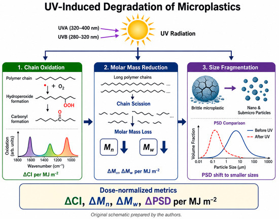
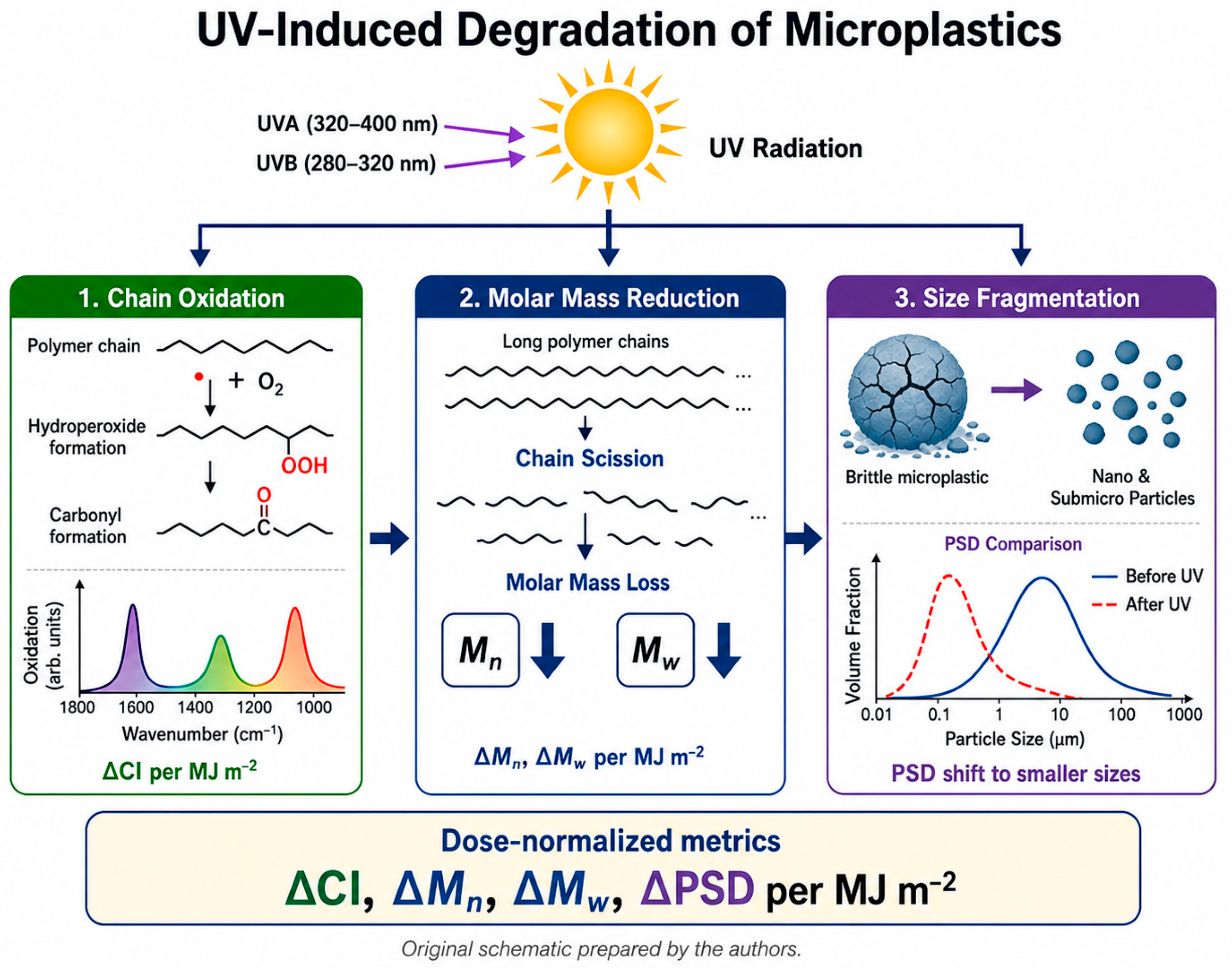
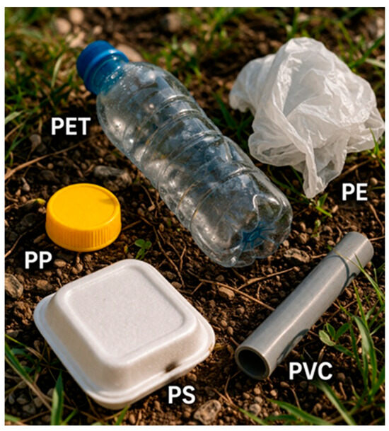
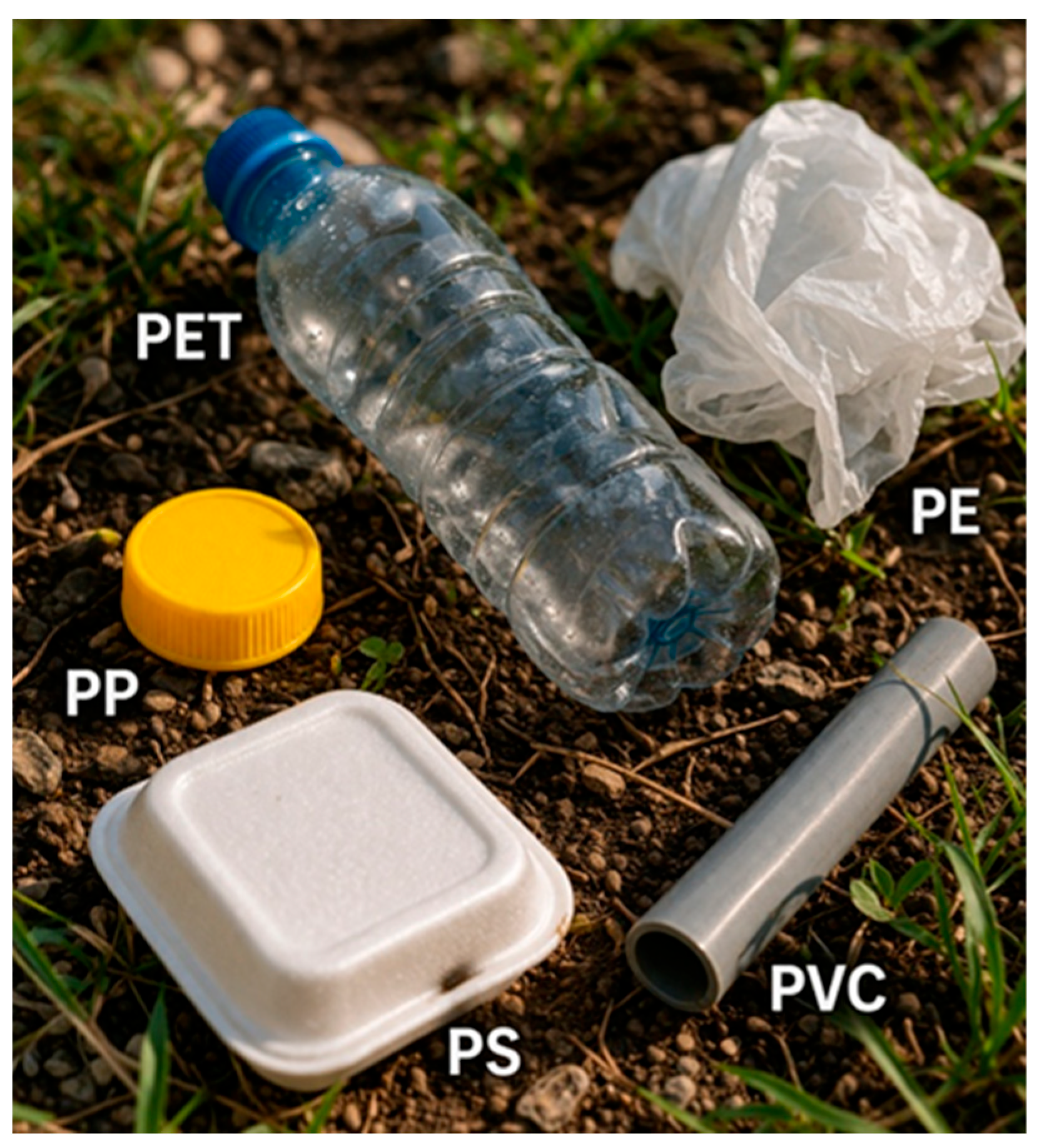
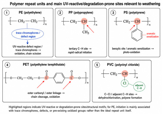
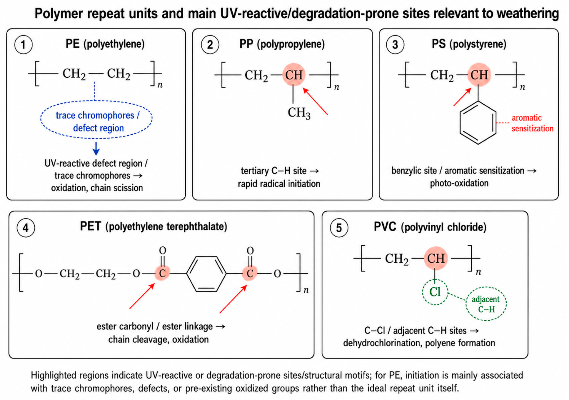

Disclaimer/Publisher’s Note: The statements, opinions and data contained in all publications are solely those of the individual author(s) and contributor(s) and not of MDPI and/or the editor(s). MDPI and/or the editor(s) disclaim responsibility for any injury to people or property resulting from any ideas, methods, instructions or products referred to in the content. |
© 2026 by the authors. Licensee MDPI, Basel, Switzerland. This article is an open access article distributed under the terms and conditions of the Creative Commons Attribution (CC BY) license.