


Abstract
Renal tubular disorders are often overlooked causes of acquired or inherited bone hypomineralization and fragility fractures in adults. The proximal tubule reabsorbs glucose, phosphate, low-molecular-weight proteins, amino acids, bicarbonate, and much of the sodium, potassium, chloride, and calcium. The distal nephron—the thick ascending limb of the loop of Henle, the distal convoluted tubule, and the collecting duct—regulates urine concentration and dilution, maintains acid-base balance via urinary proton secretion, and controls electrolytes, including sodium, potassium, magnesium, and calcium. Tubular defects may cause hyperphosphaturia (high urinary phosphate), hypercalciuria (high urinary calcium), or chronic metabolic acidosis (renal tubular acidosis, RTA). These changes weaken bone mineralization, disrupt bone turnover, and raise the risk of muscle weakness and fractures. This review summarizes acquired and genetic tubulopathies linked to hyperphosphaturia, hypercalciuria, and RTA and outlines a practical diagnostic approach for outpatients with bone fragility and suspected renal tubulopathy.
1. Introduction
Renal tubular disorders should be considered in the differential diagnosis of bone diseases, particularly given reports of increased incidence of secondary rickets due to renal tubulopathy, with a corresponding impact on mortality in recent years [1]. Their clinical manifestations can mimic those of more common bone conditions but often require different management. Bone strength depends on both bone mass and mineralization. Hypophosphatemia and renal phosphaturia directly impair mineralization, causing rickets or osteomalacia. Persistent urinary calcium loss (hypercalciuria), magnesium deficiency, or chronic metabolic acidosis can accelerate bone loss and skeletal fragility [2,3]. Tubular disorders may first present as bone pain, muscle weakness, or fractures. These conditions are sometimes misdiagnosed as ‘idiopathic osteoporosis’ if renal causes are not considered. Several case series and reviews have reported osteomalacia, recurrent low-impact fractures, and proximal muscle weakness in adults. For instance, these cases include patients with previously unrecognized Fanconi-type proximal tubulopathies. They also include drug-induced tubular dysfunction, notably from tenofovir, adefovir, and ifosfamide, as well as acquired or genetic calcium/phosphate-wasting disorders and renal tubular acidosis [4]. Early identification of these conditions permits precise interventions, such as discontinuing the causative drug, administering phosphorus or vitamin D supplementation, initiating alkali therapy, administering thiazide diuretics, and providing disease-specific treatments. These measures may help prevent further fractures [5].
2. Methods
We conducted a literature search for the present narrative review using PubMed, Cochrane, the National Library of Medicine, and Embase between October 2025 and March 2026. The search included the terms: “tubulopathy”, “renal tubular disorders”, “hyperphosphaturia”, “hypercalciuria”, “renal tubular acidosis”, “secondary osteoporosis”, “metabolic bone disease”, “bone fragility”, “bone mineralization”, “fractures and renal”, “fractures and tubulopathy”, “osteoporosis and renal”, and “bone and renal”.
First, we screened the articles based on their titles and abstracts. We considered studies published between 1995 and 2026 that focused on the diagnosis, prognosis, or treatment of bone mineral disorders associated with renal tubulopathies. Using the search terms described above individually, we initially retrieved 48,059 articles. After cross-referencing the data—primarily by linking hereditary or acquired tubulopathies to bone involvement—and prioritizing clinical trials, observational cohorts, case series, and review articles published in the last 5 years, we ultimately selected 82 articles.
3. How Does the Interaction Among Hyperphosphaturia, Hypercalciuria, and Renal Tubular Acidosis (RTA) Contribute to Complex Disruptions in Mineral Metabolism and Compromise Skeletal Integrity?
3.1. Hyperphosphaturia
Phosphate, an essential part of hydroxyapatite, is necessary for cellular metabolism. The kidneys maintain systemic phosphate homeostasis primarily by reabsorbing phosphate in the proximal tubule. In hyperphosphaturia, impaired phosphate reabsorption leads to persistent hypophosphatemia, which compromises bone mineralization and results in osteomalacia, pseudofractures, and increased bone fragility. Phosphate wasting results from downregulation of type II sodium-dependent phosphate cotransporters (NaPi-IIa, NaPi-IIc) at the apical membrane of proximal tubular cells, a process regulated by systemic factors [6]. Both parathyroid hormone (PTH) and fibroblast growth factor 23 (FGF23), along with its cofactor α-Klotho, decrease renal phosphate reabsorption. PTH and FGF23 downregulate NaPi cotransporters, whereas FGF23 also inhibits 1α-hydroxylase, reducing the synthesis of active vitamin D (1,25(OH)2D). Reduced levels of active vitamin D further limit intestinal absorption of calcium and phosphate. Disruptions at any of these regulatory points increase urinary phosphate excretion, and hypophosphatemia impairs bone health through multiple mechanisms [7]. First, defective matrix mineralization arises because phosphate is required for hydroxyapatite crystal formation in osteoid. Chronic hypophosphatemia reduces inorganic phosphate availability, leading to widened osteoid seams, decreased mineralization density, and clinical osteomalacia, characterized by bone pain, muscle weakness, pseudofractures, and fragility fractures. Osteomalacia is characterized by low bone turnover [8]. Second, FGF23 exerts direct skeletal and endocrine effects, including paracrine actions within bone, some of which are independent of α-Klotho, that modify osteocyte and osteoblast function and matrix mineralization. Elevated FGF23 is associated with poor bone mineralization and increased fragility, as demonstrated by histologic and imaging studies [8,9]. Third, FGF23-mediated suppression of 1α-hydroxylase reduces 1,25(OH)2D, thereby decreasing intestinal calcium and phosphate absorption. Lower vitamin D levels further worsen mineralization and may induce secondary hyperparathyroidism, which increases bone turnover and reduces bone mass, exacerbating fragility [10]. Finally, recent research describes extracellular matrix “stenciling,” in which local enzymes and inhibitors, such as alkaline phosphatase and pyrophosphate-generating enzymes, regulate mineral deposition. Hypophosphatemic conditions disturb this homeostasis, leading to focal defects in mineral nucleation and expansion. These microscopic alterations soften bone and increase fracture risk [11].
Chronic phosphate waste produces two overlapping skeletal phenotypes. Osteomalacia, characterized by inadequate mineralization of new osteoid, presents as wide unmineralized osteoid seams, pseudofractures, bone pain, and increased fragility. This phenotype is typical of sustained hypophosphatemia, as observed in genetic disorders such as X-linked hypophosphatemic rickets (XLH) and in acquired conditions such as tumor-induced osteomalacia (TIO) [11]. In contrast, vitamin D deficiency and secondary hyperparathyroidism increase bone turnover, reduce cortical thickness, and compromise bone microarchitecture. Although these changes differ from those of classical osteomalacia, they further increase fracture risk. Genetic alterations may additionally disrupt osteocyte-matrix interactions, leading to enthesopathies and impairing mechanical loading and skeletal integrity [12].
Multiple causes can decrease renal phosphate reabsorption or suppress active vitamin D synthesis. These include hormonal, genetic, acquired proximal tubular dysfunction, and paraneoplastic processes. All these mechanisms result in chronic hypophosphatemia, impaired bone mineralization (osteomalacia), altered bone microarchitecture, and a higher risk of fragility fractures [6]. Suppression of active vitamin D is significant in the setting of impaired proximal tubular function. Some genetic disorders cause tubular injury and phosphate loss, stimulating 1,25(OH)2D production. In other conditions, such as chronic kidney disease (CKD), active vitamin D levels are reduced. Both scenarios negatively affect bone mineralization. Certain genetic phosphate-wasting disorders, such as hypophosphatemic rickets with hypercalciuria (HHRH), are defined by high 1,25(OH)2D and hypercalciuria [5]. Other causes of renal phosphate waste involve FGF23. For example, excess circulating FGF23 from genetic upregulation—such as PHEX mutations in XLH, DMP1 or ENPP1 mutations in other hereditary hypophosphatemias—changes in NaPi cotransporters, or FGF23-secreting tumors in TIO can lead to renal phosphate wasting with skeletal consequences [13]. Advances in molecular genetics have refined the classification of FGF23-mediated and non-FGF23 hypophosphatemic disorders [12]. XLH and TIO are the principal hereditary disorders that link hyperphosphaturia to osteomalacia and fragility fractures [10,14]. Other causes include primary or tertiary hyperparathyroidism, in which chronic elevation of PTH leads to bone loss and disrupts mineral homeostasis in several ways [15]. Generalized proximal tubulopathies, such as Fanconi syndrome, are characterized by phosphaturia, aminoaciduria, glucosuria, and bicarbonaturia. Causes include inherited disorders and acquired forms from drugs (such as tenofovir or ifosfamide), heavy metals, and mitochondrial toxins [5]. Some malignancies or systemic illnesses can also indirectly affect phosphate handling [8].
Clinically, chronic hyperphosphaturia presents as bone pain, diffuse muscle weakness, delayed fracture healing, pseudofractures, rickets, and, in children, growth failure [16]. Laboratory findings include low serum phosphate, high urinary fractional excretion of phosphate (FEP > 5%), inappropriately normal or low 1,25(OH)2D, and often elevated FGF23. Bone markers, such as elevated alkaline phosphatase, and radiographs showing Looser zones or pseudofractures aid diagnosis. Dual-energy X-ray absorptiometry (DXA) may show low bone mineral density (BMD), but this can be misleading since osteomalacia may coexist with normal BMD. Differentiating FGF23-mediated causes (e.g., XLH or TIO) from non-FGF23-mediated causes (e.g., proximal tubulopathies or PTH-mediated disorders) is essential. This distinction guides subsequent tests, including genetic analysis and, when necessary, tumor localization using functional imaging [3].
3.2. Hypercalciuria
Hypercalciuria is a common metabolic problem in calcium kidney stone disease and can also occur without symptoms. Beyond its role in nephrolithiasis, hypercalciuria affects bone health. Studies show lower bone mineral density (BMD) and higher osteopenia and osteoporosis rates in people with hypercalciuria. Up to one-third of osteoporosis patients have hypercalciuria [17,18].
More than 60% of serum calcium filtered by the glomerulus is reabsorbed in the proximal tubule by passing between cells through tight junction proteins called claudins, which control which ions move through. This reabsorption is mainly driven by water, which follows sodium. Approximately 30–35% of the remaining calcium is reabsorbed in the thick ascending limb of the loop of Henle, a process mediated by intercellular proteins called claudins. Claudins also facilitate magnesium transport between cells, with their abundance increasing when magnesium is low [19]. The distal convoluted tubule and collecting ducts complete the final reabsorption of calcium via transporters such as TRPV5 on the tubular side and Na-Ca antiporters on the blood-facing side. Idiopathic hypercalciuria, the most common form, usually results from genetic changes that alter these reabsorption processes [20].
Several mechanisms have been proposed to explain the association between hypercalciuria, nephrolithiasis, and bone fragility [21]. First, increased intestinal calcium absorption, termed absorptive hypercalciuria, is often driven by upregulation of the vitamin D receptor (VDR). In specific individuals, this boosted absorption results from elevated calcitriol (1,25(OH)2D) activity and/or increased VDR expression or sensitivity. Genetic association studies implicate VDR-related pathways in the pathogenesis of absorptive hypercalciuria. Enhanced intestinal absorption increases the filtered calcium load, leading to greater urinary calcium loss if renal compensation is insufficient [22]. Second, a primary increase in bone resorption with osteoclastic activation may result from systemic biochemical changes, such as an altered parathyroid hormone (PTH) setpoint, or from intracellular signaling within bone cells. In some patients, elevated markers of bone turnover accompany urinary calcium loss, indicating that bone is the source of the released calcium. Systemic factors, including calcitriol, cytokines, and RANKL/OPG balance, as well as local osteocyte and osteoblast signaling, can promote osteoclastic activation. The concept of “bone as the buffer” suggests that when renal losses exceed intestinal absorption or when endocrine factors favor resorption, bone mineral is mobilized to maintain normal serum calcium [6,23]. Third, renal calcium wasting may result from defects in renal epithelial calcium transport, such as TRPV5/6 dysfunction or claudin abnormalities, as well as dysregulation of endocrine modulators, including FGF23/klotho and PTH. Renal distal tubular reabsorption of calcium, mediated by TRPV5 (apical entry), calbindins, and basolateral extrusion mechanisms, is essential for the reclamation of filtered calcium. Genetic and experimental disruption of TRPV5 reduces renal calcium reabsorption, leading to hypercalciuria [24]. Recent human genetic studies have shown that biallelic TRPV5 loss-of-function causes autosomal recessive renal calcium-wasting hypercalciuria, providing direct evidence linking defective renal epithelial calcium transport to systemic calcium loss and bone consequences [25,26].
Additional paracellular pathways, including claudin family proteins in the proximal nephron and thick ascending limb, and differences in sodium handling, given the close coupling of sodium and calcium transport, also influence urinary calcium excretion [27]. Polymorphisms in genes encoding proteins involved in tubular calcium reabsorption (CASR, CLDN14, TRPV6, TRPV5) or in the prevention of calcium salt precipitation (CaSR, MGP, OPN, UMOD) have been associated with idiopathic hypercalciuria and kidney stones [28]. Finally, interactions among endocrine modulators such as PTH, FGF23/Klotho, and calcitriol are critical. The endocrine axis regulates both renal and bone calcium fluxes. Elevated calcitriol increases intestinal absorption and can stimulate bone resorption. While PTH typically increases renal calcium reabsorption, chronically altered PTH dynamics, including impaired secretion set points or episodic changes, are observed in hypercalciuric populations. FGF23 and its cofactor Klotho regulate distal tubular TRPV5 abundance and, consequently, renal calcium reabsorption. Mouse models have demonstrated that FGF23 influences TRPV5 membrane expression and calcium handling. Dysregulation of these hormones can therefore increase urinary calcium excretion and affect bone remodeling [29].
Systemic disorders that are associated with both hypercalcemia and hypercalciuria include excess glucocorticoid, hyperthyroidism, hypervitaminosis D, metastatic bone tumors, paraneoplastic syndromes, milk-alkali syndrome resulting from excessive oral calcium ingestion, multiple myeloma, Paget disease, sarcoidosis, and other granulomatous disorders [6].
The identification of hypercalciuria should lead to a bone assessment, such as Dual-energy X-ray Absorptiometry (DXA), given its established association with low bone mineral density. Laboratory evaluation should include urinary calcium (24 h collection or spot urine Ca/Cr), serum calcium, parathyroid hormone (PTH), 25(OH)2D, and 1,25(OH)2D. Additional assessment of urinary lithogenic factors and bone turnover markers may be warranted in selected cases [22].
3.3. Metabolic Acidosis
Metabolic acidosis adversely affects bone, which functions as part of the intracellular buffer system by providing cations such as sodium, potassium, and calcium in exchange for protons, and by providing carbonate and phosphate to the extracellular fluid, consequently maintaining blood pH [30]. The acidic pH of the extracellular fluid also directly affects the transcriptome, promoting morphological changes in osteoclasts and increasing their activity [31]. In addition to these cellular effects, an acidic environment directly inhibits osteoblastic activity. It stimulates osteoclastic activity through proton-sensitive receptors, including the G protein-coupled receptor 1 of ovarian cancer (OGR1), and mediates this effect via phosphoinositide metabolites and increased PGE2 levels [30]. At the renal level, metabolic acidosis reduces tubular calcium reabsorption. For instance, metabolic acidosis can directly downregulate claudin-2 in the proximal tubule, TRPV5 and calbindin-D28K in the distal tubule, and, indirectly, inhibit claudins 16–19 in the thick ascending limb of Henle, thereby promoting hypercalciuria [24,32]. Metabolic acidosis should be recognized as a cause of secondary osteoporosis. Although most patients with metabolic acidosis have chronic kidney disease and reduced glomerular filtration rate (eGFR < 60 mL/min/1.73 m2), certain conditions can cause metabolic acidosis despite preserved GFR due to defects in urinary acidification. Renal tubular acidosis (RTA) illustrates this, being characterized by hyperchloremic metabolic acidosis resulting from decreased tubular capacity for urinary acidification. RTA may be hereditary or acquired through various diseases or drugs [33,34,35,36,37,38].
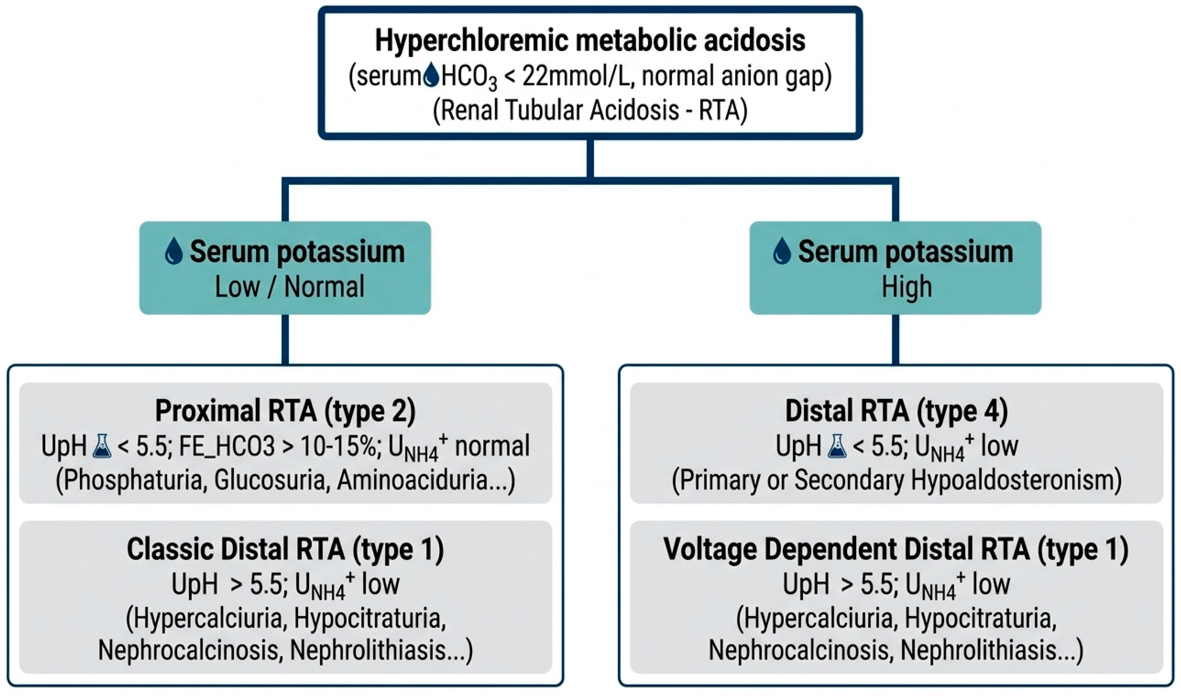
Renal tubular acidosis (RTA) is classified into different types: proximal RTA (pRTA or type 2), resulting from decreased proximal tubular reabsorption of filtered bicarbonate; classic distal RTA (dRTA or type 1), due to reduced proton secretion in the collecting duct; distal RTA with bicarbonaturia (type 3); distal RTA voltage-dependent with hyperkalemia (hyperkalemic type 1); and distal RTA with hyperkalemia from decreased aldosterone action in the collecting duct (type 4). pRTA (type 2) and dRTA (type 1) generally have a greater impact on bone, particularly in hereditary forms, and are usually associated with normokalaemia or hypokalaemia. In pRTA, there is often concurrent renal loss of other solutes, such as phosphorus, uric acid, glucose, and amino acids, as observed in Fanconi syndrome. In dRTA, hypercalciuria, hypocitraturia, nephrocalcinosis, and nephrolithiasis are common [35,39]. RTA should be considered in patients with suspected secondary osteoporosis, cognitive disorders, deformities from rickets or osteomalacia, hypokalemia, hypophosphatemia, nephrocalcinosis, recurrent nephrolithiasis, and especially metabolic acidosis with a disproportionately alkaline urinary pH in early-morning urine samples (urine pH > 6.0) [40].
In the outpatient setting, the primary test for diagnosing metabolic acidosis (excluding chronic respiratory disorders) is measurement of serum total CO2 (bicarbonate), which accounts for approximately 95% of total CO2. This test is widely available and can be performed using a standard biochemistry tube. A total CO2 value below 22 mEq/L confirms metabolic acidosis and necessitates prompt evaluation for RTA by a nephrologist. The absence of systemic metabolic acidosis characterizes an “incomplete” form of RTA. Patients with recurrent nephrolithiasis, nephrocalcinosis, hypokalemia, or other clinical and laboratory findings suggestive of RTA, even in the absence of metabolic acidosis, should be referred to a nephrologist for assessment of tubular acidification capacity using functional tests [35,41].
4. What Are the Indicators for Suspecting Impaired Tubular Renal Function?
Patients with bone fragility should be evaluated for renal tubulopathy if specific indicators are present. These include exposure to nephrotoxic medications (such as tenofovir, adefovir, cisplatin, ifosfamide, aminoglycosides, high-dose proton-pump inhibitors, and some chemotherapeutics); a history of cancer (especially tumor-induced osteomalacia) or monoclonal gammopathy (such as light-chain proximal tubulopathy); a family history of kidney stones, rickets or osteomalacia, salt wasting, or early fractures, which may suggest a genetic tubulopathy; and symptoms of proximal or distal tubulopathies, including polyuria or nocturia, polydipsia, bone pain, muscle weakness, recent nephrolithiasis, cardiac arrhythmias from electrolyte imbalance, and episodes of dehydration.
5. How to Approach Investigating Tubular Renal Function in Outpatient Settings?
A focused evaluation of tubular function combines clinical history, basic blood tests, spot and 24 h urine measurements, and selected laboratory calculations. This includes measuring fractional excretion of solutes, evaluating urinary biomarkers of tubular injury, and assessing urinary concentration and acidification capacity. The following serum and urine tests are recommended, ideally collected together:
These are the first-line laboratory tests:
- (a)
- Serum: creatinine, electrolytes (Na, K, Ca, P, Cl, HCO3−), alkaline phosphatase, 25(OH)2D, iPTH.
- (b)
- Urine single sample (spot urine): Urinalysis (dipstick), and urinary pH by potentiometry (for RTA workup).
- (c)
- Urine single sample (spot urine): Low-molecular-weight proteins: Urinary β2-microglobulin, α1-microglobulin, or retinol-binding protein (RBP) serves as a sensitive marker of proximal tubule injury (Fanconi pattern). α1-microglobulin and RBP are more stable in acidic urine than β2-microglobulin and may therefore be preferable [42,43].
- (d)
- Urine 24 h: creatinine, electrolytes (Na, K, Cl, Ca, P, HCO3−), glucose, uric acid, urea, citrate, protein, albumin.
After the initial laboratory results are available, multiple important derived indices should be calculated.
- (a)
- Fractional excretion (FE) of solutes: The fractional excretion of phosphate, potassium, magnesium, uric acid, and bicarbonate supplies important diagnostic information. FE (solute) is calculated as follows:
FE (solute) = ([Solute urine × Creatinine serum]/[Solute serum × Creatinine urine]) × 100. For FE of magnesium, serum magnesium must be multiplied by 0.7 [7].
- (b)
- TmP/GFR (tubular maximum reabsorption of phosphate per glomerular filtration rate) provides a physiologically relevant estimate of renal phosphate handling that accounts for filtered load. A low TmP/GFR indicates inappropriate renal phosphate waste relative to serum phosphate concentration. Online calculators and laboratory protocols are available. Typical ranges vary by age [44].
- (c)
- 24 h urinary calcium (or urine Ca/Cr ratio on a spot sample): These tests detect hypercalciuria (Ca > 4 mg/Kg/24 h or Ca/Cr ratio > 0.2). Calcium excretion should be treated as a continuous variable, as values exceeding 150 mg/24 h are associated with an increased risk of nephrolithiasis [22].
- (d)
- 24 h urinary glucose: In euglycemic patients, glucosuria over 500 mg/24 h strongly suggests proximal tubular dysfunction, such as partial or complete Fanconi syndrome.
- (e)
- Aminoaciduria: Detection requires specialized assays, such as thin-layer chromatography, available in select tertiary laboratories [45].
- (f)
- Acid-base testing: Assessment should include serum bicarbonate and urine pH measured by potentiometry. An inability to acidify urine in the presence of systemic acidosis suggests distal renal tubular acidosis (Type 1). Proximal RTA (Type 2), which is associated with Fanconi syndrome, presents with bicarbonaturia and, in severe cases, low serum bicarbonate. Measurement of urinary ammonium (UNH4+) further evaluates the acidification capacity of the distal nephron [35,46]. It is worth noting that the urine anion gap and osmolal gap have traditionally been used as indirect measures of UNH4+, but recent studies have shown low accuracy with this approach [47,48].
If indicated, additional specialty tests may be requested to clarify the etiology of tubulopathy:
- (a)
- 1,25(OH)2D (calcitriol) and PTHrP (PTH-related peptide): both tests are indicated in cases of hypercalcemia caused by calcitriol (for instance, granulomatous diseases) or by bone, respectively [6].
- (b)
- FGF23: This test is indicated in cases of phosphate waste without classical Fanconi features or when tumor-induced osteomalacia is suspected [10].
- (c)
- Serum and urine electrophoresis, along with free light chain analysis, are essential for detecting monoclonal gammopathies that may cause light-chain proximal tubulopathy [49].
- (d)
- Genetic testing: DNA analysis is essential in cases of unexplained lifelong renal phosphate or calcium waste, renal tubular acidosis, particularly when there is a positive family history, early onset of disease, or affection of multiple organs [50]. Contemporary panels for hereditary tubulopathies assess nearly one hundred genes (including SLC34A1, SLC34A3, PHEX, FGF23, CLCN5, OCRL, among others).
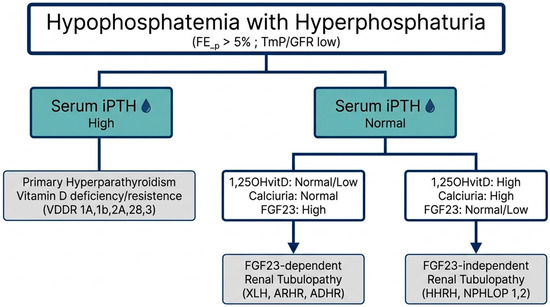
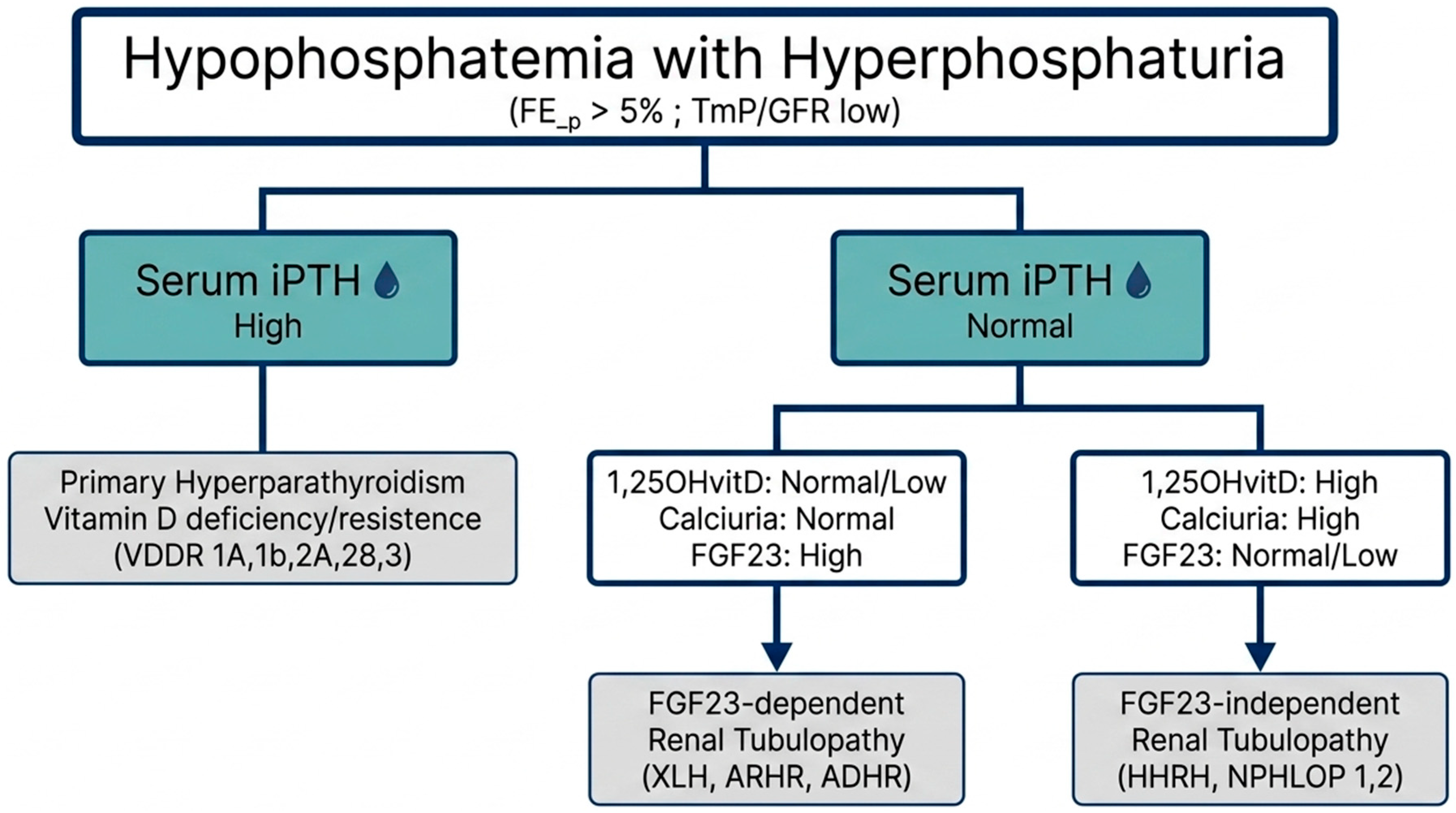
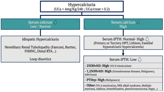
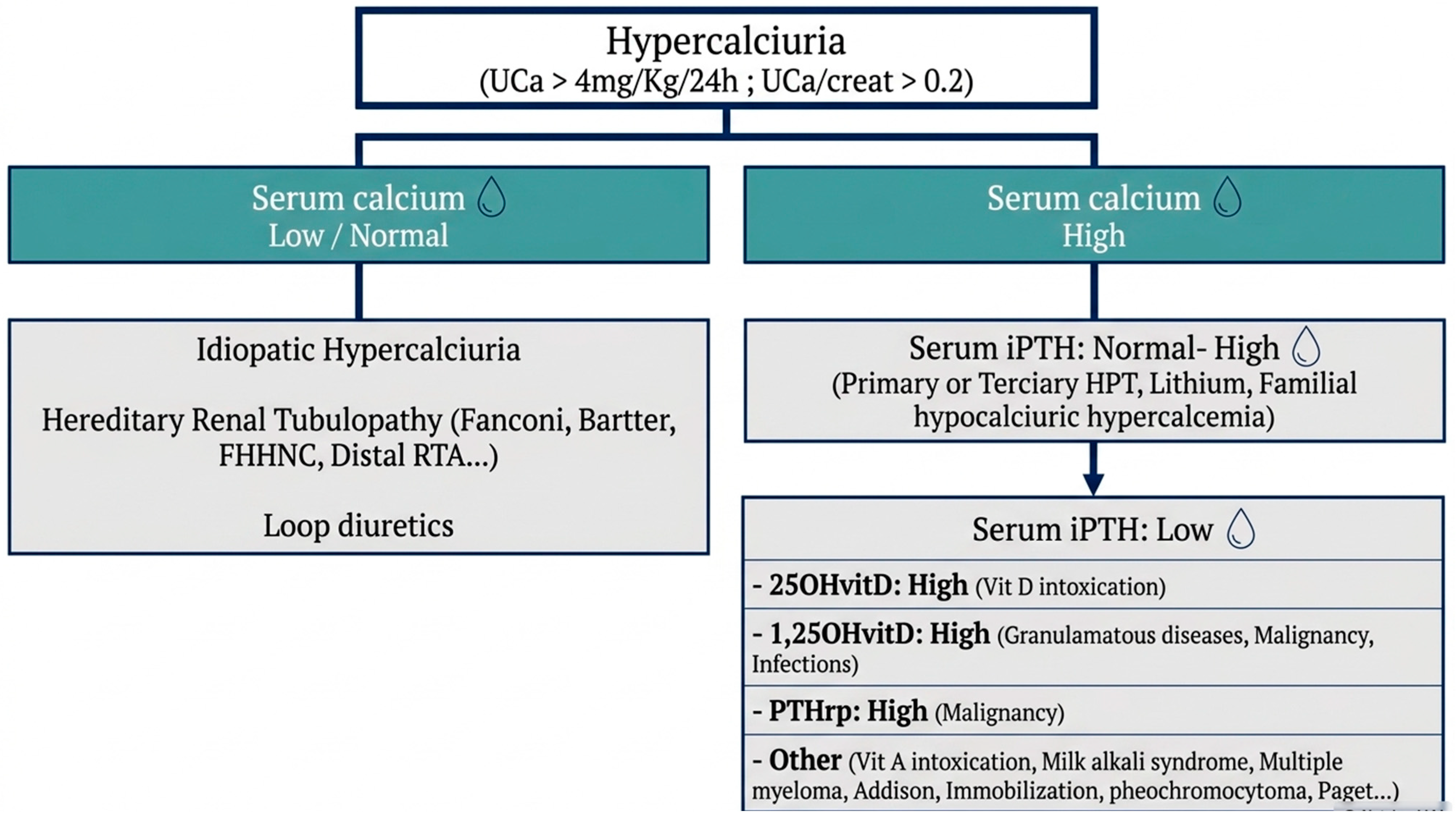
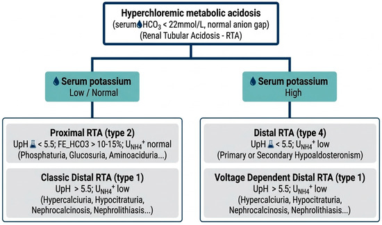
Diagnostic algorithms for the principal tubulopathies associated with bone fragility, based on the predominant renal manifestation (hyperphosphaturia, hypercalciuria, or renal tubular acidosis), are provided in Figure 1, Figure 2 and Figure 3.
Figure 1.
Flowchart for the diagnosis of Hyperphosphaturia. FEp: fractional excretion of phosphorus; TmP/GFR: tubular maximum reabsorption of phosphate per Glomerular filtration rate; VDDR: Vitamin D-dependent Rickets types 1A,1B,2A,2B,3; XLH: Hypophosphatemic Rickets X-linked; ARHR: AR Hypophosphatemic Rickets; ADHR: AD Hypophosphatemic Rickets; HHRH: Hypophosphatemic Rickets with Hypercalciuria; NPHLOP: Nephrolithiasis/Hypophosphatemic Osteoporosis types 1,2.
Figure 2.
Flowchart for the diagnosis of Hypercalciuria. Uca: urinary calcium; RTA: Renal tubular acidosis; FHHNC: Familial Hypomagnesemia with Hypercalciuria and Nephrocalcinosis; HPT: hyperparathyroidism; PTHrp: Parathyroid hormone-related protein.
Figure 3.
Flowchart for the diagnosis of Renal Tubular Acidosis. UpH: urinary pH; UNH4+: urinary ammonium.
After diagnosing renal tubular dysfunction, it is essential to determine whether the tubulopathy is acquired or hereditary to guide treatment and prognosis. In acquired cases, treating or removing the underlying systemic disease, drug, or toxin can restore tubular function and improve bone fragility [51]. In monogenic hereditary cases, some tubulopathies already have treatments targeting the metabolic or genetic defect (enzyme replacement, specific anti-receptor antibodies, RNA interference drugs, CRISPR-Cas9-based drugs) that may cure the condition [52].
6. What Are the Leading Causes of Tubulopathies Associated with Bone Fragility, Including Inherited and Acquired Disorders That Disrupt Renal Tubular Function and Mineral Homeostasis?
Numerous inherited disorders of tubular transport, detailed in Table 1, Table 2, Table 3 and Table 4, are primary genetic causes of tubulopathies and often appear in childhood. However, adult-onset or late-diagnosed cases are common and may first present with fractures or low bone mineral density (BMD), as discussed below.
Table 1.
Inherited renal tubular disorders causing Fanconi syndrome.
Table 2.
Inherited renal tubular disorders causing hyperphosphaturia.
Table 3.
Inherited renal tubular disorders causing hypercalciuria.
Table 4.
Inherited renal tubular disorders causing renal tubular acidosis (RTA).
- (a)
- Disorders causing renal phosphate waste include X-linked hypophosphatemia (XLH, PHEX mutations), the most significant FGF-23-mediated disorder. XLH leads to hypophosphatemia, rickets in children, and osteomalacia or fractures in adults, and is typically lifelong, sometimes recognized late [12]. Hereditary hypophosphatemic rickets with hypercalciuria (HHRH, SLC34A3/NaPi-IIc mutations) causes renal phosphate wasting, low serum phosphate, elevated 1,25(OH)2D, suppressed PTH, and hypercalciuria. Adults may present with bone pain and multiple fractures. HHRH is treated with phosphate replacement instead of FGF-23 antagonists [2]. Tumor-induced osteomalacia (TIO), due to FGF-23-producing mesenchymal tumors, results in severe phosphaturia, osteomalacia, and fractures; tumor localization is the primary therapeutic goal [13]. Cystinosis and other inherited proximal tubulopathies cause Fanconi syndrome with severe phosphate loss and early bone disease. Adult survivors may present with bone fragility [53].
- (b)
- Disorders with hypercalciuria, nephrocalcinosis, and bone loss include Dent disease (CLCN5 or OCRL mutations), an X-linked tubulopathy with low-molecular-weight proteinuria, hypercalciuria, nephrolithiasis or nephrocalcinosis, and progressive CKD. Bone consequences, such as BMD reduction and fractures, are increasingly recognized. Early recognition prevents nephrocalcinosis and bone complications [53]. Bartter syndrome variably affects bone, with some forms causing impaired growth and bone mass [6].
The leading causes of acquired tubulopathies resulting in bone fragility (Table 5) include:
Table 5.
Acquired renal tubular disorders and nephron involvement.
- (a)
- Drug-induced proximal tubulopathy and Fanconi syndrome: Tenofovir disoproxil fumarate (TDF) is a well-documented cause of proximal tubular dysfunction, Fanconi syndrome, hypophosphatemic osteomalacia, and fracture. Risk factors include older age, low body weight, coadministration of boosters, preexisting kidney disease, and prolonged exposure. Clinicians should suspect tenofovir-related tubulopathy in HIV/HBV patients with bone pain, hypophosphatemia, or proximal muscle weakness. Tenofovir alafenamide (TAF) causes less tubulopathy but still needs vigilance [54]. Ifosfamide is a chemotherapeutic agent known to cause delayed proximal tubular dysfunction and osteomalacia, mainly in children but also in adults post-chemotherapy, with insidious and late tubular damage. Platinum-based chemotherapy, aminoglycosides, contrast agents, and heavy metals may cause tubular injury; persistent proximal injury can lead to phosphate waste and bone effects [49]. Adefovir and other nucleotide antivirals are also associated with proximal tubular injury and phosphate waste [49].
- (b)
- Systemic diseases causing acquired tubulopathies: Multiple myeloma and monoclonal light-chain–associated proximal tubulopathy commonly present as Fanconi-type syndrome with osteomalacia and fragility fractures. Screening with serum/urine protein electrophoresis and free light chains is essential. Autoimmune diseases, such as Sjögren’s syndrome, cause distal RTA and osteomalacia; other tubulointerstitial nephritides (e.g., sarcoidosis) may affect tubular mineral handling [36].
- (c)
- Environmental toxins: Heavy metals like lead and cadmium, and other exposures, can cause tubular dysfunction and mineralization defects. Chronic exposure should be identified with a thorough patient history and investigated if suspected [49].
7. How Should Patients with Tubulopathies Associated with Bone Disease Be Treated, with a Focus on Correcting Electrolyte and Acid-Base Disturbances and Targeting the Underlying Cause?
Essential steps include removing offending drugs or toxins. Drug withdrawal may improve tubular markers and bone pain [55]. Genetic counseling and multidisciplinary care are recommended [12].
Conventional therapy for many hypophosphatemic disorders includes oral phosphate in divided doses and active vitamin D analogs (calcitriol or alfacalcidol) to promote mineralization. This calls for close monitoring to prevent secondary hyperparathyroidism and nephrocalcinosis, and for close surveillance of urinary calcium to avoid hypercalciuria [5]. Management may involve treating the underlying cause and repairing mineralization. For instance, surgical resection of FGF23-secreting tumors cures TIO, while genetic causes require long-term management. Monoclonal antibodies against FGF23, like burosumab, have been developed for XLH and FGF23-mediated hypophosphatemia, improving phosphate, 1,25(OH)2D, and skeletal outcomes [56]. This approach shows that understanding phosphate regulation enables the development of disease-modifying treatments [5].
In hypercalciuria, thiazide diuretics reduce urinary calcium by enhancing tubular calcium reabsorption, therefore improving urinary calcium excretion and, in some studies, BMD [23,38]. It is noteworthy that a recent randomized controlled clinical trial showed that indapamide had potential bone-protective effects, substantially reducing β-crosslaps compared with hydrochlorothiazide [57]. Dietary priorities include moderate sodium restriction, adequate calcium intake, and sufficient vitamin D. Low calcium intake is not recommended, as it may increase the risk of stone formation and bone loss. Vitamin D supplementation should be personalized, given the variation in stone risk [58]. In patients with osteoporosis and hypercalciuria, antiresorptive drugs (bisphosphonates, denosumab) may increase BMD; correcting hypercalciuria first may enhance antiresorptive effects [17]. Bone-directed antiresorptive drugs must be administered cautiously, only after excluding osteomalacia or treating it, as potent antiresorptive drugs are contraindicated in untreated osteomalacia. Correction of phosphate and vitamin D levels, as well as therapy of tubulopathy, must precede the use of antiresorptive drugs [5].
When treating metabolic acidosis due to renal tubular acidosis (RTA), it is essential to identify the underlying cause to guide a specific approach, such as in Sjögren’s syndrome in adults. Oral alkaline therapy—using potassium citrate, sodium bicarbonate, or SHOHL solution—is the primary treatment. Calcitriol may be used for proximal RTA (pRTA), along with other steps to support bone health [39]. When initiating osteoporosis medications, note that RTA can also lead to reduced bone remodeling, including adynamic bone or osteomalacia [30]. Because of this, as mentioned before, antiresorptive drugs such as bisphosphonates and anti-RANKL therapies ought to be administered cautiously, ideally after assessing bone remodeling with a tetracycline-labeled bone biopsy or after an anabolic agent has been used [59].
For long-term monitoring of patients with bone diseases caused by renal tubular disorders, it is important to routinely assess serum phosphate, calcium, and bicarbonate, and to evaluate urinary phosphate and calcium; if possible, also test for low-molecular-weight proteinuria. Some recent observational studies, for example, suggest an association between abnormal serum levels of uric acid and aldosterone and an increased risk of osteoporosis and fractures in middle-aged and elderly patients with hypertension [60,61]; therefore, long-term monitoring is also recommended in cases of tubulopathies involving abnormalities in these electrolytes or hormones.
After correcting all mineral imbalances, serial DXA bone scanning, including the trabecular bone score (TBS) when possible, is also recommended to monitor changes in bone mineral density in these patients [62,63].
8. Limitations
Several limitations are inherent to this review. As a narrative review, it remains susceptible to selection and publication bias, even with a structured literature search. The included studies varied in design, study type (prognostic, diagnostic, or treatment), and patient populations [64,65,66,67,68,69,70,71,72,73]. Furthermore, many studies were retrospective, conducted at single centers, or involved small sample sizes, which limits the generalizability of the findings [74,75,76,77,78,79,80,81,82]. Future research should prioritize randomized clinical trials, as they remain underrepresented in studies of renal tubulopathies.
9. Conclusions
Renal tubular disorders are a significant and often under-recognized cause of bone hypomineralization and fragility fractures in adults. Accurate diagnosis requires a systematic approach that includes clinical history; targeted laboratory testing (serum calcium, phosphate, and bicarbonate); calculated indices (fractional excretion of phosphate, tubular maximum reabsorption of phosphate per glomerular filtration rate, urinary calcium/creatinine ratio); urine pH; and low-molecular-weight protein assays. These steps help identify various tubulopathies, such as drug-induced Fanconi syndrome, genetic phosphate-wasting disorders, hypercalciuric states, and renal tubular acidosis. Early recognition and intervention, including withdrawal of nephrotoxic drugs, phosphate and vitamin D supplementation, alkali therapy, thiazide diuretics, tumor localization and removal, and disease-specific therapies, are important for preventing further fractures and renal injury. Coordinated multidisciplinary care involving nephrologists, endocrinologists, rheumatologists, metabolic bone disease specialists, and geneticists is often required for complete evaluation and management.
10. Practical Clinical Pearls
In adults with multiple low-impact fractures and bone pain, clinicians should consider causes beyond osteoporosis. Assessment of possible renal tubulopathy should include serum and urine phosphate, serum and urine calcium, serum bicarbonate, and urinalysis before starting antiresorptive therapy.
Simultaneous collection of serum and urine samples is necessary for calculating the tubular maximum reabsorption of phosphate per glomerular filtration rate and the fractional excretion of all electrolytes.
Low-molecular-weight proteinuria testing (β2-microglobulin, α1-microglobulin, or retinol-binding protein) is a sensitive early marker of proximal tubular injury. It should be performed when a suspected acquired or inherited renal tubulopathy is identified.
Hereditary renal tubulopathy may be presented in adulthood with fractures or nephrolithiasis. Clinicians should have a low threshold for genetic referral if the phenotype is suggestive.
Author Contributions
Conceptualization, C.P.G., A.d.S.P. and M.L.F.d.F.; methodology, C.P.G.; investigation, C.P.G.; resources, C.P.G.; writing—original draft preparation, C.P.G.; writing—review and editing, C.P.G., A.d.S.P. and M.L.F.d.F.; visualization, C.P.G. All authors have read and agreed to the published version of the manuscript.
Funding
This research received no external funding.
Institutional Review Board Statement
Not applicable.
Informed Consent Statement
Not applicable.
Data Availability Statement
No new data were created or analyzed in this study. Data sharing is not applicable to this article.
Conflicts of Interest
The authors declare no conflicts of interest.
Abbreviations
The following abbreviations are used in this manuscript:
| 1,25(OH)2D | Calcitriol |
| 25(OH)2D | Vitamin D3 |
| BMD | Bone Mineral Density |
| CKD | Chronic Kidney Disease |
| dRTA | Distal Renal Tubular Acidosis |
| DXA | Dual-energy X-ray Absorptiometry |
| eGFR | Glomerular Filtration Rate |
| FGF23 | Fibroblast Growth Factor 23 |
| pRTA | Proximal Renal Tubular Acidosis |
| PTH | Parathyroid hormone |
| UNH4+ | Urinary ammonium |
References
- Chu, C.H.; Chen, Y.C.; Liu, P.Y.; Hu, C.C.; Lin, Y.L.; Kuo, F.C.; Lu, C.H.; Hsu, T.J.; Hung, Y.T.; Tsai, F.J.; et al. Long-term epidemiological insights into rickets: A nationwide population-based retrospective study. Clin. Exp. Pediatr. 2025, 68, 879–891. [Google Scholar] [CrossRef] [PubMed]
- Haffner, D.; Leifheit-Nestler, M.; Grund, A.; Schnabel, D. Rickets guidance: Part I-diagnostic workup. Pediatr. Nephrol. 2022, 37, 2013–2036. [Google Scholar] [CrossRef] [PubMed]
- Sahay, M.; Sahay, R. Renal rickets-practical approach. Indian J. Endocrinol. Metab. 2013, 17, S35–S44. [Google Scholar] [CrossRef] [PubMed]
- Kim, D. Hypophosphatemic Osteomalacia Induced by Low-Dose Adefovir-Related Fanconi Syndrome. J. Med. Cases 2026, 17, 133–139. [Google Scholar] [CrossRef] [PubMed]
- Arboleya, L.; Braña, I.; Pardo, E.; Loredo, M.; Queiro, R. Osteomalacia in Adults: A Practical Insight for Clinicians. J. Clin. Med. 2023, 12, 2714. [Google Scholar] [CrossRef] [PubMed]
- Murray, S.L.; Wolf, M. Calcium and Phosphate Disorders: Core Curriculum 2024. Am. J. Kidney Dis. 2024, 83, 241–256. [Google Scholar] [CrossRef] [PubMed]
- Antonowicz, A.; Lipiński, P.; Popow, M.; Skrzypczyk, P. Approach to a Child with Hypophosphatemia. Biomolecules 2025, 15, 1321. [Google Scholar] [CrossRef] [PubMed]
- Modest, J.M.; Sheth, H.; Gohh, R.; Aaron, R.K. Osteomalacia and Renal Osteodystrophy. Rhode Isl. Med. J. 2022, 105, 22–27. [Google Scholar]
- Erben, R.G. Editorial: Endocrine and Paracrine Role of FGF23 and Klotho in Health and Disease. Front. Endocrinol. 2019, 10, 2. [Google Scholar] [CrossRef] [PubMed]
- Kurpas, A.; Supeł, K.; Idzikowska, K.; Zielińska, M. FGF23: A Review of Its Role in Mineral Metabolism and Renal and Cardiovascular Disease. Dis. Markers 2021, 2021, 8821292. [Google Scholar] [CrossRef] [PubMed]
- Buss, D.J.; Deering, J.; Reznikov, N.; McKee, M.D. Understanding the structural biology of osteomalacia through multiscale 3D X-ray and electron tomographic imaging: A review of X-linked hypophosphatemia, the. JBMR Plus 2025, 9, ziae176. [Google Scholar] [CrossRef] [PubMed]
- Hamdy, N.A.T.; Harvengt, P.; Usardi, A. X-linked hypophosphatemia: The medical expert’s challenges and the patient’s concerns on their journey with the disease. Arch. Pediatr. 2021, 28, 612–618. [Google Scholar] [CrossRef] [PubMed]
- Álvarez-Rivas, N.; Lugo-Rodríguez, G.; Maneiro, J.R.; Iñiguez-Ubiaga, C.; Melero-Gonzalez, R.B.; Iglesias-Cabo, T.; Carmona, L.; García-Porrúa, C.; de Toro-Santos, F.J. Tumor-induced osteomalacia: A systematic literature review. Bone Rep. 2024, 21, 101772. [Google Scholar] [CrossRef] [PubMed]
- Huang, X.; Jiang, Y.; Xia, W. FGF23 and Phosphate Wasting Disorders. Bone Res. 2013, 1, 120–132. [Google Scholar] [CrossRef] [PubMed]
- Walker, V. The Intricacies of Renal Phosphate Reabsorption-An Overview. Int. J. Mol. Sci. 2024, 25, 4684. [Google Scholar] [CrossRef] [PubMed]
- Kim, S.; Kim, S.W.; Lee, B.C.; Kim, D.H.; Sung, D.H. Adult-onset hypophosphatemic osteomalacia as a cause of widespread musculoskeletal pain: A retrospective case series of single center experience. World J. Clin. Cases 2023, 11, 7785–7794. [Google Scholar] [CrossRef] [PubMed]
- Nicoli, F.; Dito, G.; Guabello, G.; Longhi, M.; Corbetta, S. Hypercalciuria in Postmenopausal Women with Reduced Bone Mineral Density Is Associated with Different Mineral Metabolic Profiles: Effects of Treatment with Thiazides and Anti-resorptives. Front. Med. 2021, 8, 780087. [Google Scholar] [CrossRef] [PubMed]
- Shah, N.; Galitzer, H.; Yalamanchi, S.; Sellmeyer, D.E. High prevalence of laboratory abnormalities indicative of secondary osteoporosis detected by systematic testing. JBMR Plus 2025, 9, ziaf089. [Google Scholar] [CrossRef] [PubMed]
- Hou, J. Claudins and mineral metabolism. Curr. Opin. Nephrol. Hypertens. 2016, 25, 308–313. [Google Scholar] [CrossRef] [PubMed]
- Meher, D.; Agarwal, V.; Das, S.; Choudhury, A.; Sahoo, D.; Sahu, S.K.; Prusty, B.; Das, B. Idiopathic Hypercalciuria: A Comprehensive Review of Clinical Insights and Management Strategies. Cureus 2025, 17, e81778. [Google Scholar] [CrossRef] [PubMed]
- Penido, M.G.M.G.; Tavares, M.S. Beyond kidney stones: Why pediatricians should worry about hypercalciuria. World J. Clin. Pediatr. 2021, 10, 137–150. [Google Scholar] [CrossRef] [PubMed]
- Sella, S.; Cattelan, C.; Realdi, G.; Giannini, S. Bone disease in primary hypercalciuria. Clin. Cases Min. Bone Metab. 2008, 5, 118–126. [Google Scholar]
- Ryan, L.E.; Ing, S.W. Idiopathic hypercalciuria: Can we prevent stones and protect bones? Cleve Clin. J. Med. 2018, 85, 47–54. [Google Scholar] [CrossRef] [PubMed]
- Fluck, E.C.; Yazici, A.T.; Rohacs, T.; Moiseenkova-Bell, V.Y. Structural basis of TRPV5 regulation by physiological and pathophysiological modulators. Cell Rep. 2022, 39, 110737. [Google Scholar] [CrossRef] [PubMed]
- Guleray Lafci, N.; van Goor, M.; Cetinkaya, S.; van der Wijst, J.; Acun, M.; Kurt Colak, F.; Cetinkaya, A.; Hoenderop, J. Decreased calcium permeability caused by biallelic TRPV5 mutation leads to autosomal recessive renal calcium-wasting hypercalciuria. Eur. J. Hum. Genet. 2024, 32, 1506–1514. [Google Scholar] [CrossRef] [PubMed]
- van der Wijst, J.; van Goor, M.K.; Schreuder, M.F.; Hoenderop, J.G. TRPV5 in renal tubular calcium handling and its potential relevance for nephrolithiasis. Kidney Int. 2019, 96, 1283–1291. [Google Scholar] [CrossRef] [PubMed]
- Moor, M.B.; Bonny, O. Ways of calcium reabsorption in the kidney. Am. J. Physiol. Ren. Physiol. 2016, 310, F1337–F1350. [Google Scholar] [CrossRef] [PubMed]
- Pavlou, M.; Serbis, A.; Kostara, M.; Challa, A.; Siomou, E. Bone Metabolism Defects in Children with Idiopathic Hypercalciuria: An Update. Cureus 2025, 17, e82931. [Google Scholar] [CrossRef] [PubMed]
- Andrukhova, O.; Smorodchenko, A.; Egerbacher, M.; Streicher, C.; Zeitz, U.; Goetz, R.; Shalhoub, V.; Mohammadi, M.; Pohl, E.E.; Lanske, B.; et al. FGF23 promotes renal calcium reabsorption through the TRPV5 channel. EMBO J. 2014, 33, 229–246. [Google Scholar] [CrossRef] [PubMed]
- Bushinsky, D.A.; Krieger, N.S. Effects of acid on bone. Kidney Int. 2022, 101, 1160–1170. [Google Scholar] [CrossRef] [PubMed]
- Davies, B.K.; Skelton, A.J.; Hopkinson, M.; Lumb, S.; Holdsworth, G.; Arnett, T.R.; Orriss, I.R. Extracellular pH is a critical regulator of osteoclast fusion, size and activation. Bone 2025, 195, 117466. [Google Scholar] [CrossRef] [PubMed]
- Kim, G.H. Renal Mechanisms for Hypercalciuria Induced by Metabolic Acidosis. Am. J. Nephrol. 2022, 53, 839–846. [Google Scholar] [CrossRef] [PubMed]
- Berend, K. Review of the Diagnostic Evaluation of Normal Anion Gap Metabolic Acidosis. Kidney Dis. 2017, 3, 149–159. [Google Scholar] [CrossRef] [PubMed]
- Guo, W.; Ji, P.; Xie, Y. Genetic Diagnosis and Treatment of Inherited Renal Tubular Acidosis. Kidney Dis. 2023, 9, 371–383. [Google Scholar] [CrossRef] [PubMed]
- Bonner, R.; Hladik, G. Renal Tubular Acidosis: Core Curriculum 2025. Am. J. Kidney Dis. 2025, 85, 501–512. [Google Scholar] [CrossRef] [PubMed]
- Banhara, P.B.; Gonçalves, R.T.; Rocha, P.T.; Delgado, A.G.; Leite, M.; Gomes, C.P. Tubular dysfunction in renal transplant patients using sirolimus or tacrolimus. Clin. Nephrol. 2015, 83, 331–337. [Google Scholar] [CrossRef] [PubMed]
- Fernandes, P.C.; Silva, M.D.D.; Waddington-Cruz, M.; Gomes, C.P. Renal tubular acidosis in hereditary transthyretin amyloidosis (ATTRv). J. Bras. Nefrol. 2024, 46, e20240016. [Google Scholar] [CrossRef] [PubMed]
- Ritter, A.; Mohebbi, N. Causes and Consequences of Metabolic Acidosis in Patients after Kidney Transplantation. Kidney Blood Press Res. 2020, 45, 792–801. [Google Scholar] [CrossRef] [PubMed]
- Palmer, B.F.; Kelepouris, E.; Clegg, D.J. Renal Tubular Acidosis and Management Strategies: A Narrative Review. Adv. Ther. 2021, 38, 949–968. [Google Scholar] [CrossRef] [PubMed]
- Sobh, M.M.; Abdalbary, M.; Elnagar, S.; Nagy, E.; Elshabrawy, N.; Abdelsalam, M.; Asadipooya, K.; El-Husseini, A. Secondary Osteoporosis and Metabolic Bone Diseases. J. Clin. Med. 2022, 11, 2382. [Google Scholar] [CrossRef] [PubMed]
- Fuster, D.G.; Moe, O.W. Incomplete Distal Renal Tubular Acidosis and Kidney Stones. Adv. Chronic Kidney Dis. 2018, 25, 366–374. [Google Scholar] [CrossRef] [PubMed]
- Argyropoulos, C.P.; Chen, S.S.; Ng, Y.H.; Roumelioti, M.E.; Shaffi, K.; Singh, P.P.; Tzamaloukas, A.H. Rediscovering Beta-2 Microglobulin As a Biomarker across the Spectrum of Kidney Diseases. Front. Med. 2017, 4, 73. [Google Scholar] [CrossRef] [PubMed]
- Kang, J.; Liu, J.; Ding, H.; Li, X.; Wang, Q.; Guo, X.; Geng, W.; Shang, H. Urine alpha1-microglobulin is a better marker for early tubular dysfunction than beta2-microglobulin among tenofovir-exposed human immunodeficiency virus-infected men who have sex with men. Braz. J. Infect. Dis. 2015, 19, 410–416. [Google Scholar] [CrossRef] [PubMed]
- Payne, R.B. Renal tubular reabsorption of phosphate (TmP/GFR): Indications and interpretation. Ann. Clin. Biochem. 1998, 35, 201–206. [Google Scholar] [CrossRef] [PubMed]
- Ajayi, J.A.; Ananias, E.N.; Issa-Lawal, M.; Gambari, A.M.; Aribatise, A.B.; Ojulari, L.S.; Alagbonsi, A.I. Mechanisms involved in aminoacidurias: Impacts of genetic and environmental factors. Curr. Res. Physiol. 2025, 8, 100168. [Google Scholar] [CrossRef] [PubMed]
- Santos, F.; Ordóñez, F.A.; Claramunt-Taberner, D.; Gil-Peña, H. Clinical and laboratory approaches in the diagnosis of renal tubular acidosis. Pediatr. Nephrol. 2015, 30, 2099–2107. [Google Scholar] [CrossRef] [PubMed]
- Uribarri, J.; Oh, M.S. The Urine Anion Gap: Common Misconceptions. J. Am. Soc. Nephrol. 2021, 32, 1025–1028. [Google Scholar] [CrossRef] [PubMed]
- Ito, S.H.; Kanzaki, G.; Kuno, H.; Niikura, T.; Honda, K.; Nakada, Y.; Tsuboi, N.; Hasegawa, T.; Yokoo, T. Urine anion and osmolality gaps in the comprehensive evaluation of acid base balance across different levels of kidney function. Sci. Rep. 2025, 15, 22783. [Google Scholar] [CrossRef] [PubMed]
- Kashoor, I.; Batlle, D. Proximal renal tubular acidosis with and without Fanconi syndrome. Kidney Res. Clin. Pract. 2019, 38, 267–281. [Google Scholar] [CrossRef] [PubMed]
- Kumar, S.; Duncan, E.L.; Hayes, L.; Berman, Y.; Clifton-Bligh, R.J.; De Sousa, S.M.C. Heritable metabolic bone disorders: A guide to current genetic testing and clinical management for adult endocrinologists. Pathology 2026, 58, 189–198. [Google Scholar] [CrossRef] [PubMed]
- Joseph, A.; Cherian, K.E.; Kapoor, N.; Paul, T.V. Tenofovir-induced hypophosphatemic osteomalacia: How do bone mineral density, trabecular bone score and proximal hip geometry change with treatment? Endocrinol. Diabetes Metab. Case Rep. 2023, 2023, 22-0259. [Google Scholar] [CrossRef] [PubMed]
- Gómez-García, F.; Martínez-Pulleiro, R.; Carrera, N.; Allegue, C.; Garcia-Gonzalez, M.A. Genetic Kidney Diseases (GKDs) Modeling Using Genome Editing Technologies. Cells 2022, 11, 1571. [Google Scholar] [CrossRef] [PubMed]
- Alamilla-Sanchez, M.; Alcalá Salgado, M.A.; Ulloa Galván, V.M.; Yanez Salguero, V.; Yamá Estrella, M.B.; Morales López, E.F.; Ramos García, N.A.; Carbajal Zárate, M.O.; Salazar Hurtado, J.D.; Delgado Pineda, D.A.; et al. Understanding Renal Tubular Function: Key Mechanisms, Clinical Relevance, and Comprehensive Urine Assessment. Pathophysiology 2025, 32, 33. [Google Scholar] [CrossRef] [PubMed]
- Buti, M.; Riveiro-Barciela, M.; Esteban, R. Tenofovir Alafenamide Fumarate: A New Tenofovir Prodrug for the Treatment of Chronic Hepatitis B Infection. J. Infect. Dis. 2017, 216, S792–S796. [Google Scholar] [CrossRef] [PubMed]
- Shivakumar, Y.M.; Burra, E.; Shahid, K.; Tamene, Y.; Mody, S.P.; Sadiq, K.O.; Penumetcha, S.S. Tenofovir-Induced Renal Dysfunction Among HIV-Infected Patients: A Systematic Review. Cureus 2023, 15, e45787. [Google Scholar] [CrossRef] [PubMed]
- Kritmetapak, K.; Kumar, R. Phosphatonins: From Discovery to Therapeutics. Endocr. Pract. 2023, 29, 69–79. [Google Scholar] [CrossRef] [PubMed]
- Peraire, M.; Guimerà, J.; Costa-Bauzá, A.; Sanchís, P.; Grases, F.; Pieras, E. Low-Dose Indapamide vs. Hydrochlorothiazide in Idiopathic Hypercalciuria: A Randomized Prospective Trial. J. Clin. Med. 2025, 14, 7426. [Google Scholar] [CrossRef] [PubMed]
- Milart, J.; Lewicka, A.; Jobs, K.; Wawrzyniak, A.; Majder-Łopatka, M.; Kalicki, B. Effect of Vitamin D Treatment on Dynamics of Stones Formation in the Urinary Tract and Bone Density in Children with Idiopathic Hypercalciuria. Nutrients 2020, 12, 2521. [Google Scholar] [CrossRef] [PubMed]
- Lin, M.; Ganda, K. Treating ’osteoporosis’: A near miss in an unusual case of FGF-23-mediated hypophosphataemic osteomalacia. Endocrinol. Diabetes Metab. Case Rep. 2022, 2022, 22-0300. [Google Scholar] [CrossRef] [PubMed]
- Song, S.; Cai, X.; Hu, J.; Zhu, Q.; Shen, D.; Heizhati, M.; Yang, W.; Li, N.; Hong, J. Correlation between plasma aldosterone concentration and bone mineral density in middle-aged and elderly hypertensive patients: Potential impact on osteoporosis and future fracture risk. Front. Endocrinol. 2024, 15, 1373862. [Google Scholar] [CrossRef] [PubMed]
- Song, S.; Cai, X.; Hu, J.; Zhu, Q.; Shen, D.; Ma, H.; Zhang, Y.; Ma, R.; Zhou, P.; Yang, W.; et al. Serum Uric Acid and Bone Health in Middle-Aged and Elderly Hypertensive Patients: A Potential U-Shaped Association and Implications for Future Fracture Risk. Metabolites 2025, 15, 15. [Google Scholar] [CrossRef] [PubMed]
- Shevroja, E.; Reginster, J.Y.; Lamy, O.; Al-Daghri, N.; Chandran, M.; Demoux-Baiada, A.L.; Kohlmeier, L.; Lecart, M.P.; Messina, D.; Camargos, B.M.; et al. Update on the clinical use of trabecular bone score (TBS) in the management of osteoporosis: Results of an expert group meeting organized by the European Society for Clinical and Economic Aspects of Osteoporosis, Osteoarthritis and Musculoskeletal Diseases (ESCEO), and the International Osteoporosis Foundation (IOF) under the auspices of WHO Collaborating Center for Epidemiology of Musculoskeletal Health and Aging. Osteoporos. Int. 2023, 34, 1501–1529. [Google Scholar] [CrossRef] [PubMed]
- Haseltine, K.N.; Chukir, T.; Smith, P.J.; Jacob, J.T.; Bilezikian, J.P.; Farooki, A. Bone Mineral Density: Clinical Relevance and Quantitative Assessment. J. Nucl. Med. 2021, 62, 446–454. [Google Scholar] [CrossRef] [PubMed]
- Hoshino, Y.; Ito, N. Autoimmune osteomalacia: A novel FGF23-related hypophosphatemic osteomalacia. J. Bone Miner. Metab. 2026, 44, 269–275. [Google Scholar] [CrossRef] [PubMed]
- Qorban, G.N.; Alyami, J.; Samargandy, S.; Madani, T.A. Delayed Irreversible Fanconi Syndrome Associated with Vertebral Fracture After Tenofovir Discontinuation. Cureus 2024, 16, e53280. [Google Scholar] [CrossRef] [PubMed]
- Kamenický, P.; Houillier, P.; Vantyghem, M.C. Chapter 4: Differential diagnosis of primary hyperparathyroidism. Ann. Endocrinol. 2025, 86, 101693. [Google Scholar] [CrossRef] [PubMed]
- Thillard, E.M.; Sade, P.; Michot, J.; Bres, V.; Jonville-Bera, A.P. Drug-related hypophosphatemia: Descriptive study and case/non-case analysis of the French national pharmacovigilance database. Therapie 2024, 79, 371–378. [Google Scholar] [CrossRef] [PubMed]
- Kato, H.; Ansh, A.J.; Lester, E.R.; Kinoshita, Y.; Hidaka, N.; Hoshino, Y.; Koga, M.; Taniguchi, Y.; Uchida, T.; Yamaguchi, H.; et al. Identification of ENPP1 Haploinsufficiency in Patients with Diffuse Idiopathic Skeletal Hyperostosis and Early-Onset Osteoporosis. J. Bone Miner. Res. 2022, 37, 1125–1135. [Google Scholar] [CrossRef] [PubMed]
- Ashouri, D.; Kastoon, T. Phosphaturic Tumor-Induced Osteomalacia. Cureus 2024, 16, e54712. [Google Scholar] [CrossRef] [PubMed]
- Xu, T.; Tao, X.; Zhang, Z.; Yue, H. Clinical and genetic characteristics of 29 Chinese patients with X-linked hypophosphatemia. Front. Endocrinol. 2022, 13, 956646. [Google Scholar] [CrossRef] [PubMed]
- Trombetti, A.; Al-Daghri, N.; Brandi, M.L.; Cannata-Andía, J.B.; Cavalier, E.; Chandran, M.; Chaussain, C.; Cipullo, L.; Cooper, C.; Haffner, D.; et al. Interdisciplinary management of FGF23-related phosphate wasting syndromes: A Consensus Statement on the evaluation, diagnosis and care of patients with X-linked hypophosphataemia. Nat. Rev. Endocrinol. 2022, 18, 366–384. [Google Scholar] [CrossRef] [PubMed]
- Abdalbary, M.; Chishti, E.; Shakhashiro, M.; Mohamed, R.; Parikh, T.; Nassar, M.K.; Sayed-Ahmed, N.; Faugere, M.C.; Sawaya, B.P.; El-Husseini, A. Impact of urinary calcium excretion on kidney, bone, and cardiovascular systems in patients with bone biopsy proven osteoporosis: A longitudinal long-term follow-up study. Osteoporos. Int. 2023, 34, 763–774. [Google Scholar] [CrossRef] [PubMed]
- Boro, H.; Khatiwada, S.; Alam, S.; Kubihal, S.; Dogra, V.; Mannar, V.; Khadgawat, R. Renal Tubular Acidosis Manifesting as Severe Metabolic Bone Disease. touchREV Endocrinol. 2021, 17, 59–67. [Google Scholar] [CrossRef] [PubMed]
- Jain, G.; Kalra, S.; Vasnik, G.; Bhandari, S. Primary Sjogren’s syndrome manifesting with distal renal tubular acidosis and severe metabolic bone disease. BMJ Case Rep. 2020, 13, e234929. [Google Scholar] [CrossRef] [PubMed]
- Islam, M.S.; Mamtaz Uddin Ahmed, Q.; Ahmed, F.; Uddin, M.A.; Naher, N. Sjogren’s Syndrome Presenting with Proximal Myopathy Due to Osteomalacia Complicating Renal Tubular Acidosis: A Case Report. Cureus 2025, 17, e82206. [Google Scholar] [CrossRef] [PubMed]
- Furqan, S.; Banu, S.; Ram, N. Osteoporosis Complicating Renal Tubular Acidosis in Association with Sjogren’s Syndrome. Cureus 2021, 13, e18373. [Google Scholar] [CrossRef] [PubMed]
- Nuransoy Cengiz, A.; Yolbaş, S.; Şahi, N.; Evren, B.; Özdemi, R.Z. Type 2 renal tubular acidosis presenting with joint pain: A case report and literature review. Med. Int. 2022, 2, 11. [Google Scholar] [CrossRef] [PubMed]
- Giri, S.; Tahaseen, S.M.; Patra, S.; Roy, A. Bouquet of flower calcification in renal tubular acidosis. QJM 2025, 118, 849–850. [Google Scholar] [CrossRef] [PubMed]
- Matti, M.R.; Mohan, A.; Maralihalli, M.; Kulkarni, V. Unusual presentation of Wilson’s disease as proximal renal tubular acidosis with isolated phosphaturia. BMJ Case Rep. 2024, 17, e260027. [Google Scholar] [CrossRef] [PubMed]
- Portales-Castillo, I.; Mount, D.B.; Nigwekar, S.U.; Yu, E.W.; Rennke, H.G.; Gupta, S. Zoledronic Acid-Associated Fanconi Syndrome in Patients with Cancer. Am. J. Kidney Dis. 2022, 80, 555–559. [Google Scholar] [CrossRef] [PubMed]
- Gupta, S.; Kamat, N.; Shenoy, D.V.; Agarwal, S. Adult-Onset Fanconi Syndrome Presenting as Stress Fractures: A Case Report. J. Orthop. Case Rep. 2025, 15, 32–35. [Google Scholar] [CrossRef] [PubMed]
- Giri, S.; Roy, A.; Kumar, A.; Ghosh, S.; Bhunia, A.; Patra, S. Cadmium toxicity-related metabolic bone disease: A clinical conundrum of five cases. Osteoporos. Int. 2025, 37, 525–531. [Google Scholar] [CrossRef] [PubMed]

Disclaimer/Publisher’s Note: The statements, opinions and data contained in all publications are solely those of the individual author(s) and contributor(s) and not of MDPI and/or the editor(s). MDPI and/or the editor(s) disclaim responsibility for any injury to people or property resulting from any ideas, methods, instructions or products referred to in the content. |
© 2026 by the authors. Licensee MDPI, Basel, Switzerland. This article is an open access article distributed under the terms and conditions of the Creative Commons Attribution (CC BY) license.