


Abstract
Background/Objectives: Hysteroscopy is the gold standard for visualization and treatment of intrauterine pathology. Because hysteroscopic interpretation remains operator-dependent, artificial intelligence (AI) has been evaluated as a tool to improve consistency, lesion recognition, and decision support. We aimed to systematically review AI, machine learning (ML), deep learning (DL), or computer-aided diagnosis (CAD) applications in hysteroscopy. Methods: A systematic search of PubMed/MEDLINE and EBSCOhost was performed from database inception to 8 March 2026, supplemented by targeted searches. Risk of bias was assessed using QUADAS-2 (diagnostic), PROBAST (prognostic), RoB2, and structured technical quality domains. Results: Nineteen primary studies were included, covering five areas: diagnostic classification and object detection (n = 8), real-time lesion detection and localization (n = 4), segmentation and visual-field support (n = 3), operative guidance (n = 1), and prognostic or decision-support applications (n = 3). Performance was highest in narrowly defined binary tasks and in large multicenter systems (e.g., ECCADx: AUC 0.979 internal, 0.975 external) and in prognostic fertility-prediction models after hysteroscopic adhesiolysis (AUC up to 0.992). Broader multiclass classification of heterogeneous lesions showed uneven and lower performance. Most studies were single-center, retrospective, and lacked external validation. Only one randomized study linked AI support to measurable procedural outcomes. Conclusions: The available studies indicate good technical performance in selected hysteroscopic tasks, particularly binary classification, focal lesion detection, and postoperative fertility stratification. Current evidence, however, remains limited by retrospective design, operator-dependent image acquisition, inconsistent validation, and scarce outcome-based clinical testing. In the short term, the most likely role of these systems is to support image interpretation, improve visual quality control, highlight suspicious lesions, and integrate hysteroscopic findings with complementary clinical data.
1. Introduction
Hysteroscopy remains the reference method for direct visualization and treatment of intrauterine pathology [1,2,3]. Improvements in optics and instrumentation have further expanded its use in outpatient settings, with high patient acceptability and low complication rates [4,5].
While other minimally invasive approaches, such as conventional, robotic-assisted, or NOTES laparoscopy, replaced or refined techniques that are, in principle, also feasible by open surgery, hysteroscopy developed into a distinct diagnostic and therapeutic field that cannot be replaced by alternative access routes. A targeted biopsy or removal of focal intrauterine pathology (polypectomy, submucosal myomectomy), the systematic intrauterine adhesiolysis, the correction of a septum, T-shape or niche—none of these are achievable by any other approach [6,7,8,9,10]. In selected early-stage cases of intrauterine malignancy, hysteroscopy enables uterus-preserving surgery under direct visualization—a therapeutic option that is not possible outside this access route [11,12].
These advantages are counterbalanced by substantial operator dependence. Macroscopic interpretation of mucosal color, vascular patterns, desquamation, protrusions, edema, fibrosis, and other subtle surface features depends heavily on the operator’s experience and on image quality [13,14,15,16]. In abnormal uterine bleeding, sensitivity for endometrial cancer or atypical hyperplasia increased from 55.5% in junior observers to 86.6% in experts, but inter-observer agreement remained only poor to fair [17,18]. Providing clinical information improves agreement, yet uncertainty persists for specific cavity abnormalities [19]. In addition, the sensitivity and specificity of hysteroscopy differ depending on type of pathology [14,20,21,22]. Structured training in hysteroscopy and standardized scoring systems are not uniformly implemented, and the learning curve remains steep, especially in low-volume centers [23]. These limitations highlight a need for tools that can standardize interpretation and support less-experienced operators.
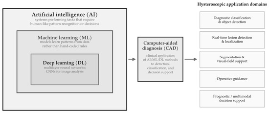
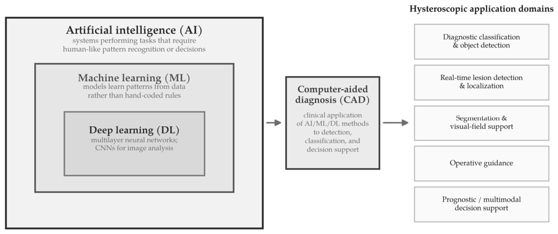
Artificial intelligence (AI) is the broad umbrella term for computational systems designed to perform tasks that usually require human-like pattern recognition or decision-making. Machine learning (ML) is a subset of AI in which models learn patterns from data instead of manually coded rules. Deep learning (DL) is a further subset of ML based on multilayer neural networks, particularly effective for image analysis. Computer-aided diagnosis (CAD) refers to the clinical application of such methods to support detection, classification, or diagnostic decision-making, but is not itself a distinct algorithmic class [24,25,26,27]. The conceptual relationship between AI, ML, DL, and CAD, and the five hysteroscopic application domains used to organize this review, is shown in Figure 1. A glossary of the main AI and computer-vision terms used in this review is provided in Table A1.
Figure 1.
Conceptual taxonomy of the methods and applications reviewed. Artificial intelligence (AI), machine learning (ML), and deep learning (DL) form nested subsets. Convolutional neural networks (CNN) are the deep-learning architecture most relevant to hysteroscopic image analysis. Computer-aided diagnosis (CAD) denotes the clinical application of these methods to detection, classification, and decision support and is not a distinct algorithmic class.
AI, and particularly DL, has been increasingly applied across diagnostic medicine over the past decade. Convolutional neural networks (CNNs) have achieved high diagnostic performance in dermatology, ophthalmology, radiology and gastrointestinal endoscopy—tasks that share key features with hysteroscopic image analysis: pattern recognition in optical images, classification of texture and color anomalies, and detection of focal lesions against complex backgrounds [25,28]. The success of AI-assisted colonoscopy polyp detection—now supported by randomized controlled trials demonstrating a significant reduction in the adenoma miss rate [29]—provides a useful analogy for AI assistance in endoscopic procedures. These developments are directly relevant to hysteroscopy, where focal lesion detection, mucosal pattern analysis, and real-time visual interpretation are likewise central tasks.
Over the same period, AI applications have also expanded across benign and oncologic gynecology, particularly in imaging-rich domains [26,27,30]. The implementation of AI to integrate augmented reality and multimodal information (imaging techniques, radiomics, and molecular diagnostics) into a virtually enhanced surgical field led to a shift from “robotic-assisted” to “robotic-guided” laparoscopic surgery [31]. Existing reviews on AI in gynecology and gynecologic cancers summarize applications across ultrasound, MRI, CT, and colposcopy, and mention hysteroscopy only tangentially or as one of many imaging modalities [26,30]. Initial applications of AI, DL, and CAD in hysteroscopy suggest that AI may improve image interpretation, support lesion recognition, and assist selected intra- or post-procedural decisions [10,32,33,34]. To date, no focused systematic review has synthesized the small and heterogeneous evidence base on AI, ML, and DL specifically applied to hysteroscopic images, videos, and hysteroscopy-based prognostic models.
The objective of this systematic review is, therefore, to identify and assess studies that apply AI, ML, or DL to hysteroscopic imaging or hysteroscopy-based prediction tasks. We aim to (1) describe model architectures, input data, reference standards and validation methods; (2) summarize diagnostic performance for intrauterine pathology (benign, premalignant, and malignant) and prognostic performance for reproductive outcomes or post-treatment recurrence; and (3) explore how these systems compare with conventional hysteroscopic assessment and existing clinical workflows.
2. Methods
2.1. Study Design and Reporting Standard
This systematic review was conducted and reported in accordance with the “Preferred Reporting Items for Systematic Reviews and Meta-Analyses (PRISMA) 2020” guidelines [35], and the completed PRISMA checklist is provided in the Supplementary Materials. Owing to the methodological and clinical heterogeneity of the available literature, the review was designed as a qualitative evidence synthesis. The review protocol was not prospectively registered.
2.2. Eligibility Criteria
Studies were eligible if they met all of the following criteria: (1) original research, (2) clear relevance to hysteroscopy, (3) application of AI, ML, DL, or computer-aided diagnostic/decision-support systems, and (4) reporting of quantitative performance or outcome data.
We included studies in which AI was applied to hysteroscopic images, hysteroscopic videos, or hysteroscopy-relevant structured clinical/imaging data in one of the following domains:
- Diagnostic lesion classification or detection;
- Segmentation or visual-field support;
- Operative guidance;
- Postoperative prognostic prediction, multimodal prediction, or hysteroscopy-related decision support.
Studies were also eligible when hysteroscopy was a core clinical component of the AI application, even if the model additionally incorporated non-hysteroscopic variables or was designed to support hysteroscopy-related management decisions.
Conference abstracts without full methods and results were excluded. However, full-text conference/proceedings papers were eligible if they reported original data, fulfilled the predefined eligibility criteria, and were not superseded by a later, more comprehensive retained publication from the same research line or overlapping cohort.
Narrative reviews, editorials, commentaries, and non-original papers were excluded. When multiple publications from the same research group reported overlapping cohorts or iterative developmental stages of the same model, only the methodologically most complete and non-redundant report was retained in the main synthesis.
2.3. Information Sources and Search Strategy
The electronic literature search was performed in PubMed/MEDLINE and via the EBSCOhost platform, including Academic Search Premier, APA PsycArticles, APA PsycInfo, CINAHL, and MEDLINE, from database inception to 8 March 2026.
The PubMed search yielded 52 records using the following search string: “(hysteroscop*[Title/Abstract]) AND (“deep learning”[Title/Abstract] OR “machine learning”[Title/Abstract] OR “artificial intelligence”[Title/Abstract] OR “computer-aided diagnosis”[Title/Abstract] OR “computer aided diagnosis”[Title/Abstract] OR “convolutional neural network*”[Title/Abstract] OR “neural network*”[Title/Abstract] OR YOLO[Title/Abstract] OR FCNN[Title/Abstract] OR “support vector machine*”[Title/Abstract] OR transformer*[Title/Abstract])”.
The EBSCOhost search yielded 42 records using the following search string: “TX hysteroscop* AND TX (“artificial intelligence” OR “machine learning” OR “deep learning” OR “computer-aided diagnosis” OR “computer aided diagnosis” OR “computer-assisted diagnosis” OR “convolutional neural network*” OR “neural network*” OR YOLO OR “support vector machine*” OR transformer*)”.
In addition to database searching, a targeted supplementary search of Google Scholar and ResearchGate was performed to identify relevant full-text reports not indexed in the core databases. This supplementary search identified one additional full-text conference paper that met the eligibility criteria and is reported in the Results section.
2.4. Selection Process
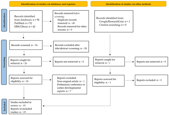
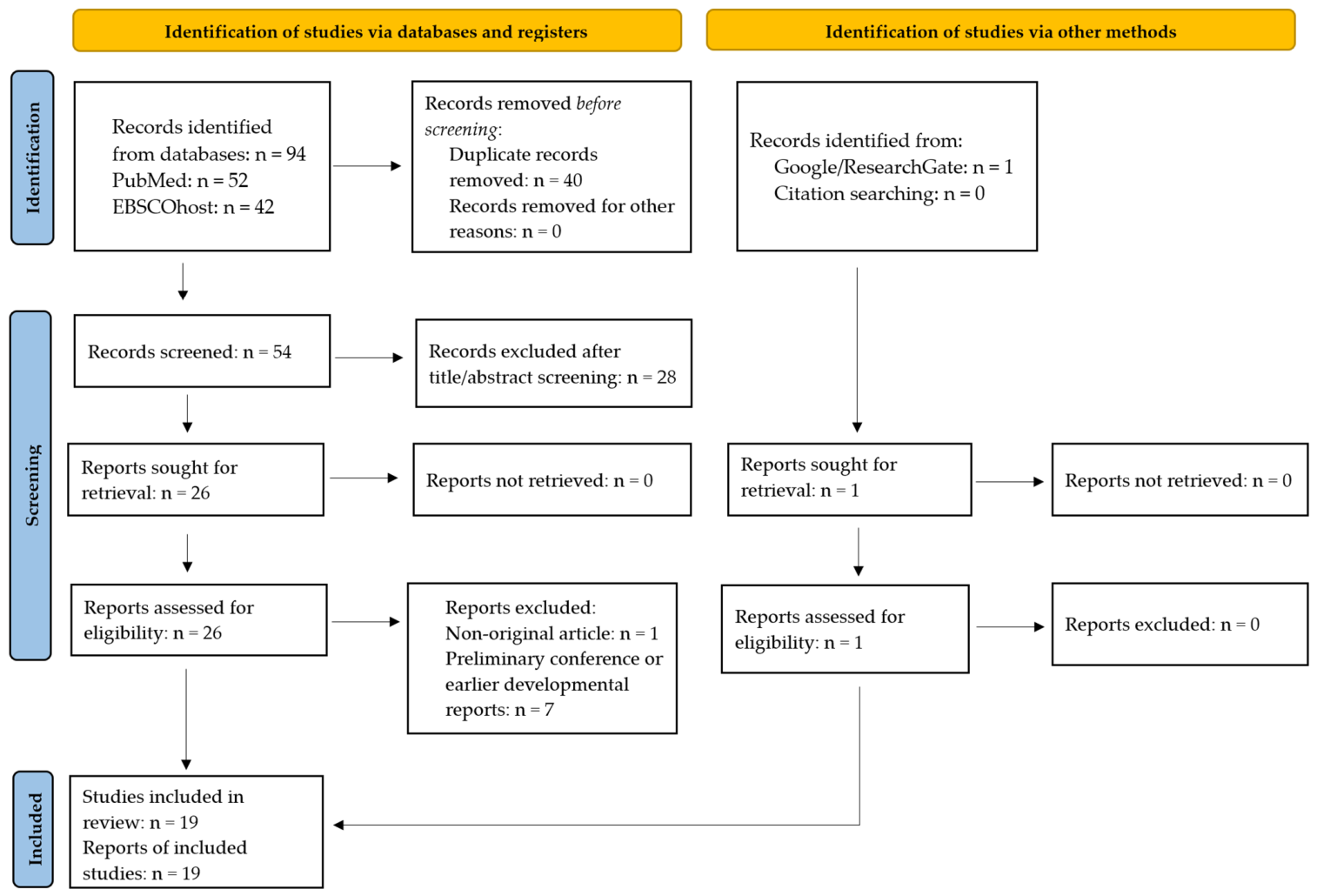
All records retrieved from PubMed and EBSCOhost were deduplicated before screening. Titles and abstracts were screened by two reviewers (R.W., S.K.), and potentially eligible reports underwent full-text assessment. Disagreements were resolved by discussion with other authors (A.R., S.G.V.). For studies published by the same research group on overlapping cohorts or representing successive developmental stages of the same model, a hierarchical inclusion strategy was applied: (1) preference for peer-reviewed journal articles over conference proceedings, (2) preference for publications with broader validation, especially external or multicenter validation, over single-center developmental reports, and (3) preference for the most recent and methodologically most complete publication when iterative model development was identified. No automation tools were used for study selection or data extraction. The selection process is visualized on the PRISMA 2020 flow diagram (Figure 2). After title/abstract screening, 27 full-text reports were assessed for eligibility. Finally, 19 primary studies were included in the final synthesis.
Figure 2.
PRISMA 2020 flowchart of study identification and selection.
2.5. Data Extraction
Data were extracted into predefined evidence tables. The following items were collected for each study: full citation, country and center(s), clinical domain, study design, prospective versus retrospective design, number of patients, number of images/frames/videos, unit of analysis, clinical setting, lesion or target type, reference standard, AI model(s), comparator(s), validation approach, and quantitative performance or clinical outcome measures.
Where applicable, extracted performance metrics included the following: area under the curve (AUC/AUROC), sensitivity, specificity, accuracy, positive and negative predictive values, precision, recall, F1 score, Dice coefficient, intersection over union (IoU), mean average precision (mAP), concordance index (c-index), kappa statistics, and runtime or deployment-related information such as frames per second (FPS) or latency. For operative and prognostic studies, relevant clinical outcomes were also extracted, including operative time, blood loss, complete resection or incomplete resection, pregnancy-related outcomes, and treatment-benefit stratification.
2.6. Risk-of-Bias Assessment
Because the included literature comprised several different study types, risk of bias was assessed using study-type-specific instruments: QUADAS-2 [36] was used for diagnostic accuracy studies, PROBAST [37] was used for prognostic or predictive modeling studies, RoB 2 [38] was used for the randomized trial. For technical segmentation or visibility-related studies that did not fit a standard diagnostic-accuracy framework, we used a structured technical assessment of dataset selection, annotation quality, unit of analysis, data-leakage risk, validation strategy, and clinical relevance. Special attention was paid to AI-specific methodological issues, including selection bias, exclusion of poor-quality images, class imbalance, image-level instead of patient-level splitting, risk of overlap between training and test material, lack of external validation, unclear reference standards, overlap of datasets between publications, and limited reporting of calibration or deployment conditions.
2.7. Data Synthesis
Because of heterogeneity in target tasks, reference standards, model architectures, validation strategies, and reported metrics, statistical pooling was not considered appropriate. The studies were therefore synthesized narratively and organized into five thematic domains:
- Diagnostic lesion classification and object detection;
- Real-time lesion detection and localization;
- Segmentation and visual-field support;
- Operative guidance;
- Prognostic, multimodal, and hysteroscopy-related decision-support applications.
Within these domains, results were interpreted in regard to study design, validation rigor, reference standard, and independence of evidence.
3. Results
3.1. Study Selection
The database search yielded 94 records, including 52 from PubMed and 42 from EBSCOhost. In addition, one further full-text report was identified through supplementary searching via Google/ResearchGate [39]. After removal of duplicates and title/abstract screening, 27 full-text reports were assessed for eligibility. Of these, eight reports were excluded after full-text review, leaving nineteen primary studies for inclusion in the final qualitative synthesis [39,40,41,42,43,44,45,46,47,48,49,50,51,52,53,54,55,56,57].
Full-text exclusions comprised one narrative review/non-original article [33], two earlier developmental reports from the intrauterine adhesion prediction line [58,59], two preliminary conference/developmental reports from the CATIA line [60,61], one prototype/integrated developmental report from the same CATIA research line [62], and two earlier developmental or conference-stage reports from research lines later represented by more comprehensive retained studies [63,64]. The final dataset therefore consisted of 19 primary studies, including both peer-reviewed journal articles and eligible full-text conference/proceedings papers.
3.2. Study Characteristics
Study characteristics are summarized in Table 1, and the study-quality/risk-of-bias overview is given in Table A2. The evidence from 19 included studies covering diagnostic lesion classification and detection, segmentation and visual-field support, operative guidance, and prognostic or decision-support applications. Most studies were retrospective or retrospective in silico; one study used a randomized prospective comparison of AI-assisted operative planning, and the fertility-prediction studies from the Beijing group were based on prospectively assembled intrauterine adhesion cohorts [50,52,56]. Dataset size varied markedly, from 52 subjects and 516 ROIs in the CAD study by Neofytou et al. [41] to 1394 patients and 55,874 hysteroscopy images in the multicenter ECCADx study by Wang et al. [54]. The technically oriented segmentation studies were generally based on comparatively small datasets. For example, Wang et al. [47] analyzed 1385 hysteroscopic images for bubble segmentation, whereas Burai et al. [43] used 28 hysteroscopic videos for uterine-wall segmentation.
Table 1.
Characteristics of the included primary studies.
The included studies did not rely on a single reference standard, reflecting their different clinical and technical aims. Histopathology was used in several lesion-classification studies [40,41,44,53,55]; Kitaya et al. evaluated expert-annotated EMiP recognition within a histologically confirmed CE cohort [51]. Expert annotation served as the reference for object-detection and segmentation studies, including fibroid, polyp, uterine-wall, and bubble detection or segmentation [42,43,46,47,49]. In the prognostic studies by Li et al., reproductive follow-up after hysteroscopic adhesiolysis was used as the outcome reference [52,56]. In Givon et al. [57], incomplete hysteroscopic myomectomy was defined as residual submucosal myoma documented at the end of surgery.
Across the nineteen studies, nine were judged at high or high/unclear risk of bias—including all three prognostic models assessed with PROBAST—and eight at moderate to moderate–high risk, while the single randomized trial carried “some concerns” (RoB 2); the recurring sources were absent of external validation, had single-center retrospective design, and used image- or frame-level rather than patient-level data splitting (Table A2).
3.3. Lesion Classification and Object Detection
Diagnostic classification and object detection studies are summarized in Table 2. Early work relied on handcrafted descriptors and conventional classifiers. Vlachokosta et al. classified three categories of hysteroscopic images, derived from 28 patients with abnormal uterine bleeding, 10 with endometrial cancer, and 39 normal subjects, and reported an accuracy of 91.2%, sensitivity of 93.6%, and specificity of 83.8% using an artificial neural network after reduction of 167 vessel and texture features to four selected features [40]. Neofytou et al. developed a CAD system for early endometrial cancer detection based on gamma-corrected red–green–blue (RGB), hue–saturation–value (HSV), and YCrCb color-space texture analysis in 516 regions of interest (ROIs) from 52 subjects; the best ROI-level result was an 81% correct classification rate with statistical features plus gray-level difference statistics (SF + GLDS) and a support vector machine (SVM) [41].
Table 2.
Diagnostic classification and object detection studies.
Later deep-learning studies expanded both lesion spectrum and dataset size. Takahashi et al. analyzed 177 patients with normal endometrium, myoma, polyp, atypical endometrial hyperplasia, or endometrial cancer and showed that diagnostic accuracy increased to 90.29% after continuity analysis and model combination; sensitivity and specificity were 91.66% and 89.36%, respectively [45]. Zhang et al. used 1851 histologically confirmed images from 454 patients and reported an overall five-class accuracy of 80.8% for endometrial hyperplasia without atypia, atypical hyperplasia, endometrial cancer, endometrial polyp, and submucous myoma. In the same study, binary classification of benign versus premalignant or malignant lesions performed better, with an accuracy of 90.8%, sensitivity of 83.0%, and specificity of 96.0%; class-specific AUCs ranged from 0.916 for atypical hyperplasia to 0.981 for endometrial polyp [44].
A similar pattern was seen in the multiclass work by Raimondo et al. [53]. In 266 patients contributing 1500 images, lesions were grouped as benign focal, benign diffuse, and preneoplastic or neoplastic. The best classification performance reached an overall accuracy of 86.74%, specificity of 90.06%, and an F1 score (harmonic mean of precision and recall) of 80.11%. However, class-wise performance was uneven. In the best-performing configuration, recall was 94.07% for benign focal lesions, 50.0% for benign diffuse lesions, and 27.59% for preneoplastic or neoplastic lesions. In the separate identification task, the best model, obtained with clinical factors, achieved detection of 85.82%, precision of 93.12%, recall of 91.63%, and F1 score of 92.37%.
Several studies focused on binary or narrower target recognition and generally reported higher performance. Kitaya et al. analyzed fluid hysteroscopic images from 208 infertile women and developed a CNN for automatic detection of endometrial micropolyps associated with chronic endometritis (CE). Sensitivity, specificity, accuracy, precision, and F1 score were 93.6%, 92.3%, 92.8%, 88.0%, and 0.907, respectively, and the AUC was 0.930. The model’s AUC was statistically comparable to that of three experienced gynecologists, whose AUCs ranged from 0.906 to 0.948 [51].
The largest and methodologically strongest diagnostic study was the multicohort ECCADx system by Wang et al. [54]. In the final report, 1394 patients contributed 55,874 hysteroscopy images, with 1204 patients in the training set, 85 in the internal MCH test set, and 105 in the external TJH/ZZSH test set. With contrastive learning, internal test performance reached an AUC of 0.979, accuracy of 94.1%, sensitivity of 95.2%, specificity of 91.3%, and F1 score of 0.959. On the external test set, the corresponding values were AUC 0.975, accuracy 93.3%, sensitivity 92.1%, specificity 100%, and F1 score 0.959. Without contrastive learning, external specificity was 62.5%, showing a marked gain after domain-adaptive training. The authors also compared ECCADx with junior, medium, and senior gynecological endoscopists and found that the model outperformed human readers on both internal and external datasets [54]. A smaller report by Kürkçü et al. [39] evaluated four YOLO variants on 1482 physician-annotated hysteroscopy images of polyps, myomas, and endometrial cancer. The prototype was intended to support clinicians by localizing suspicious hysteroscopic abnormalities for review. YOLOv9c achieved the highest mAP50, defined at an intersection-over-union (IoU) threshold of 0.50, with 0.906, and the highest precision with 0.894, whereas YOLOv8s achieved the highest recall with 0.906; expert review of 50 images yielded model accuracies of 94–98%.
Across these studies, narrowly defined binary or lesion-specific tasks generally yielded stronger and more stable performance than broader multiclass differential diagnosis. This was shown most explicitly in the five-class versus binary comparison by Zhang et al. [44] and was also reflected by the class imbalance and low minority-class recall in the three-category study by Raimondo et al. [53]. By contrast, lesion-specific systems for micropolyps or AEH/endometrial cancer generally reported higher sensitivity, AUC, or F1 values [51,54].
3.4. Real-Time Lesion Detection and Localization
Real-time lesion detection and localization studies are summarized in Table 3. Several studies focused on object detection or real-time lesion recognition in hysteroscopic images and videos. A CNN-transformer hybrid system developed for uterine fibroid recognition achieved in the test set of 2312 images sensitivity 94.21%, specificity 83.76%, accuracy 88.93%, F1 score 89.36%, and AUC 0.96 [48].
Table 3.
Real-time lesion detection and localization studies.
A modified YOLOX model was trained on 11,839 images from 323 polyp cases and evaluated on internal and external hospital datasets comprising 431 cases in total. Lesion-based sensitivity reached 100% on the internal test set and 92.0% on the external test set, compared with 95.83% and 77.33%, respectively, for the original YOLOX model [49]. The proposed system processed video at 63 frames per second. In an earlier conference study, the same group reported real-time hysteromyoma detection with a YOLOv3 plus deep convolutional generative adversarial network (DCGAN) hybrid model at 25 frames per second (FPS) and an accuracy of 91.73% [46].
Mascarenhas et al. trained a YOLOv1-based multicentric model on 65 hysteroscopies yielding 33,239 frames and 37,512 annotated objects. On the test set, object-level recall was 0.96, precision 0.95, mAP50 was 0.98, and mAP50-95 was 0.77. At frame level, mean recall was 0.75, mean precision 0.98, and mean F1 score was 0.82 [55].
In sum, these studies indicate that real-time lesion detection and localization are feasible in hysteroscopy, particularly for visually distinctive focal pathology such as fibroids and polyps, although validation remains limited in most datasets.
3.5. Segmentation and Visual-Field Assessment
Segmentation and visual-field support studies are summarized in Table 4. Török and Harangi [42] analyzed 13 videos of hysteroscopic myomectomy and trained a fully convolutional neural network (FCNN) on 4688 annotated images, with 1600 previously unseen images reserved for testing. The model aimed to identify the plane between myoma and normal myometrium and achieved a reported pixel-wise segmentation accuracy of 86.19%. Burai et al. addressed uterine-wall extraction in hysteroscopic video frames affected by bubbles, instruments, and detached mucosa. Their ensemble of FCNNs yielded a best Dice coefficient of 0.9156 and Jaccard index of 0.8443 [43]. Wang et al. [47] focused on bubble segmentation during operative hysteroscopy, using 1385 clear images. Their edge-aware network achieved accuracy of 0.859 ± 0.017, sensitivity of 0.868 ± 0.019, precision of 0.955 ± 0.005, Dice score of 0.862 ± 0.005, and mean intersection over union of 0.758 ± 0.007. Compared with U-Net, Dice improved from 0.819 to 0.862 and mean IoU from 0.713 to 0.758. Reported inference time was 0.15 s per image.
Table 4.
Segmentation and visual-field support studies.
These segmentation studies addressed different technical problems and are therefore not directly comparable, but they show that hysteroscopy-oriented AI has extended beyond diagnosis toward intraoperative scene parsing, field stabilization, and extraction of visually relevant structures [42,43,47].
3.6. Operative Planning and Guidance
The only included operative-guidance study is summarized in Table 5. Chen et al. [50] randomized 56 patients with submucosal myomas to standard MRI-based preoperative planning or AI-augmented MRI-based planning before hysteroscopic myomectomy. The AI-assisted group had a shorter operative time than the control group (32.11 ± 11.86 vs. 41.32 ± 17.83 min, p = 0.03) and a (clinically likely irrelevant) lower intraoperative blood loss (median 10.00 [5.00–15.00] vs. 10.00 [6.25–15.00] mL, p = 0.04). This was the only included study that directly linked AI support to a measurable downstream procedural effect, although the AI input in this study was MRI segmentation rather than real-time hysteroscopic image analysis.
Table 5.
Operative guidance study.
3.7. Prognostic and Preoperative Decision Support
Prognostic, multimodal, and preoperative decision-support studies are summarized in Table 6. Two studies came from the Beijing intrauterine adhesion database. Li et al. [52] developed a multimodal learning system that integrated EMR variables with 5014 revisited hysteroscopic images from 753 post-adhesiolysis patients. Using MobileNetV3 for image feature extraction and XGBoost for multimodal ensemble learning, the model achieved AUCs of 0.967 in training, 0.936 in validation, and 0.965 in the test set for one-year conception prediction. The system operated on the hysteroscopic platform with an average analysis time of 3.7 ± 0.8 s per patient. Risk stratification based on predicted conception probability was clinically informative: mid-high-risk patients had significant benefit from assisted reproductive technology, with an odds ratio of 6 (95% CI 1.27–27.8; p = 0.02), whereas low-risk patients showed no significant ART benefit.
Table 6.
Prognostic, multimodal, and hysteroscopy-related decision-support studies.
In a related image–deep-learning study [56], Li et al. analyzed 555 cases with 4922 hysteroscopic images and developed a proportional-hazard CNN system for fertility assessment after endometrial injury. Reported AUCs were 0.982, 0.992, and 0.990 across the three randomly assigned datasets in the abstract; after excluding patients who underwent ART within one year, the corresponding AUCs were 0.982, 0.992, and 0.989. Net benefit at the chosen intervention threshold reached 69.4% in the test cohort. Concordance indices for the proportional-hazard CNN were 0.940, 0.920, and 0.925 in the training, validation, and test cohorts, respectively. Agreement between the system’s quantified risk-factor panel and senior hysteroscopist assessment was high, with kappa values ranging from 0.84 to 0.89 across four intrauterine pathological domains.
Givon et al. [57] evaluated a different use case: preoperative prediction of incomplete hysteroscopic myomectomy using routine clinical variables together with ultrasound and diagnostic hysteroscopy findings. In 345 procedures from 328 women, incomplete resection occurred in 16.2%. CatBoost, trained with stratified five-fold patient-level cross-validation, achieved an AUROC of 0.72 and average precision of 0.93; at the prespecified 0.50 threshold, accuracy was 0.76, positive predictive value 0.89, sensitivity 0.81, specificity 0.50, and F1 score 0.85. In a fixed preprocessed comparator analysis, CatBoost reached accuracy 0.69, positive predictive value 0.88, sensitivity 0.73, specificity 0.50, F1 score 0.80, AUROC 0.71, and average precision 0.93, modestly outperforming logistic regression trained on identical inputs.
3.8. Performance by Task Type
Across domains, the strongest performance was reported in tightly defined tasks with clear target labels, including binary AEH/endometrial cancer discrimination, endometrial micropolyp detection, focal polyp or fibroid localization, and postoperative fertility prediction after adhesiolysis [51,54,55,56]. Broader multiclass classification of heterogeneous intrauterine lesions was less stable, and the lowest class-specific recall was reported for the preneoplastic/neoplastic category in the Raimondo study [53]. Segmentation and visual-field support studies showed technical feasibility, often with good Dice or IoU values, but did not report patient-level diagnostic or clinical endpoints. Chen et al. [50] was the only study that directly linked AI support to procedural outcomes, whereas Li et al. [52,56] directly linked hysteroscopy-based AI outputs to postoperative reproductive stratification.
4. Discussion
This review synthesized nineteen primary studies applying AI, ML, and DL to hysteroscopy across diagnostic, technical-support, operative, and prognostic tasks. Across these studies, performance is strongest in large binary diagnostic systems for AEH and endometrial cancer and in narrow prognostic models for postoperative fertility after adhesiolysis. The ECCADx system, achieving an AUC of 0.979 on internal validation and 0.975 on external multicenter data, is among the highest reported in AI-based gynecological diagnostics [54]. At the same time, the overall evidence base remains preliminary. Most studies were retrospective, most were single-center, and only one directly evaluated a clinical procedural endpoint in a comparative design [50]. The available evidence therefore establishes technical feasibility in selected tasks but does not yet demonstrate prospective clinical effectiveness or workflow superiority in routine practice, neither of which can be inferred from retrospective, single-center accuracy estimates.
A consistent pattern across the reviewed literature is the difference between performance in narrow, well-delimited tasks and performance in broader multiclass classification. In Zhang et al. [44], binary discrimination between benign and premalignant/malignant lesions outperformed five-class classification within the same dataset. In Raimondo et al. [53], overall accuracy remained acceptable (87%), but recall for the preneoplastic/neoplastic category fell to 28%, meaning that more than 70% of cases in this category were not detected as such by the model.
The best-supported hysteroscopic applications are image-based recognition tasks: lesion classification, focal lesion localization, and detection of subtle inflammatory patterns. These are also tasks in which human interpretation varies substantially with experience, image quality, and clinical context [14,16,18,19]. At the current stage, the most plausible role of AI is not “replacement” of the hysteroscopist, but support for more consistent recognition of patterns that may otherwise be overlooked, overcalled, or interpreted differently across operators. Chronic endometritis (CE) is a good example of a setting in which AI may be clinically useful despite only partially overlapping hysteroscopic, histopathological, immunohistochemical, and microbiologic reference standards [65]. The CNN for automatic detection of endometrial micropolyps associated with CE developed by Kitaya et al. [51] achieved an AUC of 0.930, comparable to experienced gynecologists.
Examples of AI narrowing the performance gap between junior and more experienced examiners are also available from gynecological ultrasonography. In the study by Xu et al., a YOLOv8-based model for deep infiltrating endometriosis improved the AUROC of two junior sonologists from 0.748 to 0.878 and from 0.713 to 0.798, with sensitivity increasing up to 94.35% [66]. This supports the broader point that AI may be particularly useful in operator-dependent visual diagnostics, where expert-level pattern recognition is difficult to acquire and immediate supervision is not always available. Deep learning has also been applied to 3D transvaginal ultrasound for intrauterine adhesions, reaching high accuracy with external validation [67]. For T-shaped uterus, marked discordance between competing 3D-ultrasound diagnostic criteria illustrates the instability of morphometric thresholds [68], while a recent machine-learning model based on quantitative 3D-TVUS parameters achieved good diagnostic discrimination [69]. At the same time, poor-to-moderate inter-observer agreement for a proposed gynecological TVS image-quality scoring system shows that variability begins already at image acquisition and labeling [70].
Hysteroscopic image quality depends heavily on the procedure itself. Distance from tissue, viewing angle, bleeding, specular reflections from the fluid-distension medium, variable cavity distension, motion blur, and the curved, partially occluded field of view all shape the input available to the model. This differs from colonoscopy and gastroscopy, where larger benchmark datasets and better-developed artifact frameworks have supported more generalizable systems [71,72,73,74]. In hysteroscopy, the studies by Wang et al. on bubble segmentation [47], by Burai et al. [43] on uterine-wall extraction, and the myomectomy plane segmentation by Török and Harangi [42] address technical conditions that are directly relevant for practical deployment.
Translational adoption also depends on clinically interpretable output and reliable uncertainty estimates at the point of care. The deep-learning models that perform best on hysteroscopic images are largely opaque, and the resulting “black-box” character, together with the need for interpretability, calibrated confidence, and demonstrable fairness, is now recognized as a principal barrier to clinical deployment of AI in medical imaging [24]. None of the included hysteroscopic studies reported calibration of predicted probabilities or provided interpretability output that a clinician could use to accept or override a given prediction. For real-time hysteroscopy, retrospective expert-level accuracy is insufficient unless the output is interpretable and its uncertainty is clearly communicated during the examination. Confidence calibration and interpretable localization are needed before these systems can support intraoperative decisions.
Hysteroscopy, at its current developmental stage, remains a manually performed procedure. Navigation, lesion exposure, tissue handling, instrument positioning, and the final operative decision still depend on the operator’s real-time judgment. This limits direct comparison with laparoscopy, especially robotic laparoscopy, where the operative interface is already digital and telemanipulated, preoperative imaging can be overlaid onto the operative field, critical structures can be annotated in real time, and workflow analysis can be integrated more directly into surgical guidance [27,75,76,77]. Hysteroscopy does not currently offer an equivalent platform. The main contribution of AI is more likely to lie in perception support, image-quality control, lesion highlighting, multimodal integration, and postoperative prediction.
The reviewed studies point to four near-term applications: (1) image processing and classification during or after the procedure, as a decision-support layer that augments interpretation without replacing manual control [53,54,55]; (2) texture and vascular-pattern analysis for subtle pathology, including chronic endometritis [51], early hyperplasia [44,54], and structural anomalies; (3) multimodal integration with 3D ultrasound or sonohysterography for pre-hysteroscopic planning in T-shaped uterus, uterine septa, and Asherman syndrome [52,59], where cavity-level and panoramic imaging provide complementary information; and (4) AI-assisted postoperative outcome prediction, as demonstrated by the Beijing group [52,56,57,59], in which hysteroscopic findings contribute to probabilistic clinical decision-support tools.
4.1. Limitations
Risk-of-bias assessments consistently show that technically sound model development is undermined by limited validation. This pattern is not specific to hysteroscopy, as several clinical studies evaluating AI in surgery rely on sparse multicenter data, heterogeneous annotation standards, limited external validation, and insufficient outcome-oriented evaluation [78]. Fourteen of the nineteen included studies were retrospective and single-center. Many used internal train–test splits from the same institutional archive. Under these conditions, performance may be inflated by shared acquisition settings, equipment, annotation habits, or patient mix [78,79,80,81,82]. This problem is compounded when the unit of analysis is the image or frame rather than the patient. If multiple images from the same patient are distributed across training and test sets, effective sample size is overstated and leakage may remain undetected.
A further constraint concerns the provenance of the datasets themselves. Most included studies drew on curated retrospective archives in which low-quality frames were excluded before model development, so that the images used for training and testing represent favorable acquisition conditions instead of routine hysteroscopic environment, in which glare, bleeding, mucus, debris, motion blur, and incomplete cavity distension are common. Annotation reference standards were heterogeneous and frequently rested on a single expert reader or on the interpreting clinician, without reported inter-annotator agreement, so that label noise cannot be quantified. This combination of curated inputs and single-source labels limits ecological validity, as reported performance reflects development conditions rather than real-world deployment.
The heterogeneity of reporting across studies makes cross-study comparison unreliable. Studies report different primary metrics (AUC, accuracy, F1, Dice, C-statistic), use different reference standards (histopathology from directed biopsy, expert hysteroscopic annotation, blinded panel review), and employ different units of analysis (per-image, per-frame, per-patient). The choice of per-image instead of per-patient analysis artificially inflates effective sample sizes and can mask patient-level confounding: when multiple images from the same patient are split across training and test sets—a form of data leakage whose performance-inflating effects have been quantified at 30–55% in CNN-based classification tasks [81,82]—reported performance metrics are optimistic in ways that are practically impossible to detect without access to the original data. For the detection and localization studies specifically, the unit of analysis, the intersection-over-union threshold defining a true detection, and whether negative frames or videos were included in the test set were reported inconsistently; this incomplete reporting limits comparison across studies. Inter-observer variation in histopathological diagnosis of endometrial hyperplasia remains substantial even among specialist pathologists [83], compounding the difficulty of constructing unambiguous reference standards for AI training. The study by Raimondo et al. [53] illustrates this limitation: overall classification accuracy of 86.74% coexisted with a recall of only 27.59% for the preneoplastic/neoplastic class, precisely the category of greatest clinical relevance and the one for which a missed diagnosis carries the highest patient risk.
The same limitation appears differently in the prognostic studies. Their reported AUCs are high, but validation remains internal to a single cohort line [52,56]. Calibration of predicted probabilities was rarely reported. Li et al. [56] included decision-curve analysis and reported a net benefit of 69.4% for the fertility-assessment system, whereas Li et al. [52] reported risk-stratified ART benefit without formal probability calibration. Earlier Cyprus CAD work provided an internally validated texture-based proof of concept [41]. The Beijing intrauterine-adhesion models report high AUCs, but validation remains internal to one cohort line [52,56]. Wang et al. [54] provide the strongest diagnostic validation with independent multicenter test datasets; Chen et al. [50] remains the only study with a comparative procedural endpoint. The imbalance between tasks where AI implementation produces statistically significant numbers and their clinical utility can be illustrated by two studies addressing hysteroscopic myomectomy [50,57]. The work of Chen et al. [50] remains the only randomized comparison linking AI support to a procedural endpoint. It reports a shorter operative time (around 10 min) and statistically lower blood loss despite identical median values in both groups and only slightly different interquartile ranges. The clinical relevance, however, remains speculative. First, the almost identical and extremely low blood loss contrasted with a difference of approximately 25% in surgical time. It is surprising, because surgical time is a strong predictor of blood loss at least in, e.g., laparoscopic myomectomy [84,85]. By contrast, Givon et al. [57] addressed the clinically relevant problem of incomplete myomectomy. In real-world settings, incomplete myomectomy has been reported in 36.69% of hysteroscopic approaches and is associated with complications (PR: 2.77; 95% CI: 1.43–5.38) [86]. The model performance in [57] was clearly lower than in the Beijing fertility studies or ECCADx, but the results suggest that AI can help identify cases in which complete hysteroscopic treatment is less likely.
4.2. Future Directions
Progress toward clinical use requires prospective multicenter validation. The primary outcomes should include, beyond image-level accuracy, patient-relevant measures, such as pregnancy, detection of (pre)malignant lesions, recurrence, or completeness of treatment. Where possible, analyses should also be stratified by operator experience, because support systems may be most useful for less-experienced hysteroscopists, as demonstrated by Xu et al. in the ultrasound setting [66]. Reporting standards for early-stage clinical evaluation of AI decision-support systems, as formalized in the DECIDE-AI guideline, should be adopted to prospective hysteroscopic AI studies [87].
The parallel development of standardized hysteroscopic image databases will be needed for generalizable models and cross-study comparison. Such databases require standardized image-acquisition protocols and agreed annotation standards for specific intrauterine pathologies. Coordinated international annotation initiatives, analogous to those that underpinned the CVC-ClinicDB and REAL-Colon colonoscopy benchmarks, are necessary for progress [71,72,73].
Multimodal AI, combining hysteroscopic images with 3D ultrasonography, MRI findings, hormonal profiles, and clinical variables, is a plausible next step. The success of the Beijing multimodal system [52] suggests that the discriminative power of hysteroscopic images is substantially amplified when contextualized by complementary data modalities. Extending this principle to diagnostic tasks, for example by integrating imaging studies (MRI, 2D- or 3D-TVS) or electronic medical records with real-time hysteroscopic assessment, could address the limits of image-only approaches [56,75].
Finally, AI-driven training simulators that provide real-time feedback on instrument handling and pattern recognition may help shorten the steep learning curve in hysteroscopy and reduce interobserver variability, particularly in settings with limited expert supervision [24,88].
These training and decision-support roles raise an ethical question that cannot be separated from the technical one. Pope Leo XIV’s first encyclical, Magnifica Humanitas, addresses the place of the human person in the age of artificial intelligence and warns that ready-made answers may “weaken personal creativity and judgment” and that easy retrieval can “risk extinguishing the desire to ask questions” [89]. Medical research expresses a related concern through co-intelligence, a model of human–AI collaboration that preserves clinician judgment and treats AI literacy as a core competence [90]; medical imaging literature similarly identifies interpretability and trust as prerequisites for adoption [24]. In hysteroscopy, AI should therefore be judged by diagnostic performance and by its effect on operator judgment, procedural skill, and the questioning that real-time intrauterine decision-making still requires [50,87].
5. Conclusions
The existing literature shows that machine-learning and deep-learning systems can classify endometrial lesions, detect focal pathology in real time, segment relevant intraoperative structures, and support selected prognostic decisions. The strongest evidence currently concerns binary diagnostic classification for AEH and endometrial cancer, focal lesion detection, and fertility-related risk stratification after hysteroscopic treatment of intrauterine adhesions. At the same time, the field remains constrained by retrospective design, operator-dependent image generation, limited external validation, heterogeneous reference standards, and scarce evaluation of patient-centered outcomes. Unlike laparoscopy, where AI-robotics integration is moving toward real-time guidance and image overlay, hysteroscopy remains a manually performed procedure. Near-term use is therefore likely to remain concentrated in image processing, texture analysis, multimodal data integration, and decision-support. These systems are most likely to support perception by improving interpretation consistency, highlighting suspicious lesions, assisting biopsy targeting, and integrating hysteroscopic findings with clinical data. Translating these technical achievements into clinical practice requires prospective multicenter validation, standardized datasets, and workflow integration.
Supplementary Materials
The following supporting information can be downloaded at: https://www.mdpi.com/article/10.3390/diagnostics16121899/s1, PRISMA 2020 Checklist.
Author Contributions
Conceptualization, R.W. and S.G.V.; methodology, R.W., A.R., S.G.V., and I.A.; software, R.W. and P.T.; formal analysis, R.W., S.K., and A.R.; investigation, R.W. and P.T., data curation, R.W. and S.K.; writing—original draft preparation, R.W.; writing—review and editing, R.W., S.K., S.G.V., and I.A.; visualization, R.W.; supervision, S.K., A.D.S.S., I.A., and S.G.V.; project administration, R.W. All authors have read and agreed to the published version of the manuscript.
Funding
This research received no external funding.
Institutional Review Board Statement
Not applicable.
Informed Consent Statement
Not applicable.
Data Availability Statement
No new data were created or analyzed in this study. Data sharing is not applicable to this article.
Conflicts of Interest
The authors declare no conflicts of interest.
Appendix A
Table A1.
Practical definitions of key technical terms used in this review.
Table A2.
Risk of bias and quality assessment of included studies.
References
- The Use of Hysteroscopy for the Diagnosis and Treatment of Intrauterine Pathology: ACOG Committee Opinion, Number 800. Obstet. Gynecol. 2020, 135, e138–e148. [CrossRef]
- Andrić, B.Ž.; Perović, M.; Hanželič, E.T.; Garcia, M.A.; Casadio, P.; De Angelis, C.; Van Kerrebroeck, H.; Vitagliano, A.; Catena, U. A European Society for Gynaecological Endoscopy Survey of Hysteroscopic Practice. Facts Views Vis. ObGyn 2025, 17, 391–401. [Google Scholar] [CrossRef]
- Vitale, S.G.; Watrowski, R.; Barra, F.; D’Alterio, M.N.; Carugno, J.; Sathyapalan, T.; Kahramanoglu, I.; Reyes-Muñoz, E.; Lin, L.-T.; Urman, B.; et al. Abnormal Uterine Bleeding in Perimenopausal Women: The Role of Hysteroscopy and Its Impact on Quality of Life and Sexuality. Diagnostics 2022, 12, 1176. [Google Scholar] [CrossRef] [PubMed]
- Vitale, S.G.; Haimovich, S.; Riemma, G.; Ludwin, A.; Zizolfi, B.; De Angelis, M.C.; Carugno, J. Innovations in Hysteroscopic Surgery: Expanding the Meaning of “in-Office”. Minim. Invasive Ther. Allied Technol. 2021, 30, 125–132. [Google Scholar] [CrossRef] [PubMed]
- Breitkopf, D.M.; Green, I.C. Key Innovations in the Development of Hysteroscopy. J. Minim. Invasive Gynecol. 2026, 33, 637–641. [Google Scholar] [CrossRef]
- Di Spiezio Sardo, A.; Saccone, G.; Carugno, J.; Pacheco, L.A.; Zizolfi, B.; Haimovich, S.; Clark, T.J. Endometrial Biopsy under Direct Hysteroscopic Visualisation versus Blind Endometrial Sampling for the Diagnosis of Endometrial Hyperplasia and Cancer: Systematic Review and Meta-Analysis. Facts Views Vis. ObGyn 2022, 14, 103–110. [Google Scholar] [CrossRef]
- Vitale, S.G.; Buzzaccarini, G.; Riemma, G.; Pacheco, L.A.; Di Spiezio Sardo, A.; Carugno, J.; Chiantera, V.; Török, P.; Noventa, M.; Haimovich, S.; et al. Endometrial Biopsy: Indications, Techniques and Recommendations. An Evidence-Based Guideline for Clinical Practice. J. Gynecol. Obstet. Hum. Reprod. 2023, 52, 102588. [Google Scholar] [CrossRef] [PubMed]
- Vitale, S.G.; Haimovich, S.; Laganà, A.S.; Alonso, L.; Di Spiezio Sardo, A.; Carugno, J.; From the Global Community of Hysteroscopy Guidelines Committee. Endometrial Polyps. An Evidence-Based Diagnosis and Management Guide. Eur. J. Obstet. Gynecol. Reprod. Biol. 2021, 260, 70–77. [Google Scholar] [CrossRef]
- Garzon, S.; Laganà, A.S.; Di Spiezio Sardo, A.; Alonso Pacheco, L.; Haimovich, S.; Carugno, J.; Vitale, S.G.; Casarin, J.; Raffaelli, R.; Andrisani, A.; et al. Hysteroscopic Metroplasty for T-Shaped Uterus: A Systematic Review and Meta-Analysis of Reproductive Outcomes. Obstet. Gynecol. Surv. 2020, 75, 431–444. [Google Scholar] [CrossRef]
- Riemma, G.; Vitale, S.G.; Manchanda, R.; Rathore, A.; Török, P.; De Angelis, C.; Urman, B.; Iraci Sareri, M.; La Verde, M.; Carugno, J.; et al. The Role of Hysteroscopy in Reproductive Surgery: Today and Tomorrow. J. Gynecol. Obstet. Hum. Reprod. 2022, 51, 102350. [Google Scholar] [CrossRef]
- Gallo, A.; Catena, U.; Saccone, G.; Di Spiezio Sardo, A. Conservative Surgery in Endometrial Cancer. J. Clin. Med. 2021, 11, 183. [Google Scholar] [CrossRef] [PubMed]
- Watrowski, R.; Jäger, C.; Möckel, J.; Kurz, P.; Schmidt, D.; Freudenberg, N. Hysteroscopic Treatment of Uterine Tumor Resembling Ovarian Sex Cord-like Tumor (UTROSCT). Gynecol. Endocrinol. 2015, 31, 856–859. [Google Scholar] [CrossRef]
- Abrahamsen, K.H.; Nielsen, M.S.; Brandt, N.B.; Nielsen, A.B.; Konge, L.; Dueholm, M.; Ravn, P. Training and Assessment of Competencies in Hysteroscopy—A Systematic Review. Eur. J. Obstet. Gynecol. Reprod. Biol. 2026, 318, 114890. [Google Scholar] [CrossRef] [PubMed]
- Matsumoto, Y.; Yamaguchi, K.; Maruyama, M.; Sone, K.; Mori-Uchino, M.; Hirota, Y.; Wada-Hiraike, O.; Osuga, Y. Characteristic Hysteroscopy Appearance Considerations for Detecting Uterine Endometrial Malignancies. J. Obstet. Gynaecol. Res. 2024, 50, 566–571. [Google Scholar] [CrossRef]
- Pérez-Cejuela, B.A.; Vitale, S.G.; Pérez-Medina, T.; Rios-Vallejo, M.; Della Corte, L.; Vicente, A.R.; Angioni, S.; Calles-Sastre, L. Hysteroscopic versus Histopathological Agreement in the Diagnosis of Chronic Endometritis: Results from a Retrospective Observational Study. Arch. Gynecol. Obstet. 2023, 308, 1817–1822. [Google Scholar] [CrossRef]
- Halouani, A.; Aloui, H.; Hamdaoui, R.; Masmoudi, Y.; Triki, A.; Ben Amor, A.; Halouani, L. Performance of Hysteroscopy in Diagnosing Chronic Endometritis and the Role of Intra- and Inter-Observer Variability: A Prospective Study of 70 Cases. AJOG Glob. Rep. 2025, 5, 100515. [Google Scholar] [CrossRef]
- De Franciscis, P.; Riemma, G.; Schiattarella, A.; Cobellis, L.; Guadagno, M.; Vitale, S.G.; Mosca, L.; Cianci, A.; Colacurci, N. Concordance between the Hysteroscopic Diagnosis of Endometrial Hyperplasia and Histopathological Examination. Diagnostics 2019, 9, 142. [Google Scholar] [CrossRef]
- Bourdel, N.; Modaffari, P.; Tognazza, E.; Pertile, R.; Chauvet, P.; Botchorishivili, R.; Savary, D.; Pouly, J.L.; Rabischong, B.; Canis, M. Does Experience in Hysteroscopy Improve Accuracy and Inter-Observer Agreement in the Management of Abnormal Uterine Bleeding? Surg. Endosc. 2016, 30, 5558–5564. [Google Scholar] [CrossRef]
- Martinho, M.S.L.; da Costa Santos, C.M.N.; Silva Carvalho, J.L.M.; Bernardes, J.F.M.A.L. Effect of Clinical Information and Previous Exam Execution on Observer Agreement and Reliability in the Analysis of Hysteroscopic Video-Recordings. Arch. Gynecol. Obstet. 2018, 297, 393–400. [Google Scholar] [CrossRef] [PubMed]
- Tsonis, O.; Gkrozou, F.; Dimitriou, E.; Paschopoulos, M. Comparative Retrospective Study on Transvaginal Sonography versus Office Hysteroscopy in the Diagnosis of Endometrial Pathology among Different Subgroups. J. Obstet. Gynaecol. Res. 2021, 47, 669–678. [Google Scholar] [CrossRef]
- Vasileva, R.; Meherzi, M.; Bucau, M.; Courcier, H.; Gonthier, C.; Koskas, M. Diagnostic Performances of Hysteroscopy in Post-Remission Surveillance of Patients Treated Conservatively for Endometrial Cancer and Atypical Hyperplasia: A Cohort Study. Eur. J. Obstet. Gynecol. Reprod. Biol. 2025, 312, 114108. [Google Scholar] [CrossRef]
- Pérez Milán, F.; Caballero Campo, M.; Domínguez Arroyo, J.A.; Carrera Roig, M.; Moratalla Bartolomé, E.; Haimovich, S.; Pérez de la Blanca, E.; Barrenetxea Ziarrusta, G.; Puente Águeda, J.M.; Alcázar Zambrano, J.L.; et al. Diagnostic Accuracy of Hysteroscopy for Chronic Endometritis in Reproductive-Age and Infertile Women: A Systematic Review and Latent Class Meta-Analysis. J. Minim. Invasive Gynecol. 2026, S1553-4650(26)00128-7. [Google Scholar] [CrossRef] [PubMed]
- Savran, M.M.; Sørensen, S.M.D.; Konge, L.; Tolsgaard, M.G.; Bjerrum, F. Training and Assessment of Hysteroscopic Skills: A Systematic Review. J. Surg. Educ. 2016, 73, 906–918. [Google Scholar] [CrossRef]
- Avanzo, M.; Stancanello, J.; Pirrone, G.; Drigo, A.; Retico, A. The Evolution of Artificial Intelligence in Medical Imaging: From Computer Science to Machine and Deep Learning. Cancers 2024, 16, 3702. [Google Scholar] [CrossRef]
- LeCun, Y.; Bengio, Y.; Hinton, G. Deep Learning. Nature 2015, 521, 436–444. [Google Scholar] [CrossRef]
- Akazawa, M.; Hashimoto, K. Artificial Intelligence in Gynecologic Cancers: Current Status and Future Challenges—A Systematic Review. Artif. Intell. Med. 2021, 120, 102164. [Google Scholar] [CrossRef] [PubMed]
- Pipes, G.M.; Hung, A.J.; Kim, K.H. Artificial Intelligence and Gynecologic Surgery. Obstet. Gynecol. 2025, 146, 672–678. [Google Scholar] [CrossRef]
- Bang, C.S.; Lee, J.J.; Baik, G.H. Computer-Aided Diagnosis of Diminutive Colorectal Polyps in Endoscopic Images: Systematic Review and Meta-Analysis of Diagnostic Test Accuracy. J. Med. Internet Res. 2021, 23, e29682. [Google Scholar] [CrossRef]
- Yagnik, V.D.; Choudhary, P.R.; Garg, P. Artificial Intelligence in Gastrointestinal Endoscopy: Current Advances, Clinical Integration, and Future Directions: A Narrative Review. Clin. Exp. Gastroenterol. 2026, 19, 573930. [Google Scholar] [CrossRef]
- Brandão, M.; Mendes, F.; Martins, M.; Cardoso, P.; Macedo, G.; Mascarenhas, T.; Mascarenhas Saraiva, M. Revolutionizing Women’s Health: A Comprehensive Review of Artificial Intelligence Advancements in Gynecology. J. Clin. Med. 2024, 13, 1061. [Google Scholar] [CrossRef] [PubMed]
- Watrowski, R.; Sparić, R. Editorial: Changing Backgrounds and Groundbreaking Changes: Gynecological Surgery in the Third Decade of the 21st Century Volume II. Front. Surg. 2025, 12, 1587048. [Google Scholar] [CrossRef]
- Tanos, V.; Neofytou, M.; Soliman, A.S.A.; Tanos, P.; Pattichis, C.S. Is Computer-Assisted Tissue Image Analysis the Future in Minimally Invasive Surgery? A Review on the Current Status of Its Applications. J. Clin. Med. 2021, 10, 5770. [Google Scholar] [CrossRef]
- Tanos, V.; Neofytou, M.; Tanos, P.; Pattichis, C.S.; Pattichis, M.S. Computer-Aided Diagnosis by Tissue Image Analysis as an Optical Biopsy in Hysteroscopy. Int. J. Mol. Sci. 2022, 23, 12782. [Google Scholar] [CrossRef]
- Gkrozou, F.; Bais, V.; Skentou, C.; Kalaitzopoulos, D.R.; Grigoriadis, G.; Vatopoulou, A.; Paschopoulos, M.; Daniilidis, A. Applications of Deep Learning Models in Laparoscopy for Gynecology. Medicina 2025, 61, 1460. [Google Scholar] [CrossRef] [PubMed]
- Page, M.J.; McKenzie, J.E.; Bossuyt, P.M.; Boutron, I.; Hoffmann, T.C.; Mulrow, C.D.; Shamseer, L.; Tetzlaff, J.M.; Akl, E.A.; Brennan, S.E.; et al. The PRISMA 2020 statement: An updated guideline for reporting systematic reviews. BMJ 2021, 372, n71. [Google Scholar] [CrossRef] [PubMed]
- Whiting, P.F.; Rutjes, A.W.S.; Westwood, M.E.; Mallett, S.; Deeks, J.J.; Reitsma, J.B.; Leeflang, M.M.G.; Sterne, J.A.C.; Bossuyt, P.M.M.; QUADAS-2 Group. QUADAS-2: A Revised Tool for the Quality Assessment of Diagnostic Accuracy Studies. Ann. Intern. Med. 2011, 155, 529–536. [Google Scholar] [CrossRef] [PubMed]
- Wolff, R.F.; Moons, K.G.M.; Riley, R.D.; Whiting, P.F.; Westwood, M.; Collins, G.S.; Reitsma, J.B.; Kleijnen, J.; Mallett, S.; PROBAST Group. PROBAST: A Tool to Assess the Risk of Bias and Applicability of Prediction Model Studies. Ann. Intern. Med. 2019, 170, 51–58. [Google Scholar] [CrossRef]
- Sterne, J.A.C.; Savović, J.; Page, M.J.; Elbers, R.G.; Blencowe, N.S.; Boutron, I.; Cates, C.J.; Cheng, H.-Y.; Corbett, M.S.; Eldridge, S.M.; et al. RoB 2: A Revised Tool for Assessing Risk of Bias in Randomised Trials. BMJ 2019, 366, l4898. [Google Scholar] [CrossRef] [PubMed]
- Kürkçü, R.A.; Baylan, I.Y.Y.; Attar, E.; Selçuk, B.; Şerif, T. AI-Enhanced Endometrial Cancer Diagnosis System. In Proceedings of the 9th International Conference on Computer Science and Engineering (UBMK), Antalya, Turkey, 26–28 October 2024; pp. 1–5. [Google Scholar] [CrossRef]
- Vlachokosta, A.A.; Asvestas, P.A.; Gkrozou, F.; Lavasidis, L.; Matsopoulos, G.K.; Paschopoulos, M. Classification of Hysteroscopical Images Using Texture and Vessel Descriptors. Med. Biol. Eng. Comput. 2013, 51, 859–867. [Google Scholar] [CrossRef]
- Neofytou, M.S.; Tanos, V.; Constantinou, I.; Kyriacou, E.C.; Pattichis, M.S.; Pattichis, C.S. Computer-Aided Diagnosis in Hysteroscopic Imaging. IEEE J. Biomed. Health Inform. 2015, 19, 1129–1136. [Google Scholar] [CrossRef]
- Török, P.; Harangi, B. Digital Image Analysis with Fully Connected Convolutional Neural Network to Facilitate Hysteroscopic Fibroid Resection. Gynecol. Obstet. Investig. 2018, 83, 615–619. [Google Scholar] [CrossRef]
- Burai, P.; Hajdu, A.; Manuel, F.-R.E.; Harangi, B. Segmentation of the Uterine Wall by an Ensemble of Fully Convolutional Neural Networks. In Proceedings of the 2018 40th Annual International Conference of the IEEE Engineering in Medicine and Biology Society (EMBC), Honolulu, HI, USA, 18–21 July 2018; pp. 49–52. [Google Scholar]
- Zhang, Y.; Wang, Z.; Zhang, J.; Wang, C.; Wang, Y.; Chen, H.; Shan, L.; Huo, J.; Gu, J.; Ma, X. Deep Learning Model for Classifying Endometrial Lesions. J. Transl. Med. 2021, 19, 10. [Google Scholar] [CrossRef]
- Takahashi, Y.; Sone, K.; Noda, K.; Yoshida, K.; Toyohara, Y.; Kato, K.; Inoue, F.; Kukita, A.; Taguchi, A.; Nishida, H.; et al. Automated System for Diagnosing Endometrial Cancer by Adopting Deep-Learning Technology in Hysteroscopy. PLoS ONE 2021, 16, e0248526. [Google Scholar] [CrossRef]
- Zhao, A.; Zhang, J.; Wang, S.; Wang, Y.; Zhu, X.; Shen, W.; Wang, W. Detection of Hysteroscopic Hysteromyoma in Real-Time Based on Deep Learning. J. Phys. Conf. Ser. 2021, 1861, 012079. [Google Scholar] [CrossRef]
- Wang, D.; Dai, W.; Tang, D.; Liang, Y.; Ouyang, J.; Wang, H.; Peng, Y. Deep Learning Approach for Bubble Segmentation from Hysteroscopic Images. Med. Biol. Eng. Comput. 2022, 60, 1613–1626. [Google Scholar] [CrossRef]
- Zhao, A.; Du, X.; Wang, S.; Wang, W.; Yuan, S.; Ma, W.; Yan, W.; Shen, W.; Zhu, X. A CNN-Transformer Hybrid Network for Recognizing Uterine Fibroids. In Proceedings of the 2022 IEEE International Conference on Consumer Electronics-Asia (ICCE-Asia), Yeosu, Republic of Korea, 26–28 October 2022; pp. 1–4. [Google Scholar]
- Zhao, A.; Du, X.; Yuan, S.; Shen, W.; Zhu, X.; Wang, W. Automated Detection of Endometrial Polyps from Hysteroscopic Videos Using Deep Learning. Diagnostics 2023, 13, 1409. [Google Scholar] [CrossRef]
- Chen, M.; Kong, W.; Li, B.; Tian, Z.; Yin, C.; Zhang, M.; Pan, H.; Bai, W. Revolutionizing Hysteroscopy Outcomes: AI-Powered Uterine Myoma Diagnosis Algorithm Shortens Operation Time and Reduces Blood Loss. Front. Oncol. 2023, 13, 1325179. [Google Scholar] [CrossRef]
- Kitaya, K.; Yasuo, T.; Yamaguchi, T.; Morita, Y.; Hamazaki, A.; Murayama, S.; Mihara, T.; Mihara, M. Construction of Deep Learning-Based Convolutional Neural Network Model for Automatic Detection of Fluid Hysteroscopic Endometrial Micropolyps in Infertile Women with Chronic Endometritis. Eur. J. Obstet. Gynecol. Reprod. Biol. 2024, 297, 249–253. [Google Scholar] [CrossRef]
- Li, B.; Chen, H.; Lin, X.; Duan, H. Multimodal Learning System Integrating Electronic Medical Records and Hysteroscopic Images for Reproductive Outcome Prediction and Risk Stratification of Endometrial Injury: A Multicenter Diagnostic Study. Int. J. Surg. 2024, 110, 3237–3248. [Google Scholar] [CrossRef]
- Raimondo, D.; Raffone, A.; Salucci, P.; Raimondo, I.; Capobianco, G.; Galatolo, F.A.; Cimino, M.G.C.A.; Travaglino, A.; Maletta, M.; Ferla, S.; et al. Detection and Classification of Hysteroscopic Images Using Deep Learning. Cancers 2024, 16, 1315. [Google Scholar] [CrossRef] [PubMed]
- Wang, W.; Cai, Y.; Guo, Z.; Zhao, A.; Ma, W.; Wang, W.; Wang, S.; Zhu, X.; Du, X.; Shen, W. A Deep Learning-Based Computer-Aided Diagnosis System for Detecting Atypical Endometrial Hyperplasia and Endometrial Cancer through Hysteroscopy. iScience 2025, 28, 113045. [Google Scholar] [CrossRef] [PubMed]
- Mascarenhas, M.; Peixoto, C.; Freire, R.; Cavaco Gomes, J.; Cardoso, P.; Castro, I.; Martins, M.; Mendes, F.; Mota, J.; Almeida, M.J.; et al. Artificial Intelligence and Hysteroscopy: A Multicentric Study on Automated Classification of Pleomorphic Lesions. Cancers 2025, 17, 2559. [Google Scholar] [CrossRef]
- Li, B.; Chen, H.; Duan, H. Visualized Hysteroscopic Artificial Intelligence Fertility Assessment System for Endometrial Injury: An Image-Deep-Learning Study. Ann. Med. 2025, 57, 2478473. [Google Scholar] [CrossRef]
- Givon, I.; Sabag, D.N.; Yacobi, B.; Chaim, O.B.; Bor, N.; Matot, R.; Nassie, D.; Borovich, A. Machine Learning Prediction of Incomplete Hysteroscopic Myomectomy Using Preoperative Clinical and Imaging Variables. J. Minim. Invasive Gynecol. 2026, S1553-4650(26)00065-8. [Google Scholar] [CrossRef]
- Li, B.; Chen, H.; Duan, H. Development of a One-Step Office Hysteroscopic Lightweight Artificial Intelligence Application for Subfertility Risk Stratification of Intrauterine Adhesions. Fundam. Res. 2024, S2667325824001134. [Google Scholar] [CrossRef]
- Li, B.; Chen, H.; Duan, H. Artificial Intelligence-Driven Prognostic System for Conception Prediction and Management in Intrauterine Adhesions Following Hysteroscopic Adhesiolysis: A Diagnostic Study Using Hysteroscopic Images. Front. Bioeng. Biotechnol. 2024, 12, 1327207. [Google Scholar] [CrossRef]
- Neofytou, M.S.; Tanos, V.; Pattichis, M.S.; Pattichis, C.S.; Kyriacou, E.C.; Pavlopoulos, S. Color Based Texture—Classification of Hysteroscopy Images of the Endometrium. In Proceedings of the 2007 29th Annual International Conference of the IEEE Engineering in Medicine and Biology Society, Lyon, France, 22–26 August 2007; pp. 864–867. [Google Scholar]
- Neofytou, M.S.; Tanos, V.; Pattichis, M.S.; Kyriacou, E.C.; Pattichis, C.S.; Schizas, C.N. Color Multiscale Texture Classification of Hysteroscopy Images of the Endometrium. In Proceedings of the 2008 30th Annual International Conference of the IEEE Engineering in Medicine and Biology Society, Vancouver, BC, Canada, 20–25 August 2008; pp. 1226–1229. [Google Scholar] [CrossRef]
- Constantinou, I.P.; Koumourou, C.A.; Neofytou, M.S.; Tanos, V.; Pattichis, C.S.; Kyriakou, E.C. An Integrated CAD System Facilitating the Endometrial Cancer Diagnosis. In Proceedings of the 2009 9th International Conference on Information Technology and Applications in Biomedicine, Larnaka, Cyprus, 4–7 November 2009; pp. 1–5. [Google Scholar]
- Wang, W.; Guo, Z.; Zhao, A.; Ma, W.; Wang, Y.; Du, X.; Wang, W.; Zhu, X.; Wang, S. Computer-Aided Diagnosis System Based on Deep Learning for Diagnosis of Atypical Endometrial Hyperplasia and Endometrial Cancer on Hysteroscopy. J. Oncol. 2024, 4, 1130. [Google Scholar] [CrossRef]
- Zhao, A.; Zhu, X.; Du, X.; Wang, W.; Ma, W.; Wang, S. Deep Learning for Diagnosis of Endometrial Cancer and Atypical Endometrial Hyperplasia. In Proceedings of the 2023 IEEE 4th International Conference on Pattern Recognition and Machine Learning (PRML), Urumqi, China, 4–6 August 2023; pp. 307–312. [Google Scholar]
- Cicinelli, E.; Vitagliano, A.; Kumar, A.; Lasmar, R.B.; Bettocchi, S.; Haimovich, S.; International Working Group for Standardization of Chronic Endometritis Diagnosis. Unified Diagnostic Criteria for Chronic Endometritis at Fluid Hysteroscopy: Proposal and Reliability Evaluation through an International Randomized-Controlled Observer Study. Fertil. Steril. 2019, 112, 162–173.e2. [Google Scholar] [CrossRef]
- Xu, J.; Zhang, A.; Zheng, Z.; Cao, J.; Zhang, X. Development and Validation an AI Model to Improve the Diagnosis of Deep Infiltrating Endometriosis for Junior Sonologists. Ultrasound Med. Biol. 2025, 51, 1143–1147. [Google Scholar] [CrossRef]
- Zhao, X.; Liu, M.; Wu, S.; Zhang, B.; Burjoo, A.; Yang, Y.; Xu, D. Artificial Intelligence Diagnosis of Intrauterine Adhesion by 3D Ultrasound Imaging: A Prospective Study. Quant. Imaging Med. Surg. 2023, 13, 2314–2327. [Google Scholar] [CrossRef] [PubMed]
- Monaco, G.; Nocita, E.; Selntigia, A.; Russo, C.; Farsetti, D.; Reppuccia, S.; De Angelis, C.; Zizolfi, B.; Di Spiezio Sardo, A.; Exacoustos, C. T-Shaped Dysmorphic Uterus: Discrepancies between Current 3D-Ultrasound Diagnostic Criteria. Arch. Gynecol. Obstet. 2025, 311, 1657–1666. [Google Scholar] [CrossRef] [PubMed]
- Li, S.J.; Wang, Y.; Huang, R.; Yang, L.M.; Lyu, X.D.; Huang, X.W.; Peng, X.B.; Song, D.M.; Ma, N.; Xiao, Y.; et al. [Development of a machine learning-based diagnostic model for T-shaped uterus using transvaginal 3D ultrasound quantitative parameters]. Zhonghua Yi Xue Za Zhi 2025, 105, 2551–2557. (In Chinese) [Google Scholar] [CrossRef]
- Deslandes, A.; Avery, J.; Sjoblom, N.; Bezak, E.; Hull, M.L. Intra- and Interobserver Agreement of a Proposed Objective Transvaginal Ultrasound Image-Quality Scoring System for Use in Artificial Intelligence Algorithm Development. Ultrasound Obstet. Gynecol. 2025, 65, 364–371. [Google Scholar] [CrossRef]
- Biffi, C.; Antonelli, G.; Bernhofer, S.; Hassan, C.; Hirata, D.; Iwatate, M.; Maieron, A.; Salvagnini, P.; Cherubini, A. REAL-Colon: A Dataset for Developing Real-World AI Applications in Colonoscopy. Sci. Data 2024, 11, 539. [Google Scholar] [CrossRef]
- Ali, S.; Zhou, F.; Bailey, A.; Braden, B.; East, J.E.; Lu, X.; Rittscher, J. A Deep Learning Framework for Quality Assessment and Restoration in Video Endoscopy. Med. Image Anal. 2021, 68, 101900. [Google Scholar] [CrossRef] [PubMed]
- Ali, S.; Zhou, F.; Braden, B.; Bailey, A.; Yang, S.; Cheng, G.; Zhang, P.; Li, X.; Kayser, M.; Soberanis-Mukul, R.D.; et al. An Objective Comparison of Detection and Segmentation Algorithms for Artefacts in Clinical Endoscopy. Sci. Rep. 2020, 10, 2748. [Google Scholar] [CrossRef] [PubMed]
- Zhang, Y.-Y.; Xie, D. Detection and Segmentation of Multi-Class Artifacts in Endoscopy. J. Zhejiang Univ. Sci. B 2019, 20, 1014–1020. [Google Scholar] [CrossRef]
- Bourdel, N.; Collins, T.; Pizarro, D.; Debize, C.; Grémeau, A.-S.; Bartoli, A.; Canis, M. Use of Augmented Reality in Laparoscopic Gynecology to Visualize Myomas. Fertil. Steril. 2017, 107, 737–739. [Google Scholar] [CrossRef]
- Pavone, M.; Goglia, M.; Rosati, A.; Innocenzi, C.; Bizzarri, N.; Seeliger, B.; Mascagni, P.; Ferrari, F.A.; Forgione, A.; Testa, A.C.; et al. Unveiling the Real Benefits of Robot-Assisted Surgery in Gynaecology: From Telesurgery to Image-Guided Surgery and Artificial Intelligence. Facts Views Vis. ObGyn 2025, 17, 50–60. [Google Scholar] [CrossRef]
- Leaf, M.-C.; Musselman, K.; Wang, K.C. Cutting-Edge Care: Unleashing Artificial Intelligence’s Potential in Gynecologic Surgery. Curr. Opin. Obstet. Gynecol. 2024, 36, 255–259. [Google Scholar] [CrossRef]
- Maier-Hein, L.; Eisenmann, M.; Sarikaya, D.; März, K.; Collins, T.; Malpani, A.; Fallert, J.; Feussner, H.; Giannarou, S.; Mascagni, P.; et al. Surgical Data Science—From Concepts toward Clinical Translation. Med. Image Anal. 2022, 76, 102306. [Google Scholar] [CrossRef]
- Nagendran, M.; Chen, Y.; Lovejoy, C.A.; Gordon, A.C.; Komorowski, M.; Harvey, H.; Topol, E.J.; Ioannidis, J.P.A.; Collins, G.S.; Maruthappu, M. Artificial Intelligence versus Clinicians: Systematic Review of Design, Reporting Standards, and Claims of Deep Learning Studies. BMJ 2020, 368, m689. [Google Scholar] [CrossRef]
- Varoquaux, G.; Cheplygina, V. Machine Learning for Medical Imaging: Methodological Failures and Recommendations for the Future. npj Digit. Med. 2022, 5, 48. [Google Scholar] [CrossRef] [PubMed]
- Kaufman, S.; Rosset, S.; Perlich, C.; Stitelman, O. Leakage in Data Mining: Formulation, Detection, and Avoidance. ACM Trans. Knowl. Discov. Data 2012, 6, 15. [Google Scholar] [CrossRef]
- Yagis, E.; Atnafu, S.W.; García Seco De Herrera, A.; Marzi, C.; Scheda, R.; Giannelli, M.; Tessa, C.; Citi, L.; Diciotti, S. Effect of Data Leakage in Brain MRI Classification Using 2D Convolutional Neural Networks. Sci. Rep. 2021, 11, 22544. [Google Scholar] [CrossRef]
- McCoy, C.A.; Coleman, H.G.; McShane, C.M.; McCluggage, W.G.; Wylie, J.; Quinn, D.; McMenamin, Ú.C. Factors Associated with Interobserver Variation amongst Pathologists in the Diagnosis of Endometrial Hyperplasia: A Systematic Review. PLoS ONE 2024, 19, e0302252. [Google Scholar] [CrossRef]
- Watrowski, R.; Jäger, C.; Forster, J. Predictors of Postoperative Hemoglobin Drop after Laparoscopic Myomectomy. Videosurg. Miniinv. 2017, 12, 81–87. [Google Scholar] [CrossRef]
- Ye, M.; Zhou, J.; Chen, J.; Yan, L.; Zhu, X. Analysis of Hidden Blood Loss and Its Influential Factors in Myomectomy. J. Int. Med. Res. 2020, 48, 300060520920417. [Google Scholar] [CrossRef]
- Lima, M.P.J.d.S.; Costa-Paiva, L.; Brito, L.G.O.; Baccaro, L.F. Factors Associated with the Complications of Hysteroscopic Myomectomy. Rev. Bras. Ginecol. Obs. 2020, 42, 476–485. [Google Scholar] [CrossRef]
- Vasey, B.; Nagendran, M.; Campbell, B.; Clifton, D.A.; Collins, G.S.; Denaxas, S.; Denniston, A.K.; Faes, L.; Geerts, B.; Ibrahim, M.; et al. Reporting Guideline for the Early Stage Clinical Evaluation of Decision Support Systems Driven by Artificial Intelligence: DECIDE-AI. BMJ 2022, 377, e070904. [Google Scholar] [CrossRef] [PubMed]
- Vitale, S.G.; Caruso, S.; Vitagliano, A.; Vilos, G.; Di Gregorio, L.M.; Zizolfi, B.; Tesarik, J.; Cianci, A. The Value of Virtual Reality Simulators in Hysteroscopy and Training Capacity: A Systematic Review. Minim. Invasive Ther. Allied Technol. 2020, 29, 185–193. [Google Scholar] [CrossRef]
- Leo, P., XIV. Magnifica Humanitas: Encyclical Letter on Safeguarding the Human Person in the Time of Artificial Intelligence; Libreria Editrice Vaticana: Vatican City, 2026; Available online: https://www.vatican.va/content/leo-xiv/en/encyclicals/documents/20260515-magnifica-humanitas.html (accessed on 29 May 2026).
- Ong, A.Y.; Merle, D.A.; Shah, N.H.; Tham, Y.C.; Wong, T.Y.; Keane, P.A. Co-intelligence: A proposal for human–artificial intelligence collaboration for large language models in medical research. Lancet Digit. Health 2026, 100982. [Google Scholar] [CrossRef] [PubMed]

Disclaimer/Publisher’s Note: The statements, opinions and data contained in all publications are solely those of the individual author(s) and contributor(s) and not of MDPI and/or the editor(s). MDPI and/or the editor(s) disclaim responsibility for any injury to people or property resulting from any ideas, methods, instructions or products referred to in the content. |
© 2026 by the authors. Licensee MDPI, Basel, Switzerland. This article is an open access article distributed under the terms and conditions of the Creative Commons Attribution (CC BY) license.