


Abstract
Rising plastic production worldwide is contributing to the increasing amounts of micro- and nanoplastics found in the environment. The consumption of microplastics by humans is plausible due to the presence of plastic particles in various food commodities, yet the potential impact of microplastics on human health remains unknown. Several studies have detected microplastics in human tissues and research using mammalian in vivo and in vitro models have noted toxicity after exposure to microplastics. Using both mono- and co-culture liver cell models, we assessed the impact of environmentally relevant, cryo-milled plastic particles on hepatotoxicity. We observed that only cryo-milled polyethylene terephthalate and polystyrene altered mitochondrial energy metabolism, while the other plastic particles did not. The pristine, spherical polystyrene particles were taken up at all sizes and cryo-milled polystyrene was taken up by cells. Evidently, polymer type and shape play a critical role in hepatotoxicity. Further research is required to fully elucidate the effect the physiochemical properties of plastic particles may have on toxicity.
1. Introduction
Plastic is a persistent environmental pollutant with 1.5–4% of annual global plastic production released into the environment as waste [1,2,3,4,5]. Exposure of plastic debris to various environmental conditions causes plastics to further deteriorate into smaller microplastics (<5 mm, MPs) and nanoplastics (<100 nm, NPs) [2,3,4,6]. Small-sized plastic particles intentionally manufactured for industrial use are called primary microplastics, whereas secondary microplastics are the result of degradation or fragmentation of larger plastic waste in the environment by UV radiation, microbial degradation, and physical forces [3,4,7,8,9,10,11,12]. Plastic packaging material consists mainly of thermoplastics, which include polystyrene (PS), polyethylene terephthalate (PET), polyethylene (PE), polypropylene (PP) and polyvinyl chloride (PVC) [13,14,15]. Plastics are frequently used for packaging food materials due to their low cost, durability, and versatility [13]. Hence, it is of great importance to investigate the potential toxicity of different microplastics due to their differing properties and widespread usage.
The ubiquitous presence of micro- and nanoplastics (MNPLs) in different environmental matrices [1,16,17,18] and various food products [6,12,19,20,21] raises a concern due to their potential impact on human health. Human exposure may occur through inhalation or ingestion [3,7,8,22,23] and multiple studies have detected MNPLs in human bodily fluids and tissues [15,24,25,26,27,28,29,30,31,32,33,34,35,36,37]. The ubiquity of MNPLs in the environment and the probability of frequent exposure warrants further research on MNPL absorption and their potential impact on human health.
MNPLs found in human tissues comprised various polymer types with irregular shapes [38]. As a result of environmental weathering, secondary MNPLs tend to have varied shapes, size profiles and surface chemistry [39]. The size, shape and polymer composition of MNPLs may affect their impact on cellular physiology and biodistribution and may result in different toxicological outcomes [20,40,41]. Toxicological information about “environmentally weathered” MNPLs is limited in the literature, with a large majority of research focused on manufactured, pristine particles [38]. Pristine plastic particles of a single polymer type and size are important for elucidating the fundamentals of MNPL toxicity, but these do no accurately reflect environmental exposure or the polymer milieu that humans may be exposed to. Additionally, weathered MNPLs are laborious to generate and there is a lack of scientific consensus on what “weathering” involves (methods) and when a weathered state has been achieved.
Following oral ingestion, a small percentage of MNPLs < 150 µm are thought to be able to translocate through the gut epithelium and enter the circulatory system, with only very small particles < 1.5 µm expected to accumulate in various organs [2,3,4,20,40]. Several in vitro and in vivo studies have revealed that MNPLs cross biological barriers and enter a multitude of cells and organs/tissues [3,5,22,41,42,43,44,45,46,47,48,49]. These MNPL particles have the potential to cause physical and mechanical damage to cells, induce oxidative stress, metabolism and energy homeostasis alterations, cause inflammation, and alter gene expression [3,9,22,49,50,51].
As a primary detoxification organ, the liver plays a major role in removing xenobiotics and waste products [13,51,52,53,54]. The liver comprises several cell types, 70–80% parenchymal cells (hepatocytes) and the rest are non-parenchymal cells (Kupffer cells, sinusoidal endothelial cells and stellate cells) [13,55]. Hepatocytes are predominantly involved in metabolism, bile production, and detoxification of xenobiotics [54,55]. Blood from the gastrointestinal tract and spleen travels through the hepatic portal vein to the liver, where nutrients and toxins are filtered and metabolized [13,54]. The location of the Kupffer cells in the sinusoids allow them to act as immune sentinels [13,55]. These highly specialized macrophages are continuously exposed to foreign materials and once activated they initiate and modulate the hepatic immune response [13]. Therefore, given its critical role in detoxification and metabolism, the liver is considered an important target of microplastic toxicity. Understanding the effects of various MNPLs on human liver cells is essential in assessing the risks associated with MNPL exposure on human health.
Generally, the gold standard hepatic in vitro mono-culture systems do not fully replicate in vivo toxicological responses, as cross-talk between cell types is lacking [13]. Many studies have shown that the in vitro co-cultivation of hepatocytes and Kupffer cells (or Kupffer cell-like models) can be used to more reliably characterize tissue responses upon exposure to hepatotoxic substances than the mono-culture system [56,57,58,59,60]. In order to capture the immune component of a toxicological response, co-culturing HepG2 cells with differentiated THP-1 cells, mimicking Kupffer cells, should provide a more physiologically relevant model than the HepG2 mono-culture. The purpose of this study is to examine the toxicity of pristine PS and cryo-milled MNPLs on mono-cultured (HepG2) and co-cultured (HepG2:THP-1) liver cells in order to elucidate potential MNPL toxicity.
2. Materials and Methods
2.1. Polystyrene Microspheres
Fluoresbrite® YG and Polybead® polystyrene microspheres were obtained from Polysciences (Polysciences, Inc., Warrington, PA, USA). The 50 nm, 500 nm and 1 µm microspheres were supplied as a 2.5% (w/v) aqueous suspension. Detailed specifications can be found on the manufacturer’s website. The microspheres were vortexed for 30 s and diluted in complete media at final concentrations of 0.01 µg/mL–100 µg/mL. The mean particle size and particle distribution were measured in distilled water using a Zetasizer (PN3702, Malvern Instruments Ltd., Malvern, UK) (Table 1).
Table 1.
PS-NP and -MP characterization.
2.2. Cryo-Milled Microplastics
The plastic polymers were sourced from commercially available items used in food packaging or for food consumption. Plastic particles were generated from tea filter bags, disposable beer cups and disposable water bottles to create nylon, polystyrene (PS) and polyethylene terephthalate (PET) microplastics, respectively. The plastic materials were washed thoroughly with tap water, followed by sparkleen detergent, and then rinsed with deionized water. The plastics were cut into ~1 cm2 pieces using scissors, lightly sprayed with distilled water and frozen overnight at −80 °C. Plastic was pulverized in methanol (1:2 plastic to methanol) using a hand blender (Braun GmbH, Kronberg im Taunus, Germany) over dry ice in 5–10 min cycles until the plastic solution appeared homogenous. Methanol was evaporated in a fume hood for 24–36 h or until the plastics had dried. Prior to milling, the plastics were placed in jar and frozen at −80 °C overnight. Plastics were milled using a Retsch MM400 cryo-mill (Retsch, ATS Scientific, Burlington, ON, Canada). The plastics were cryo-milled in a zirconium jar using 10 mm grinding balls (Retsch, ATS Scientific) at 30 cycles per second for 4 min intervals with cooling in liquid nitrogen for 2 min for the duration of 7 h. The cryo-milled MNPL samples were characterized by SEM. Briefly, powdered MNPLs were coated in a sputter coater with a gold layer to induce conductivity. The samples were then imaged on a TESCAN Vega II XMU (Brno, Czech Republic) electron microscope at 1000–3000× magnification. The particle counts and shapes were analyzed in ImageJ (1.52p, NIH, Bethesda, MD, USA) using the built-in particle analyzer script. The threshold and watershed values were kept consistent between samples to prevent counting bias. Sample polymer composition was confirmed using Raman spectroscopy and the sample plastics were identified as expected.
2.3. Cell Culture
The human hepatocellular carcinoma cell line HepG2 (ATCC HB-8065) was cultured in Eagle’s Minimum Essential Medium (EMEM; Wisent Bioproducts, St-Bruno, QC, Canada) supplemented with 10% fetal bovine serum (FBS; Wisent Bioproducts), 1% penicillin/streptomycin (ThermoFisher, Burlington, ON, Canada) and 1% Glutamax (ThermoFisher). Leukemic monocyte cells THP-1 (ATCC TIB-202) were cultured in Roswell Park Memorial Institute (RPMI) 1640 (ThermoFisher) supplemented with 10% FBS and 1% Glutamax. All media were supplemented with MycoZap™ Prophylactics (Lonza, Morristown, NJ, USA). Co-culture was established by plating THP-1 cells in 150 nM PMA (phorbol 12-myristate 13-acetate; MilliporeSigma, Oakville, ON, Canada) containing RMPI medium and allowing them to differentiate for 72 h. HepG2 cells were plated on top of the differentiated cells in a 10:1 ratio (HepG2:THP-1), which is representative of liver physiology [57], and allowed to establish for 24 h.
All cells were routinely checked for mycoplasma using a MycoAlert® Mycoplasma Detection Kit (Lonza). All cell cultures were incubated at 37 °C, with 5% CO2.
2.4. Cytotoxicity Assay
A CyQuant™ MTT Cell Viability Assay (ThermoFisher) was used according to the manufacturers’ quick protocol instructions and was used in previous studies [61,62]. Cells were plated in triplicate on black 96-well plates (Corning, Corning, NY, USA). Cells were treated with non-fluorescent microspheres or cryo-milled MPs for 24 h and 72 h at concentrations ranging from 0 to 100 µg/mL. Absorbance was read at 540 nm and background at 670 nm on a plate reader (Cytation 10; Agilent Technologies, Santa Clara, CA, USA). Viability was normalized to the mean of the control cells (not exposed to MPs) and was set to 100%.
A CyQuant™ LDH Cytotoxicity Assay (ThermoFisher) was used according to the manufacturers’ instructions. Cells were plated and treated as described above. Absorbance was read at 490 nm and background at 680 nm on a plate reader (Cytation 10). Relative absorbance was calculated by subtracting the background (680 nm) from the 490 nm absorbance reading.
2.5. Flow Cytometry
The cells were plated on 6- or 12-well plates (Greiner Bio-One, Monroe, NC, USA) 24 h before microsphere addition. The cells were exposed to fluorescent microspheres for 24 h. The cells were washed with PBS and collected using trypsin/EDTA (ThermoFisher) at 37 °C. The cells were stained as in previous studies [62] and analyzed on a BD LSRFortessa™ flow cytometer (BD, Franklin Lakes, NJ, USA). The fluorescence of each cell was assessed after exposure to nano- and micro-YG-PS-beads. Control cells not exposed to microbeads were used to set the threshold of the fluorescent signal. Data analysis was performed using FlowJo™ v10.8 software (BD Life Science, Ashland, OR, USA).
2.6. GC-MS Pyrolysis
Gas chromatography/mass spectrometry–pyrolysis of cell culture lysates exposed to PS was carried out as described in our previous methodology publication with minor alterations [63].
2.7. Oxygen Consumption Rate
An Extracellular Oxygen Consumption Assay (Abcam, Montreal, QC, Canada) was used according to the manufacturers’ protocol instructions. The cells were plated in triplicate in black 96-well plates (Corning). The cells were treated with cryo-milled PS or PET for 72 h at concentrations ranging from 0 to 100 µg/mL. Fluorescence intensity was read at 650 nm on a plate reader (Cytation 10 Agilent Technologies). Statistical analysis was carried out by applying a sigmoidal, least-squares regression to create a line of best fit. The slopes of the lines of best fit were then compared against each other using a one-way ANOVA as described below.
2.8. Statistical Analysis
All experiments were performed with 3 or more biological replicates unless stated otherwise. Data are presented as mean ± standard error of the mean. Data was analyzed with one-way analysis of variance (ANOVA) followed by Dunnett’s multiple comparison post hoc test in GraphPad Software (10.8, Boston, MA, USA). Significance was set at p ≤ 0.05.
3. Results
3.1. Characterization of Cryo-Milled MNPLs
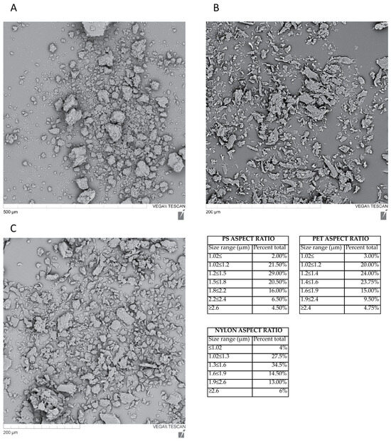
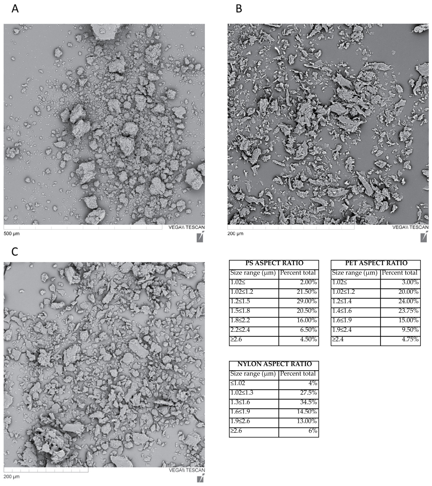
The morphology and sizing of the cryo-milled plastics was determined by SEM. PET and nylon MPs were irregular, elongated fragments (Figure 1B,C), while PS was somewhat of a rounded shape (Figure 1A). Sizing was done using Image J (Figure 1). Additionally, z-potential was measured to assess their charge and stability in colloidal suspension. As seen in Table 2, cryo-milled PS and PET resuspended in water showed a z-potential of −47 ± 7.05 mV and −32.8 ± 3.92 mV, respectively, indicating good dispersion. However, when resuspended in media, the z-potential decreased to −10.9 ± 0 and −6.43 ± 0 for PS and PET, respectively. The drop in z-potential indicates the MNPL colloidal suspension is less stable than water, likely due to the more complex nature of the culture medium and the additives present. Despite the decrease in z-potential, no precipitation of MNPLs was observed when they were resuspended in the culture medium.
Figure 1.
Physical characterization of cryo-milled MNPLs. (A) PS, (B) PET and (C) nylon and size distribution tables.
Table 2.
Z-potential of cryo-milled PS and PET.
3.2. Cell Viability
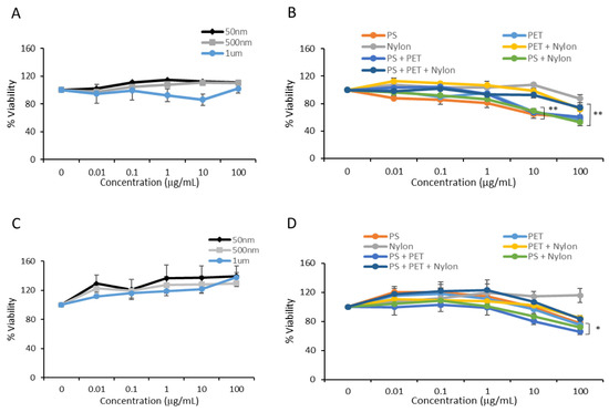
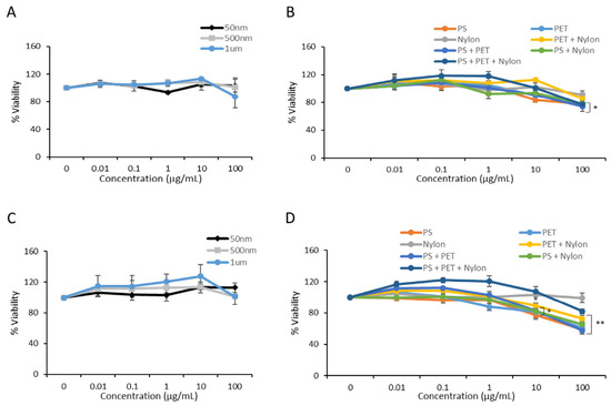
HepG2 cells (henceforth referred to as mono-culture) and HepG2:THP1 co-cultured cells (henceforth referred to as co-culture) were exposed to 50 nm, 500 nm, and 1 µm PS microspheres at concentrations ranging from 0.01 to 100 µg/mL and analyzed by MTT assay for cell viability after 24 h. No significant difference in cell viability was observed after 24 h (Figure 2A,C). However, when the mono- and co-cultured cells were exposed to cryo-milled PS, PET and nylon microplastics alone and in combinations, significant reductions ranging from 20 to 40% in cell viability were observed after 24 h (Figure 2B,D). Specifically, mono-cultured cells showed significance when exposed to PS at 10 µg/mL (p ≤ 0.01 **) and 100 µg/mL (p ≤ 0.001 ***) and PET at 10 µg/mL and 100 µg/mL (p ≤ 0.0001 ****), but no significance was observed in cells exposed to nylon MPs. Exposure to multiple plastics in combination (1:1 and 1:1:1) resulted in significant reductions in cell viability, but not in a synergistic manner. Reduced cell viability was seen in exposure to PET + nylon at 10 µg/mL (p ≤ 0.01 **), PS + PET at 10 µg/mL (p ≤ 0.01 **) and 100 µg/mL (p ≤ 0.001 ***), PS + nylon at 10 µg/mL (p ≤ 0.01 **) and 100 µg/mL (p ≤ 0.001 ***) and all polymers (PS + PET + nylon) applied together at 100 µg/mL (p ≤ 0.001 ***). Co-cultured cells showed significant changes to cell viability following exposure to PET + nylon at 100 µg/mL (p ≤ 0.01 **), PS + PET at 100 µg/mL (p ≤ 0.05 *), PS + nylon at 10 µg/mL (p ≤ 0.05 *) and 100 µg/mL (p ≤ 0.001 ***).
Figure 2.
Cell viability decreased after exposure to cryo-milled MNPLs. Cell viability was measured in mono- and co-cultured cells exposed to PS microspheres (A,C) and cryo-milled MNPLs either alone or in combination (B,D) for 24 h. MTT assay was used to assess viability. The quantification of cell viability is expressed as a percentage relative to CTL (unexposed cells). Data are the mean ±SEM from three independent experiments (n = 3). Significance in mono-cultured cells is seen at exposures to PS at 10 µg/mL (p ≤ 0.01 **) and 100 µg/mL (p ≤ 0.001 ***), PET at 10 µg/mL and 100 µg/mL (p ≤ 0.0001 ****), as well PET + nylon at 10 µg/mL (p ≤ 0.01 **), PS + PET at 10 µg/mL (p ≤ 0.01 **) and 100 µg/mL (p ≤ 0.001 ***), PS + nylon at 10 µg/mL (p ≤ 0.01 **) and 100 µg/mL (p ≤ 0.001 ***), and all polymers (PS + PET + nylon) at 100 µg/mL (p ≤ 0.001 ***). Co-cultured cells showed significant changes for PET + nylon at 100 µg/mL (p ≤ 0.01 **), PS+PET at 100 µg/mL (p ≤ 0.05 *), PS + nylon at 10 µg/mL (p ≤ 0.05 *) and 100 µg/mL (p ≤ 0.001 ***). Significance was determined by one-way ANOVA with Dunnett’s multiple comparisons test where * is p ≤ 0.05, ** is p ≤ 0.001.
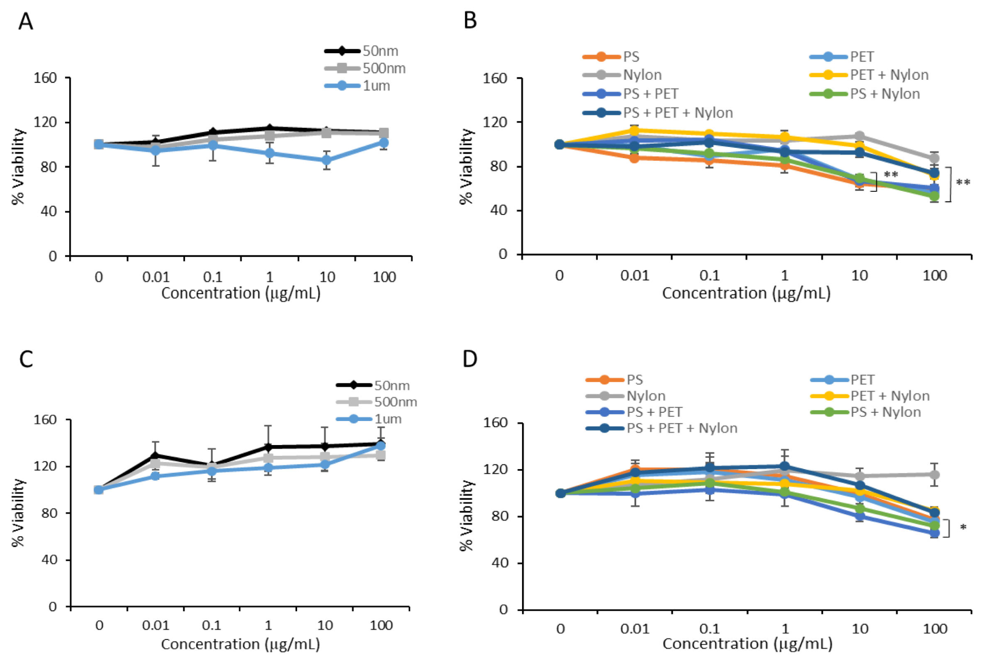
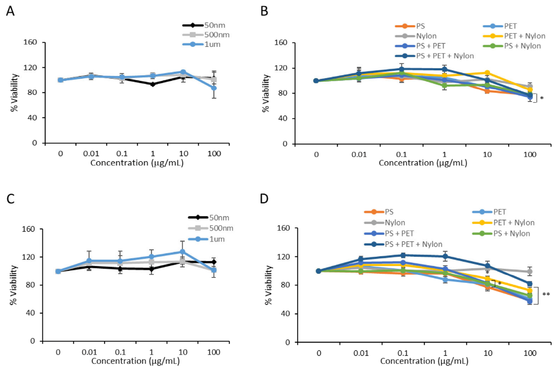
Next, cells were exposed to microplastics for 72 h in order to determine whether longer exposure resulted in further decreases in cell viability. Comparable to 24 h PS microsphere exposure, mono- and co-cultured cells exposed to PS microspheres for 72 h showed no changes in cell viability (Figure 3A,C). However, there were significant changes in cell viability in cells exposed to milled plastics. Specifically, mono-cultured cells showed significant viability changes when exposed to PS at 100 µg/mL (p ≤ 0.05 *) and PET at 100 µg/mL (p ≤ 0.001 ***) and no change when exposed to nylon MPs (Figure 3B). Furthermore, PS + PET at 100 µg/mL (p ≤ 0.05 *) had decreased cell viability in all of the various plastic exposure combinations. Moreover, co-cultured cells showed decreases in viability after 72 h of exposure to PS at 10 µg/mL (p ≤ 0.01 **) and 100 µg/mL (p ≤ 0.0001 ****) and PET at 100 µg/mL (p ≤ 0.01 **) for the single-polymer exposure conditions. Meanwhile, PET + nylon at 100 µg/mL (p ≤ 0.0001 ****), PS + PET 10 µg/mL (p ≤ 0.01 **) and 100 µg/mL (p ≤ 0.0001 ****), and PS + nylon 10 µg/mL (p ≤ 0.05 *) and 100 µg/mL (p ≤ 0.001 ***) all had reduced viability in combined plastic exposure conditions (Figure 3D). Microplastics in media with no cells were measured to observe any interference in signal detection, but none was detected. These results demonstrate that milled PS and PET MPs have an impact on cell viability or metabolism at higher concentrations, while pristine manufactured MNPLs appear to be inert.
Figure 3.
Cell viability decreased after exposure to cryo-milled MNPLs. Cell viability was measured in mono- and co-cultured cells exposed to PS microspheres (A,C) and cryo-milled MNPLs either alone or in combination (B,D) for 72 h. MTT assay was used to assess viability. The quantification of cell viability is expressed as a percentage relative to CTL (unexposed cells). Data are the mean ± SEM from three independent experiments (n = 3). Mono-cultured cells showed significance when exposed to PS at 100 µg/mL (p ≤ 0.05 *) and PET at 100 µg/mL (p ≤ 0.001 ***). Co-cultured cells showed decreases in viability after exposure to PS at 10 µg/mL (p ≤ 0.01 **) and 100 µg/mL (p ≤ 0.0001 ****), PET at 100 µg/mL (p ≤ 0.01 **), as well as PET + nylon at 100 µg/mL (p ≤ 0.0001 ****), PS + PET at 10 µg/mL (p ≤ 0.01 **) and 100 µg/mL (p ≤ 0.0001 ****), and PS + nylon at 10 µg/mL (p ≤ 0.05 *) and 100 µg/mL (p ≤ 0.001 ***). Significance was determined by one-way ANOVA with Dunnett’s multiple comparisons test where * is p ≤ 0.05, ** is p ≤ 0.001.
In order to verify if the decrease in cell viability via MTT assay was due to cellular damage or impaired metabolic activity, mono- and co-cultured cells were exposed to pristine or milled MNPLs in the same manner as described above and analyzed for cell damage via LDH release assay. Neither mono- nor co-cultured cells showed any significant changes in LDH release when exposed to the microplastics after 24 h or 72 h (Supplemental Figures S1 and S2). In summary, these results collectively suggest that milled MNPLs appear to inhibit cellular metabolism at high concentrations.
3.3. Cellular Uptake
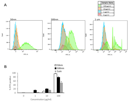
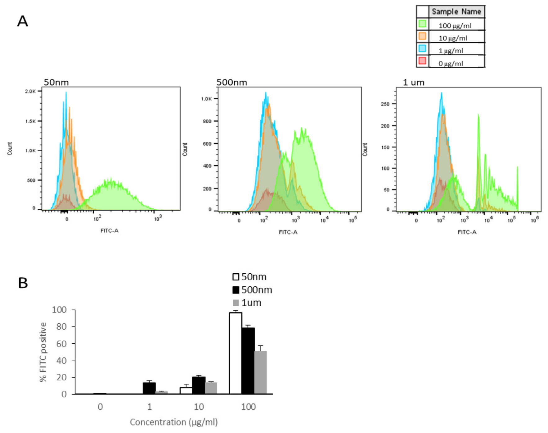
Since hepatocytes are largely responsible for metabolism and are present in the co-cultured model at a prevalence of 10:1 compared to macrophage-like cells, we verified the microplastic internalized by HepG2 cells using flow cytometry and GC-MS pyrolysis. Fluorescently labelled microspheres were used to confirm the uptake of pristine PS. The cells were exposed to 50 nm, 500 nm and 1 µm PS-MPs in a dose-dependent manner ranging from 0 to 100 µg/mL. Following 24 h of exposure, the cells were subjected to flow cytometry to determine internalization rates. HepG2 cells appeared to internalize fluorescently labelled PS of all diameters in a dose-dependent manner with internalization observed primarily at 100 µg/mL and to a lesser extent at 10 µg/mL (Figure 4A). Additional analysis of FITC+ cells appeared to confirm limited internalization at 10 µg/mL with approximately ~7–20% cells testing positive, while cells exposed to 100 µg/mL PS-MPs showed positivity rates in the range of ~50–95% with a clear trend of increasing internalization correlated with decreasing MNPL size (Figure 4B). These results demonstrate that HepG2 cells are capable of internalizing pristine PS-MPs and internalization rates appear to be size-dependent.
Figure 4.
HepG2 internalization of PS-NPs and PS-MPs after 24 h. Cellular uptake was analyzed by flow cytometry following 24 h incubation with fluorescently labelled PS microspheres (50 nm, 500 nm and 1 µm). (A) Representative histograms showing internalization. (B) Quantification of the percentage of FITC-positive cells (n = 3).
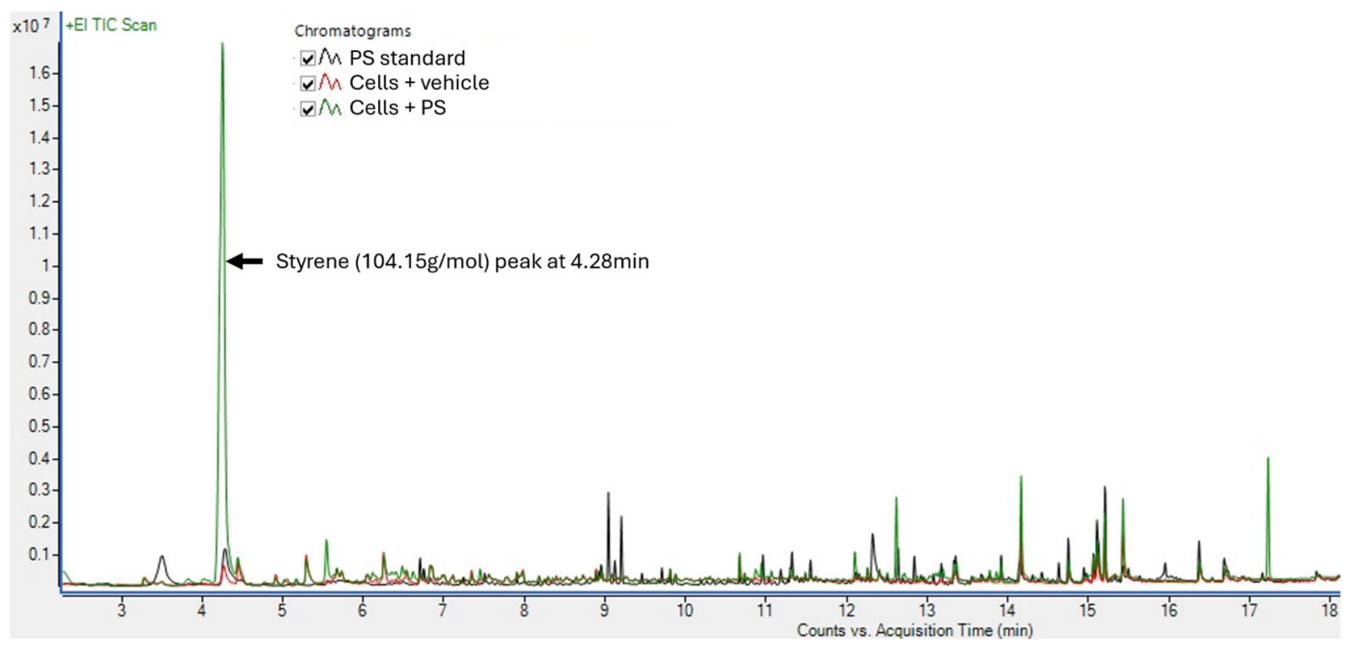
GC-MS pyrolysis was used to quantify the uptake of cryo-milled PS, since it has an impact on cellular metabolism. PET also impacted cellular metabolism, but detection methodology is currently unavailable to us. Cells were exposed to cryo-milled PS-MPs at 100 µg/mL for 24 h. Cell lysates were digested in KOH and microplastics were isolated and analyzed with GC-MS pyrolysis (Figure 5). Cryo-milled PS was taken up by cells quite readily, as it was detected in the cell lysate after isolation. (Table 3).
Figure 5.
Chromatogram of PS standards. Representative chromatograph of styrene, a pyrolysis by-product of polystyrene combustion.
Table 3.
Uptake of cryo-milled PS in HepG2 cells.
3.4. Oxygen Consumption Rates
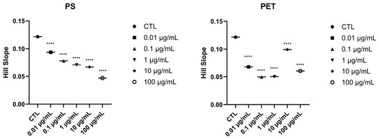
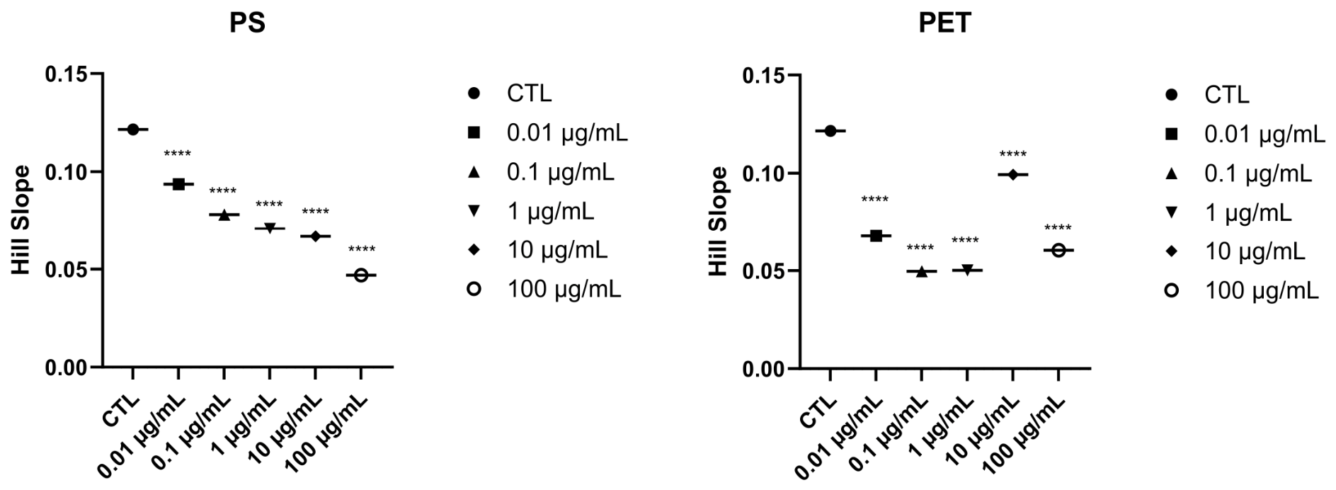
To further investigate the potential metabolic disruption observed in this study, we exposed HepG2:THP-1 co-cultured cells to cryo-milled PS and PET for 72 h. Following incubation, the oxygen consumption rates of the cells were measured for 120 min to observe any changes in mitochondrial respiration. Increased cellular respiration from assay initiation to expansion and saturation showed noticeable lags in the initiation phase and saturation phase compared to control samples (Figure S3). Further analysis of the data revealed that both PS and PET exposure at all concentrations significantly impaired mitochondrial respiration (Figure 6). OCR analysis suggests that exposure to both PS and PET MNPLs significantly slows down oxygen consumption in a primarily dose-dependent manner. Furthermore, to corroborate the metabolic changes observed by the MTT assay and OCR assay, we analyzed cell proliferation rates using trypan blue exclusion after exposure to cryo-milled PS and PET for both 24 and 72 h at 0 to 100 µg/mL. No changes in cell proliferation were observed at any exposure dose for either time point (unpublished data). This confirms that the decrease in MTT conversion and decrease in OCR are due to metabolic changes caused by cryo-milled PS and PET.
Figure 6.
Oxygen consumption rate of co-cultured cells exposed to cryo-milled PS and PET. Plotted Hill slope values representing changes in OCRs over a 120 min post 7 h incubation with PS and PET. Significance was determined by one-way ANOVA with Dunnett’s multiple comparisons test where **** is p ≤ 0.00001.
4. Discussion
Microplastics have been detected across the globe and their presence in the environment and food products has raised potential concern. The reported presence of microplastics in human tissues warrants further research into the potential toxicity of microplastics for human health. The liver, as the primary detoxifying organ, may be subject to MNPL bioaccumulation and subsequently MNPL-driven toxicity [52]. In that respect, the HepG2 cell line serves as a method for studying the hypothetical hepatotoxicity of MNPLs [51,64]. In addition, to overcome the limitations of a single-cell-culture in vitro model, a co-culture model with the addition of hepatic immune cells is used to assess toxicological responses to MNPLs that better reflect in vivo physiology. Kupffer cells are the first immune cell in the liver to come into contact with xenobiotic material transported through the hepatic portal vein [13]. Accordingly, the co-culture model that includes Kupffer-like cells is better able to recapitulate an in vivo toxicological response. Hence, we assessed the cytotoxicity of individual and combinations of MNPLs in both mono-culture and co-culture models. Unfortunately, 2D models are unable to replicate exact cell interactions because they lack the 3D architecture of the cells in vivo. Additionally, HepG2 cells have lower metabolic activity than primary cell cultures, but their accessibility and affordability make them a viable option [58]. However, our results do not show any differences in the viability of mono-cultured vs. co-cultured cells, suggesting that perhaps the immune cells do not play a large response to microplastic exposure in the measured outcomes tested.
Furthermore, the plastic’s properties can influence how the particles interact with the biological surroundings. Size, shape and composition all may impact particle internalization and thereby their potential toxicity [38,51,52,64,65]. Thus, in this study we analyzed the toxicity of various environmentally relevant plastics on both mono-culture and co-culture liver models. MTT assays revealed that cryo-milled PS and PET affected mitochondrial metabolism at some of the higher doses (10 µg/mL and 100 µg/mL) in both cell models at 24 h and 72 h. However, spherical PS (50 nm, 500 nm and 1 µm) and cryo-milled nylon did not. Unlike Goodman et al., He et al., Li et al., and Duo et al., we did not see a decrease in metabolic activity of HepG2 cells exposed to PS spheres, but they noted changes in cells when exposed to 1 µm PS spheres up to 100 µg/mL at both 24 h or 72 h, 50 nm PS spheres up to 100 µg/mL for 24 h, and 20 nm PS spheres up to 50 µg/mL at 24 h, respectively [66,67,68,69]. Also, Ying et al. saw a decrease in HepG2 viability after cells were treated with 100 nm PS for 24 h, but only at 2000 µg/cm2 [14]. Comparable to our results, no toxicity was observed in HepG2 cells treated with various sizes of spherical PS-MPs for 24 h up to 100 µg/mL [70], nor PS-MPs up to 1000 µg/mL for 24 and 48 h [71]. Additionally, no changes in cytotoxicity were noted for any of the plastic types or shapes using LDH assays. Other studies have shown that HepG2 cells exposed to PET for 24 h displayed no cytotoxicity [72,73]. However, Manoochehri et al. saw decreases in cell viability in HepG2 cells at 72 h exposed to PET-NPs up to 500 µg/mL, but no changes in LDH were noted; Najahi et al. saw increased cell viability in PET-exposed HepG2 cells and Ma et al. noted decreased cell viability in HepG2 cells exposed to PET-NPs and increased LDH leakage, but only at >100 µg/mL for 24 h [51,52,64]. This suggests that there may be an impact on mitochondrial function rather than cytotoxicity via the decreased MTT assay. Likewise, milled PS had no notable effect on HepG2 cytotoxicity after 24 h [65].
Cellular uptake of spherical PS-NPs and -MPs across various cell lines has been well documented [1,3,49,66,67,70,74,75,76,77,78,79]. Expectedly, we confirmed that HepG2 cells were able to internalize PS microspheres, with smaller-sized particles having preferential uptake. We also confirmed that cryo-milled PS particles were internalized by HepG2 cells. However, we are currently unable to measure the uptake of cryo-milled PET. A study by Manoochehri et al. was able to detect PET-NPs (~500 nm) internalized in HepG2 cells after 72 h of exposure [51] and a study by Ma et al. was able to detect PET-MPs in HepG2 cells after 12 h [64].
Since no changes in cell proliferation or cell damage (cell death) were noted, the reduction in cell viability observed by MTT assays is attributed to decreased cellular metabolism. As only cryo-milled PET and PS showed mitochondrial metabolomic changes, and there was significant evidence of MNPL interaction with cell metabolism, oxygen consumption rate (OCR) was measured in HepG2:THP-1 co-culture after 72 h of PET and PS exposure. Exposure to both cryo-milled PS and PET reduced OCR in co-cultured cells. A previous study has reported minimal changes in mitochondrial respiration in HepG2 cells treated with 2 µm PS microspheres for 48 h [80], while our results show that 72 h incubation with cryo-milled PS particles induces significant reduction in mitochondrial metabolism. Additionally, cryo-milled PET particle exposure on HepG2 cells for 72 h reduced mitochondrial metabolism, which has been documented by Najahi et al. on mono-cultured HepG2 cells exposed to PET for 72 h [52]. Additionally, Ma et al. noted decreased mitochondrial membrane potential in HepG2 cells treated with 100 µg/mL of PET-NPs for 24 h [64]. The longer incubation time combined with the milling process are likely critical factors in inducing HepG2 metabolic disruption. Other studies utilizing kidney cells [66] and lung cells [81] also showed mitochondrial membrane disruption and metabolic slowdown after MNPL exposure. Further work needs to be done to verify if glycolytic pathways may be compensating for the oxidative phosphorylation disruption or if there are additional factors responsible for these observations, such as mitochondrial membrane depolarization/integrity or proton pump efficiency deficits.
The physiochemical parameters of microplastics play a large role in toxicity-related outcomes. These properties (size, shape/surface properties and polymer type) all affect cellular uptake and biological processes [82]. Hence, we used a variety of plastic types and mimicked an element of environmental weathering by subjecting these plastics to cryogenic milling. However, a caveat of the current research is that there is no standardized methodology for the degradation of real-life plastics into micro- and nanoplastics, with a wide variety of “weathering” methods being used [72,83]. Additionally, reference materials that come in a narrow size range and are of a similar shape for various polymer types are difficult to obtain or do not exist yet. These differences could contribute to the difference in toxicological profiles observed across studies. Although this study contributes data on the impact of environmentally relevant MNPLs in potential human hepatotoxicity, further research is needed to comprehensively elucidate how the physiochemical parameters of MNPLs affect cellular physiology and various biological processes.
5. Conclusions
In this study we used both mono- and co-cultured liver cell models to assess the impact of environmentally relevant, cryo-milled plastic particles. We observed that only cryo-milled PET and PS altered mitochondrial energy metabolism, while the other plastic particles had no effects. No combinatorial effects were observed with different combinations of MNLPs in the co-culture. Furthermore, weathered particles are more sensitive than their pristine counterparts, due to the physicochemical modifications occurring during persistence in the environment.
Supplementary Materials
The following supporting information can be downloaded at https://www.mdpi.com/article/10.3390/microplastics5020086/s1, Figure S1: Cell cytotoxicity not impacted by microplastic 24h exposure; Figure S2: Cell cytotoxicity not impacted by microplastic 72h exposure; Figure S3: Data representing OCR consumption of HepG2:THP-1 co-culture cells exposed to either PS or PET.
Author Contributions
K.A.M.: Conceptualization, Data curation, Formal analysis, Investigation, Methodology, Project administration, Visualization, Writing (original draft, review and editing); M.S.: Methodology, Data curation, Visualization, Writing (original draft, review and editing); M.G.W.: Methodology; S.S.G.: Supervision, Methodology, Writing (review and editing). All authors have read and agreed to the published version of the manuscript.
Funding
This research received no external funding.
Institutional Review Board Statement
Not applicable.
Data Availability Statement
The data will be made available upon request.
Acknowledgments
We would like to thank Emily Dupuis (Scientific Services Division, Health Canada) for her help with the flow cytometry; Christopher Mason and Gurmit Singh (Food Research Division, Health Canada) for their help with the GC-MS pyrolysis; David Prescott (Regulatory Toxicology Research Division, Health Canada) and Catherine Smith (Chemical Health Hazard Assessment Division, Health Canada) for the review of this manuscript.
Conflicts of Interest
The authors declare no conflicts of interest.
Abbreviations
The following abbreviations are used in this manuscript:
| MPs | Microplastics |
| NPs | Nanoplastics |
| MNPLs | Micro- and nanoplastics |
| PS | Polystyrene |
| PET | Polyethylene terephthalate |
| OCR | Oxygen consumption rate |
References
- Cortés, C.; Domenech, J.; Salazar, M.; Pastor, S.; Marcos, R.; Hernández, A. Nanoplastics as a Potential Environmental Health Factor: Effects of Polystyrene Nanoparticles on Human Intestinal Epithelial Caco-2 Cells. Environ. Sci. Nano 2020, 7, 272–285. [Google Scholar] [CrossRef]
- Rubio-Armendáriz, C.; Alejandro-Vega, S.; Paz-Montelongo, S.; Gutiérrez-Fernández, Á.J.; Carrascosa-Iruzubieta, C.J.; Hardisson-de La Torre, A. Microplastics as Emerging Food Contaminants: A Challenge for Food Safety. Int. J. Environ. Res. Public Health 2022, 19, 1174. [Google Scholar] [CrossRef]
- Banerjee, A.; Shelver, W.L. Micro- and Nanoplastic Induced Cellular Toxicity in Mammals: A Review. Sci. Total Environ. 2021, 755, 142518. [Google Scholar] [CrossRef]
- Barbosa, F.; Adeyemi, J.A.; Bocato, M.Z.; Comas, A.; Campiglia, A. A Critical Viewpoint on Current Issues, Limitations, and Future Research Needs on Micro- and Nanoplastic Studies: From the Detection to the Toxicological Assessment. Environ. Res. 2020, 182, 109089. [Google Scholar] [CrossRef]
- Yong, C.; Valiyaveettil, S.; Tang, B. Toxicity of Microplastics and Nanoplastics in Mammalian Systems. Int. J. Environ. Res. Public Health 2020, 17, 1509. [Google Scholar] [CrossRef]
- Rahman, A.; Sarkar, A.; Yadav, O.P.; Achari, G.; Slobodnik, J. Potential Human Health Risks Due to Environmental Exposure to Nano- and Microplastics and Knowledge Gaps: A Scoping Review. Sci. Total Environ. 2021, 757, 143872. [Google Scholar] [CrossRef]
- Goodman, K.E.; Hare, J.T.; Khamis, Z.I.; Hua, T.; Sang, Q.-X.A. Exposure of Human Lung Cells to Polystyrene Microplastics Significantly Retards Cell Proliferation and Triggers Morphological Changes. Chem. Res. Toxicol. 2021, 34, 1069–1081. [Google Scholar] [CrossRef] [PubMed]
- Hou, J.; Lei, Z.; Cui, L.; Hou, Y.; Yang, L.; An, R.; Wang, Q.; Li, S.; Zhang, H.; Zhang, L. Polystyrene Microplastics Lead to Pyroptosis and Apoptosis of Ovarian Granulosa Cells via NLRP3/Caspase-1 Signaling Pathway in Rats. Ecotoxicol. Environ. Saf. 2021, 212, 112012. [Google Scholar] [CrossRef]
- Jeong, J.; Choi, J. Adverse Outcome Pathways Potentially Related to Hazard Identification of Microplastics Based on Toxicity Mechanisms. Chemosphere 2019, 231, 249–255. [Google Scholar] [CrossRef]
- Luo, T.; Wang, C.; Pan, Z.; Jin, C.; Fu, Z.; Jin, Y. Maternal Polystyrene Microplastic Exposure during Gestation and Lactation Altered Metabolic Homeostasis in the Dams and Their F1 and F2 Offspring. Environ. Sci. Technol. 2019, 53, 10978–10992. [Google Scholar] [CrossRef] [PubMed]
- Persiani, E.; Cecchettini, A.; Ceccherini, E.; Gisone, I.; Morales, M.A.; Vozzi, F. Microplastics: A Matter of the Heart (and Vascular System). Biomedicines 2023, 11, 264. [Google Scholar] [CrossRef]
- Blackburn, K.; Green, D. The Potential Effects of Microplastics on Human Health: What Is Known and What Is Unknown. Ambio 2022, 51, 518–530. [Google Scholar] [CrossRef]
- Guraka, A.; Souch, G.; Duff, R.; Brown, D.; Moritz, W.; Kermanizadeh, A. Microplastic-Induced Hepatic Adverse Effects Evaluated in Advanced Quadruple Cell Human Primary Models Following Three Weeks of Repeated Exposure. Chemosphere 2024, 364, 143032. [Google Scholar] [CrossRef] [PubMed]
- Ying, M.; Shao, N.; Dong, C.; Sha, Y.; Li, C.; Hong, X.; Ding, Y.; Xu, J.; Qian, K.; Tao, G.; et al. PS-MPs Induced Inflammation and Phosphorylation of Inflammatory Signalling Pathways in Liver. Toxics 2024, 12, 932. [Google Scholar] [CrossRef]
- Dzierżyński, E.; Gawlik, P.J.; Puźniak, D.; Flieger, W.; Jóźwik, K.; Teresiński, G.; Forma, A.; Wdowiak, P.; Baj, J.; Flieger, J. Microplastics in the Human Body: Exposure, Detection, and Risk of Carcinogenesis: A State-of-the-Art Review. Cancers 2024, 16, 3703. [Google Scholar] [CrossRef] [PubMed]
- DeLoid, G.M.; Cao, X.; Bitounis, D.; Singh, D.; Llopis, P.M.; Buckley, B.; Demokritou, P. Toxicity, Uptake, and Nuclear Translocation of Ingested Micro-Nanoplastics in an in Vitro Model of the Small Intestinal Epithelium. Food Chem. Toxicol. 2021, 158, 112609. [Google Scholar] [CrossRef]
- Evangeliou, N.; Grythe, H.; Klimont, Z.; Heyes, C.; Eckhardt, S.; Lopez-Aparicio, S.; Stohl, A. Atmospheric Transport Is a Major Pathway of Microplastics to Remote Regions. Nat. Commun. 2020, 11, 3381. [Google Scholar] [CrossRef]
- Parolini, M.; Antonioli, D.; Borgogno, F.; Gibellino, M.C.; Fresta, J.; Albonico, C.; De Felice, B.; Canuto, S.; Concedi, D.; Romani, A.; et al. Microplastic Contamination in Snow from Western Italian Alps. Int. J. Environ. Res. Public Health 2021, 18, 768. [Google Scholar] [CrossRef]
- Da Costa Filho, P.A.; Andrey, D.; Eriksen, B.; Peixoto, R.P.; Carreres, B.M.; Ambühl, M.E.; Descarrega, J.B.; Dubascoux, S.; Zbinden, P.; Panchaud, A.; et al. Detection and Characterization of Small-Sized Microplastics (≥ 5 Μm) in Milk Products. Sci. Rep. 2021, 11, 24046. [Google Scholar] [CrossRef] [PubMed]
- Liang, B.; Zhong, Y.; Huang, Y.; Lin, X.; Liu, J.; Lin, L.; Hu, M.; Jiang, J.; Dai, M.; Wang, B.; et al. Underestimated Health Risks: Polystyrene Micro- and Nanoplastics Jointly Induce Intestinal Barrier Dysfunction by ROS-Mediated Epithelial Cell Apoptosis. Part. Fibre Toxicol. 2021, 18, 20. [Google Scholar] [CrossRef]
- Li, Q.; Feng, Z.; Zhang, T.; Ma, C.; Shi, H. Microplastics in the Commercial Seaweed Nori. J. Hazard. Mater. 2020, 388, 122060. [Google Scholar] [CrossRef]
- Prata, J.C.; Da Costa, J.P.; Lopes, I.; Duarte, A.C.; Rocha-Santos, T. Environmental Exposure to Microplastics: An Overview on Possible Human Health Effects. Sci. Total Environ. 2020, 702, 134455. [Google Scholar] [CrossRef] [PubMed]
- Choi, D.; Bang, J.; Kim, T.; Oh, Y.; Hwang, Y.; Hong, J. In Vitro Chemical and Physical Toxicities of Polystyrene Microfragments in Human-Derived Cells. J. Hazard. Mater. 2020, 400, 123308. [Google Scholar] [CrossRef]
- Amato-Lourenço, L.F.; Carvalho-Oliveira, R.; Júnior, G.R.; Dos Santos Galvão, L.; Ando, R.A.; Mauad, T. Presence of Airborne Microplastics in Human Lung Tissue. J. Hazard. Mater. 2021, 416, 126124. [Google Scholar] [CrossRef]
- Braun, T.; Ehrlich, L.; Henrich, W.; Koeppel, S.; Lomako, I.; Schwabl, P.; Liebmann, B. Detection of Microplastic in Human Placenta and Meconium in a Clinical Setting. Pharmaceutics 2021, 13, 921. [Google Scholar] [CrossRef]
- Ibrahim, Y.S.; Tuan Anuar, S.; Azmi, A.A.; Wan Mohd Khalik, W.M.A.; Lehata, S.; Hamzah, S.R.; Ismail, D.; Ma, Z.F.; Dzulkarnaen, A.; Zakaria, Z.; et al. Detection of Microplastics in Human Colectomy Specimens. JGH Open 2021, 5, 116–121. [Google Scholar] [CrossRef] [PubMed]
- Jenner, L.C.; Rotchell, J.M.; Bennett, R.T.; Cowen, M.; Tentzeris, V.; Sadofsky, L.R. Detection of Microplastics in Human Lung Tissue Using μFTIR Spectroscopy. Sci. Total Environ. 2022, 831, 154907. [Google Scholar] [CrossRef]
- Leslie, H.A.; Van Velzen, M.J.M.; Brandsma, S.H.; Vethaak, A.D.; Garcia-Vallejo, J.J.; Lamoree, M.H. Discovery and Quantification of Plastic Particle Pollution in Human Blood. Environ. Int. 2022, 163, 107199. [Google Scholar] [CrossRef] [PubMed]
- Montano, L.; Giorgini, E.; Notarstefano, V.; Notari, T.; Ricciardi, M.; Piscopo, M.; Motta, O. Raman Microspectroscopy Evidence of Microplastics in Human Semen. Sci. Total Environ. 2023, 901, 165922. [Google Scholar] [CrossRef]
- Qin, X.; Cao, M.; Peng, T.; Shan, H.; Lian, W.; Yu, Y.; Shui, G.; Li, R. Features, Potential Invasion Pathways, and Reproductive Health Risks of Microplastics Detected in Human Uterus. Environ. Sci. Technol. 2024, 58, 10482–10493. [Google Scholar] [CrossRef]
- Ragusa, A.; Matta, M.; Cristiano, L.; Matassa, R.; Battaglione, E.; Svelato, A.; De Luca, C.; D’Avino, S.; Gulotta, A.; Rongioletti, M.C.A.; et al. Deeply in Plasticenta: Presence of Microplastics in the Intracellular Compartment of Human Placentas. Int. J. Environ. Res. Public Health 2022, 19, 11593. [Google Scholar] [CrossRef] [PubMed]
- Ragusa, A.; Notarstefano, V.; Svelato, A.; Belloni, A.; Gioacchini, G.; Blondeel, C.; Zucchelli, E.; De Luca, C.; D’Avino, S.; Gulotta, A.; et al. Raman Microspectroscopy Detection and Characterisation of Microplastics in Human Breastmilk. Polymers 2022, 14, 2700. [Google Scholar] [CrossRef]
- Ragusa, A.; Svelato, A.; Santacroce, C.; Catalano, P.; Notarstefano, V.; Carnevali, O.; Papa, F.; Rongioletti, M.C.A.; Baiocco, F.; Draghi, S.; et al. Plasticenta: First Evidence of Microplastics in Human Placenta. Environ. Int. 2021, 146, 106274. [Google Scholar] [CrossRef]
- Schwabl, P.; Köppel, S.; Königshofer, P.; Bucsics, T.; Trauner, M.; Reiberger, T.; Liebmann, B. Detection of Various Microplastics in Human Stool: A Prospective Case Series. Ann. Intern. Med. 2019, 171, 453–457. [Google Scholar] [CrossRef]
- Yang, Y.; Xie, E.; Du, Z.; Peng, Z.; Han, Z.; Li, L.; Zhao, R.; Qin, Y.; Xue, M.; Li, F.; et al. Detection of Various Microplastics in Patients Undergoing Cardiac Surgery. Environ. Sci. Technol. 2023, 57, 10911–10918. [Google Scholar] [CrossRef]
- Zhang, J.; Wang, L.; Trasande, L.; Kannan, K. Occurrence of Polyethylene Terephthalate and Polycarbonate Microplastics in Infant and Adult Feces. Environ. Sci. Technol. Lett. 2021, 8, 989–994. [Google Scholar] [CrossRef]
- Zhao, Q.; Zhu, L.; Weng, J.; Jin, Z.; Cao, Y.; Jiang, H.; Zhang, Z. Detection and Characterization of Microplastics in the Human Testis and Semen. Sci. Total Environ. 2023, 877, 162713. [Google Scholar] [CrossRef] [PubMed]
- Janiga-MacNelly, A.; Hoang, T.C.; Lavado, R. Comparative Toxicity of Microplastics Obtained from Human Consumer Products on Human Cell-Based Models. Food Chem. Toxicol. 2025, 196, 115194. [Google Scholar] [CrossRef]
- Kim, H.-Y.; Ashim, J.; Park, S.; Kim, W.; Ji, S.; Lee, S.-W.; Jung, Y.-R.; Jeong, S.W.; Lee, S.-G.; Kim, H.-C.; et al. A Preliminary Study about the Potential Risks of the UV-Weathered Microplastic: The Proteome-Level Changes in the Brain in Response to Polystyrene Derived Weathered Microplastics. Environ. Res. 2023, 233, 116411. [Google Scholar] [CrossRef]
- Da Silva Brito, W.A.; Mutter, F.; Wende, K.; Cecchini, A.L.; Schmidt, A.; Bekeschus, S. Consequences of Nano and Microplastic Exposure in Rodent Models: The Known and Unknown. Part. Fibre Toxicol. 2022, 19, 28. [Google Scholar] [CrossRef] [PubMed]
- Rubio, L.; Marcos, R.; Hernández, A. Potential Adverse Health Effects of Ingested Micro- and Nanoplastics on Humans. Lessons Learned from in Vivo and in Vitro Mammalian Models. J. Toxicol. Environ. Health Part B 2020, 23, 51–68. [Google Scholar] [CrossRef] [PubMed]
- Lee, C.-W.; Hsu, L.-F.; Wu, I.-L.; Wang, Y.-L.; Chen, W.-C.; Liu, Y.-J.; Yang, L.-T.; Tan, C.-L.; Luo, Y.-H.; Wang, C.-C.; et al. Exposure to Polystyrene Microplastics Impairs Hippocampus-Dependent Learning and Memory in Mice. J. Hazard. Mater. 2022, 430, 128431. [Google Scholar] [CrossRef]
- Deng, Y.; Zhang, Y.; Qiao, R.; Bonilla, M.M.; Yang, X.; Ren, H.; Lemos, B. Evidence That Microplastics Aggravate the Toxicity of Organophosphorus Flame Retardants in Mice (Mus Musculus). J. Hazard. Mater. 2018, 357, 348–354. [Google Scholar] [CrossRef]
- Jin, Y.; Lu, L.; Tu, W.; Luo, T.; Fu, Z. Impacts of Polystyrene Microplastic on the Gut Barrier, Microbiota and Metabolism of Mice. Sci. Total Environ. 2019, 649, 308–317. [Google Scholar] [CrossRef]
- Walczak, A.P.; Hendriksen, P.J.M.; Woutersen, R.A.; Van Der Zande, M.; Undas, A.K.; Helsdingen, R.; Van Den Berg, H.H.J.; Rietjens, I.M.C.M.; Bouwmeester, H. Bioavailability and Biodistribution of Differently Charged Polystyrene Nanoparticles upon Oral Exposure in Rats. J. Nanoparticle Res. 2015, 17, 231. [Google Scholar] [CrossRef]
- Yang, Y.-F.; Chen, C.-Y.; Lu, T.-H.; Liao, C.-M. Toxicity-Based Toxicokinetic/Toxicodynamic Assessment for Bioaccumulation of Polystyrene Microplastics in Mice. J. Hazard. Mater. 2019, 366, 703–713. [Google Scholar] [CrossRef] [PubMed]
- Zhang, Z.; Chen, W.; Chan, H.; Peng, J.; Zhu, P.; Li, J.; Jiang, X.; Zhang, Z.; Wang, Y.; Tan, Z.; et al. Polystyrene Microplastics Induce Size-Dependent Multi-Organ Damage in Mice: Insights into Gut Microbiota and Fecal Metabolites. J. Hazard. Mater. 2024, 461, 132503. [Google Scholar] [CrossRef]
- Wang, Y.-L.; Lee, Y.-H.; Hsu, Y.-H.; Chiu, I.-J.; Huang, C.C.-Y.; Huang, C.-C.; Chia, Z.-C.; Lee, C.-P.; Lin, Y.-F.; Chiu, H.-W. The Kidney-Related Effects of Polystyrene Microplastics on Human Kidney Proximal Tubular Epithelial Cells HK-2 and Male C57BL/6 Mice. Environ. Health Perspect. 2021, 129, 057003. [Google Scholar] [CrossRef] [PubMed]
- Hesler, M.; Aengenheister, L.; Ellinger, B.; Drexel, R.; Straskraba, S.; Jost, C.; Wagner, S.; Meier, F.; Von Briesen, H.; Büchel, C.; et al. Multi-Endpoint Toxicological Assessment of Polystyrene Nano- and Microparticles in Different Biological Models in Vitro. Toxicol. In Vitro 2019, 61, 104610. [Google Scholar] [CrossRef]
- Hu, M.; Palić, D. Micro- and Nano-Plastics Activation of Oxidative and Inflammatory Adverse Outcome Pathways. Redox Biol. 2020, 37, 101620. [Google Scholar] [CrossRef]
- Manoochehri, Z.; Etebari, M.; Pannetier, P.; Ebrahimpour, K. In Vitro Toxicity of Polyethylene Terephthalate Nanoplastics (PET-NPs) in Human Hepatocarcinoma (HepG2) Cell Line. Toxicol. Environ. Health Sci. 2024, 16, 203–215. [Google Scholar] [CrossRef]
- Najahi, H.; Alessio, N.; Venditti, M.; Oliveri Conti, G.; Ferrante, M.; Di Bernardo, G.; Galderisi, U.; Minucci, S.; Banni, M. Impact of Environmental Microplastic Exposure on Caco-2 Cells: Unraveling Proliferation, Apoptosis, and Autophagy Activation. Int. J. Environ. Res. Public Health 2025, 22, 922. [Google Scholar] [CrossRef]
- Qian, X.; Jin, P.; Fan, K.; Pei, H.; He, Z.; Du, R.; Cao, C.; Yang, Y. Polystyrene Microplastics Exposure Aggravates Acute Liver Injury by Promoting Kupffer Cell Pyroptosis. Int. Immunopharmacol. 2024, 126, 111307. [Google Scholar] [CrossRef] [PubMed]
- Li, P.; Miyamoto, D.; Adachi, T.; Hara, T.; Soyama, A.; Matsushima, H.; Imamura, H.; Kanetaka, K.; Gu, W.; Eguchi, S. Mitigation of Polystyrene Microplastic-Induced Hepatotoxicity in Human Hepatobiliary Organoids through Bile Extraction. Ecotoxicol. Environ. Saf. 2024, 288, 117330. [Google Scholar] [CrossRef] [PubMed]
- Ding, C.; Li, Y.; Guo, F.; Jiang, Y.; Ying, W.; Li, D.; Yang, D.; Xia, X.; Liu, W.; Zhao, Y.; et al. A Cell-Type-Resolved Liver Proteome. Mol. Cell. Proteom. 2016, 15, 3190–3202. [Google Scholar] [CrossRef] [PubMed]
- Boran, T.; Zengin, O.S.; Seker, Z.; Akyildiz, A.G.; Kara, M.; Oztas, E.; Özhan, G. An Evaluation of a Hepatotoxicity Risk Induced by the Microplastic Polymethyl Methacrylate (PMMA) Using HepG2/THP-1 Co-Culture Model. Environ. Sci. Pollut. Res. Int. 2024, 31, 28890–28904. [Google Scholar] [CrossRef]
- Granitzny, A.; Knebel, J.; Müller, M.; Braun, A.; Steinberg, P.; Dasenbrock, C.; Hansen, T. Evaluation of a Human in Vitro Hepatocyte-NPC Co-Culture Model for the Prediction of Idiosyncratic Drug-Induced Liver Injury: A Pilot Study. Toxicol. Rep. 2017, 4, 89–103. [Google Scholar] [CrossRef] [PubMed]
- Padberg, F.; Hering, H.; Luch, A.; Zellmer, S. Indirect Co-Cultivation of HepG2 with Differentiated THP-1 Cells Induces AHR Signalling and Release of pro-Inflammatory Cytokines. Toxicol. In Vitro 2020, 68, 104957. [Google Scholar] [CrossRef]
- Wewering, F.; Jouy, F.; Wissenbach, D.K.; Gebauer, S.; Blüher, M.; Gebhardt, R.; Pirow, R.; von Bergen, M.; Kalkhof, S.; Luch, A.; et al. Characterization of Chemical-Induced Sterile Inflammation in Vitro: Application of the Model Compound Ketoconazole in a Human Hepatic Co-Culture System. Arch. Toxicol. 2017, 91, 799–810. [Google Scholar] [CrossRef]
- Rose, K.A.; Holman, N.S.; Green, A.M.; Andersen, M.E.; LeCluyse, E.L. Co-Culture of Hepatocytes and Kupffer Cells as an In Vitro Model of Inflammation and Drug-Induced Hepatotoxicity. J. Pharm. Sci. 2016, 105, 950–964. [Google Scholar] [CrossRef]
- Marcellus, K.A.; Bugiel, S.; Nunnikhoven, A.; Curran, I.; Gill, S.S. Polystyrene Nano- and Microplastic Particles Induce an Inflammatory Gene Expression Profile in Rat Neural Stem Cell-Derived Astrocytes In Vitro. Nanomaterials 2024, 14, 429. [Google Scholar] [CrossRef]
- Marcellus, K.A.; Prescott, D.; Scur, M.; Ross, N.; Gill, S.S. Exposure of Polystyrene Nano- and Microplastics in Increasingly Complex In Vitro Intestinal Cell Models. Nanomaterials 2025, 15, 267. [Google Scholar] [CrossRef]
- Singh, G.; Velasquez, L.; Mason, C.; Scur, M.; Marcellus, K.A.; Gill, S. Detection and Identification of Non-Labeled Polystyrene Nanoplastics in Rodent Tissues Using Asymmetric Flow Field-Flow Fractionation (AF4) Combined with UV–Vis, Dynamic Light Scattering (DLS) Detectors and Offline Pyrolysis–GCMS (Pyro-GCMS). Microplastics 2026, 5, 2. [Google Scholar] [CrossRef]
- Ma, L.; Wu, Z.; Lu, Z.; Yan, L.; Dong, X.; Dai, Z.; Sun, R.; Hong, P.; Zhou, C.; Li, C. Differences in Toxicity Induced by the Various Polymer Types of Nanoplastics on HepG2 Cells. Sci. Total Environ. 2024, 918, 170664. [Google Scholar] [CrossRef] [PubMed]
- Jeon, S.; Lee, D.-K.; Jeong, J.; Yang, S.I.; Kim, J.-S.; Kim, J.; Cho, W.-S. The Reactive Oxygen Species as Pathogenic Factors of Fragmented Microplastics to Macrophages. Environ. Pollut. 2021, 281, 117006. [Google Scholar] [CrossRef]
- Goodman, K.E.; Hua, T.; Sang, Q.-X.A. Effects of Polystyrene Microplastics on Human Kidney and Liver Cell Morphology, Cellular Proliferation, and Metabolism. ACS Omega 2022, 7, 34136–34153. [Google Scholar] [CrossRef]
- He, Y.; Li, J.; Chen, J.; Miao, X.; Li, G.; He, Q.; Xu, H.; Li, H.; Wei, Y. Cytotoxic Effects of Polystyrene Nanoplastics with Different Surface Functionalization on Human HepG2 Cells. Sci. Total Environ. 2020, 723, 138180. [Google Scholar] [CrossRef]
- Guo, M.; Li, Y.; Niu, S.; Zhang, R.; Shen, X.; Ma, Y.; Wu, L.; Wu, T.; Zhang, T.; Tang, M.; et al. Oxidative Stress-Activated Nrf2 Remitted Polystyrene Nanoplastic-Induced Mitochondrial Damage and Inflammatory Response in HepG2 Cells. Environ. Toxicol. Pharmacol. 2024, 106, 104385. [Google Scholar] [CrossRef] [PubMed]
- Li, Y.; Guo, M.; Niu, S.; Shang, M.; Chang, X.; Sun, Z.; Zhang, R.; Shen, X.; Xue, Y. ROS and DRP1 Interactions Accelerate the Mitochondrial Injury Induced by Polystyrene Nanoplastics in Human Liver HepG2 Cells. Chem. Biol. Interact. 2023, 379, 110502. [Google Scholar] [CrossRef]
- Banerjee, A.; Billey, L.O.; McGarvey, A.M.; Shelver, W.L. Effects of Polystyrene Micro/Nanoplastics on Liver Cells Based on Particle Size, Surface Functionalization, Concentration and Exposure Period. Sci. Total Environ. 2022, 836, 155621. [Google Scholar] [CrossRef]
- Mognetti, B.; Cecone, C.; Fancello, K.; Saraceni, A.; Cottone, E.; Bovolin, P. Interaction of Polystyrene Nanoplastics with Biomolecules and Environmental Pollutants: Effects on Human Hepatocytes. Int. J. Mol. Sci. 2025, 26, 2899. [Google Scholar] [CrossRef]
- Roursgaard, M.; Hezareh Rothmann, M.; Schulte, J.; Karadimou, I.; Marinelli, E.; Møller, P. Genotoxicity of Particles From Grinded Plastic Items in Caco-2 and HepG2 Cells. Front. Public Health 2022, 10, 906430. [Google Scholar] [CrossRef] [PubMed]
- Stock, V.; Laurisch, C.; Franke, J.; Dönmez, M.H.; Voss, L.; Böhmert, L.; Braeuning, A.; Sieg, H. Uptake and Cellular Effects of PE, PP, PET and PVC Microplastic Particles. Toxicol. In Vitro 2021, 70, 105021. [Google Scholar] [CrossRef]
- Domenech, J.; Hernández, A.; Rubio, L.; Marcos, R.; Cortés, C. Interactions of Polystyrene Nanoplastics with in Vitro Models of the Human Intestinal Barrier. Arch. Toxicol. 2020, 94, 2997–3012. [Google Scholar] [CrossRef]
- Domenech, J.; De Britto, M.; Velázquez, A.; Pastor, S.; Hernández, A.; Marcos, R.; Cortés, C. Long-Term Effects of Polystyrene Nanoplastics in Human Intestinal Caco-2 Cells. Biomolecules 2021, 11, 1442. [Google Scholar] [CrossRef]
- Xu, D.; Ma, Y.; Han, X.; Chen, Y. Systematic Toxicity Evaluation of Polystyrene Nanoplastics on Mice and Molecular Mechanism Investigation about Their Internalization into Caco-2 Cells. J. Hazard. Mater. 2021, 417, 126092. [Google Scholar] [CrossRef]
- Ding, Y.; Zhang, R.; Li, B.; Du, Y.; Li, J.; Tong, X.; Wu, Y.; Ji, X.; Zhang, Y. Tissue Distribution of Polystyrene Nanoplastics in Mice and Their Entry, Transport, and Cytotoxicity to GES-1 Cells. Environ. Pollut. 2021, 280, 116974. [Google Scholar] [CrossRef] [PubMed]
- Bonanomi, M.; Salmistraro, N.; Porro, D.; Pinsino, A.; Colangelo, A.M.; Gaglio, D. Polystyrene Micro and Nano-Particles Induce Metabolic Rewiring in Normal Human Colon Cells: A Risk Factor for Human Health. Chemosphere 2022, 303, 134947. [Google Scholar] [CrossRef] [PubMed]
- Johnston, H.J.; Semmler-Behnke, M.; Brown, D.M.; Kreyling, W.; Tran, L.; Stone, V. Evaluating the Uptake and Intracellular Fate of Polystyrene Nanoparticles by Primary and Hepatocyte Cell Lines in Vitro. Toxicol. Appl. Pharmacol. 2010, 242, 66–78. [Google Scholar] [CrossRef]
- Peng, M.; Vercauteren, M.; Grootaert, C.; Rajkovic, A.; Boon, N.; Janssen, C.; Asselman, J. Cellular and Bioenergetic Effects of Polystyrene Microplastic in Function of Cell Type, Differentiation Status and Post-Exposure Time. Environ. Pollut. 2023, 337, 122550. [Google Scholar] [CrossRef]
- Halimu, G.; Zhang, Q.; Liu, L.; Zhang, Z.; Wang, X.; Gu, W.; Zhang, B.; Dai, Y.; Zhang, H.; Zhang, C.; et al. Toxic Effects of Nanoplastics with Different Sizes and Surface Charges on Epithelial-to-Mesenchymal Transition in A549 Cells and the Potential Toxicological Mechanism. J. Hazard. Mater. 2022, 430, 128485. [Google Scholar] [CrossRef]
- Zheng, M.; Yu, J. The Effect of Particle Shape and Size on Cellular Uptake. Drug Deliv. Transl. Res. 2016, 6, 67–72. [Google Scholar] [CrossRef] [PubMed]
- Hong, J.; Hengelbrok, O.; Gigault, J.; Ghoshal, S.; Moores, A. Accelerated Weathering of Microplastics: A Systematic Approach to Model Microplastic Production. Environ. Sci. Technol. 2025, 59, 15956–15965. [Google Scholar] [CrossRef]

Disclaimer/Publisher’s Note: The statements, opinions and data contained in all publications are solely those of the individual author(s) and contributor(s) and not of MDPI and/or the editor(s). MDPI and/or the editor(s) disclaim responsibility for any injury to people or property resulting from any ideas, methods, instructions or products referred to in the content. |
© 2026 by the authors. Licensee MDPI, Basel, Switzerland. This article is an open access article distributed under the terms and conditions of the Creative Commons Attribution (CC BY) license.