


Abstract
The plastic pieces of synthetic polymers, which were previously regarded as primary pollutants of the environment, are increasingly being discovered as internal pollutants of the human body. This review provides a comprehensive overview of the available evidence on human exposure, tissue distribution, and associated biological effects of micro- and nanoplastics. Ingesting contaminated food and water is the major exposure pathway, with inhalation and dermal contact being secondary routes. Various organ systems have been identified as containing polymer particles through the use of advanced analytical methods, including blood, liver, lungs, placenta, breast milk, and brain tissue. Experimental animal studies suggest associations with tissue injury, metabolic illness, and neurotoxicity. Polyethylene, polypropylene, polystyrene, and polyethylene terephthalate are the most frequently found polymers in human samples. New clinical findings indicate potential health implications, though current human evidence remains largely associative rather than causal: a cardiovascular study observed more than a two-fold rise in mortality among patients with polymer-containing arterial plaques, and recent evidence demonstrates over-accumulation of polymers in brain tissue, raising questions about neuroinflammatory processes. Detection technologies have advanced substantially, with deep learning-based polymer classification achieving 95–99% accuracy and ultrasensitive electrochemical and surface plasmon resonance biosensors reaching detection limits approaching 10−11 M. Despite these advances, critical issues remain, including lack of standardized analytical procedures, absence of chronic exposure models for humans, and insufficient longitudinal epidemiological data. To address these gaps, physiologically relevant experimental systems including organoids and organ-on-chip platforms will be required, in addition to well-designed prospective cohort studies.
1. Introduction
Synthetic polymers have become interwoven with virtually every aspect of contemporary existence from packaging materials and medical devices to textile fibers and construction components. Global production volumes increased from approximately 1.7 million metric tonnes annually in 1950 to around 460 million tonnes by 2019; current trajectories suggest that output may approach 884 million tonnes by mid-century [1,2]. Waste management has not kept pace. Perhaps 21–30% of post-consumer plastic undergoes recycling or thermal treatment [3]. The rest accumulates in the environment.
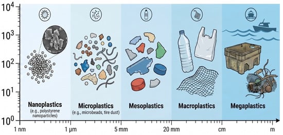
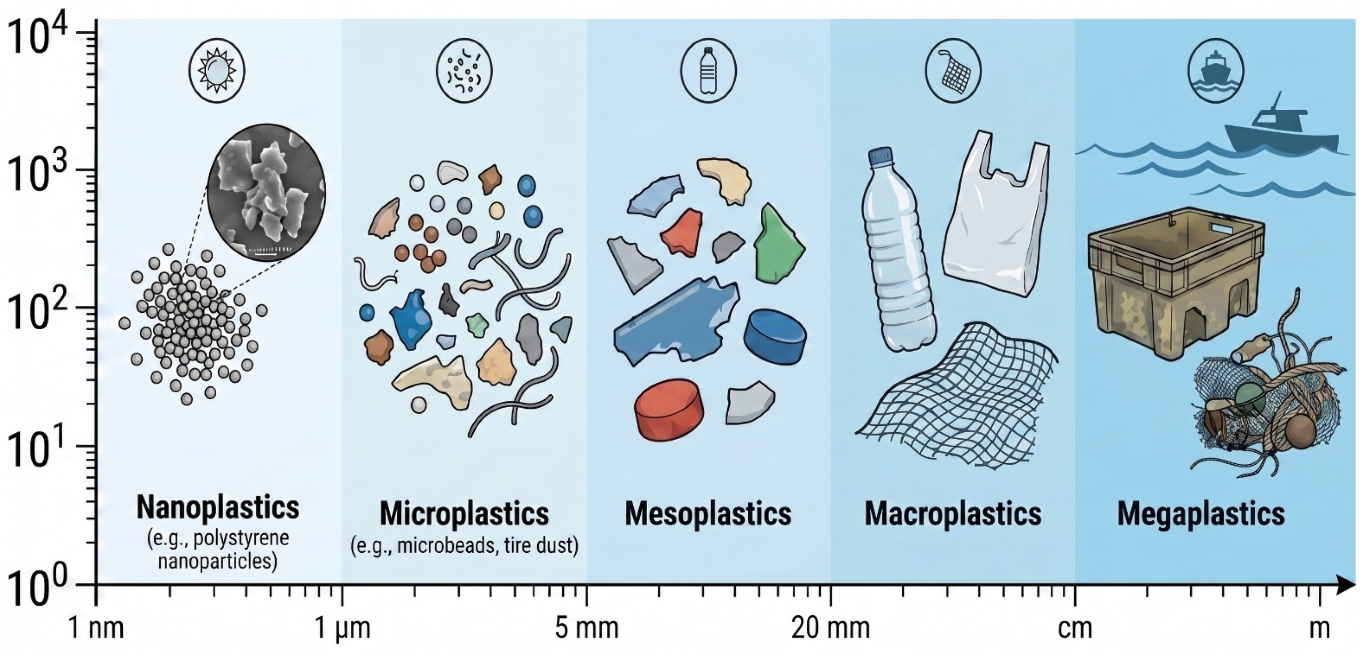
Plastic does not biodegrade. It breaks down slowly through UV light, physical wear, and some microbial action. Macroplastic debris gradually fragments into progressively smaller pieces: microplastics (conventionally defined as particles smaller than 5 mm) and nanoplastics (those below 100 nm or, by some definitions, 1 μm), as illustrated in Figure 1 [4]. Estimates suggest more than 170 trillion plastic particles collectively exceeding one million tonnes currently float within oceanic waters [4]. These particles are ubiquitous—in farm soil, rivers and lakes, the air, even in the Arctic and Antarctic [5,6].
Figure 1.
Illustration of the size range of plastic debris, from nanoplastics to mega-sized fragments. (created using Adobe Illustrator, Illustrae, and SciDraw).
For this review, we searched PubMed, Scopus, and Web of Science for papers from 2016 to 2025 using the following search terms: “Microplastics,” “Nanoplastics,” “Human Health,” “Epidemiological,” “Neurotoxicity,” “Toxicology,” “Cardiovascular disease,” “Bioaccumulation,” “Environmental contaminants,” “Risk assessment,” and “Endocrine disruption.” While the primary inclusion window covers 2016–2025 to capture contemporary human health evidence, foundational studies published before 2016 were selectively retained to provide essential background on polymer properties, environmental distribution, and early analytical methodologies. Inclusion criteria required peer-reviewed publications in English reporting original data or systematic synthesis on microplastic/nanoplastic exposure, detection, or health effects in humans, animals, or cell models. Studies were excluded if they lacked analytical verification of particle identity, reported only macro-plastic contamination, or focused exclusively on non-health environmental endpoints. Quality assessment prioritized studies with appropriate contamination controls, reported detection thresholds, and defined sample sizes. No formal PRISMA-style screening flowchart was applied, consistent with the narrative review format.
Several high-impact reviews published between 2022 and 2026 have addressed overlapping topics [7,8,9]. The present work advances beyond these by (1) integrating the most recent human biomonitoring evidence from blood, placenta, and cardiovascular tissue; (2) synthesizing emerging clinical data (including the 2024 NEJM cardiovascular study and the 2025 Nature Medicine brain accumulation findings); (3) proposing a structured multi-hit hypothesis connecting cellular mechanisms to clinical outcomes; (4) comprehensively reviewing AI/ML-based detection advances alongside biosensor platforms; and (5) critically discussing analytical uncertainty and translational relevance within a single unified framework.
Until recently, scientific attention focused predominantly on ecological consequences—marine organism mortality, soil degradation, food web disruption. A decisive shift occurred over the past five years: microplastics and nanoplastics are now recognized not merely as environmental pollutants but as internal human contaminants, increasingly supported by empirical detection across diverse human tissues and fluids. They enter the body through ingestion, inhalation, and dermal contact [10]. Study after study finds them in blood, stool, placenta, breast milk, semen, and organs including lungs, liver, kidneys, heart, and brain [10,11,12,13]. The first definitive detection in human blood was reported in 2022 [12]. Research has moved rapidly since then. A 2024 New England Journal of Medicine paper reported that people with plastic in their artery plaques had more than double the risk of stroke, heart attack, or death compared to those without [14]. A 2025 Nature Medicine study found unexpectedly high plastic concentrations in postmortem brain tissue, suggesting possible links to neuroinflammatory processes and prompting further investigation into cognitive health implications [15].
2. Classification, Sources, and Physicochemical Characteristics
Meaningful health risk evaluation requires acknowledging that “microplastic” encompasses a heterogeneous class of materials rather than a single entity. Biological effects depend upon complex interactions among particle dimension, morphology, polymer chemistry, surface properties, and adsorbed co-contaminants. This variability explains why different studies yield different results and makes risk assessment challenging [16].
2.1. Defining Microplastics and Nanoplastics
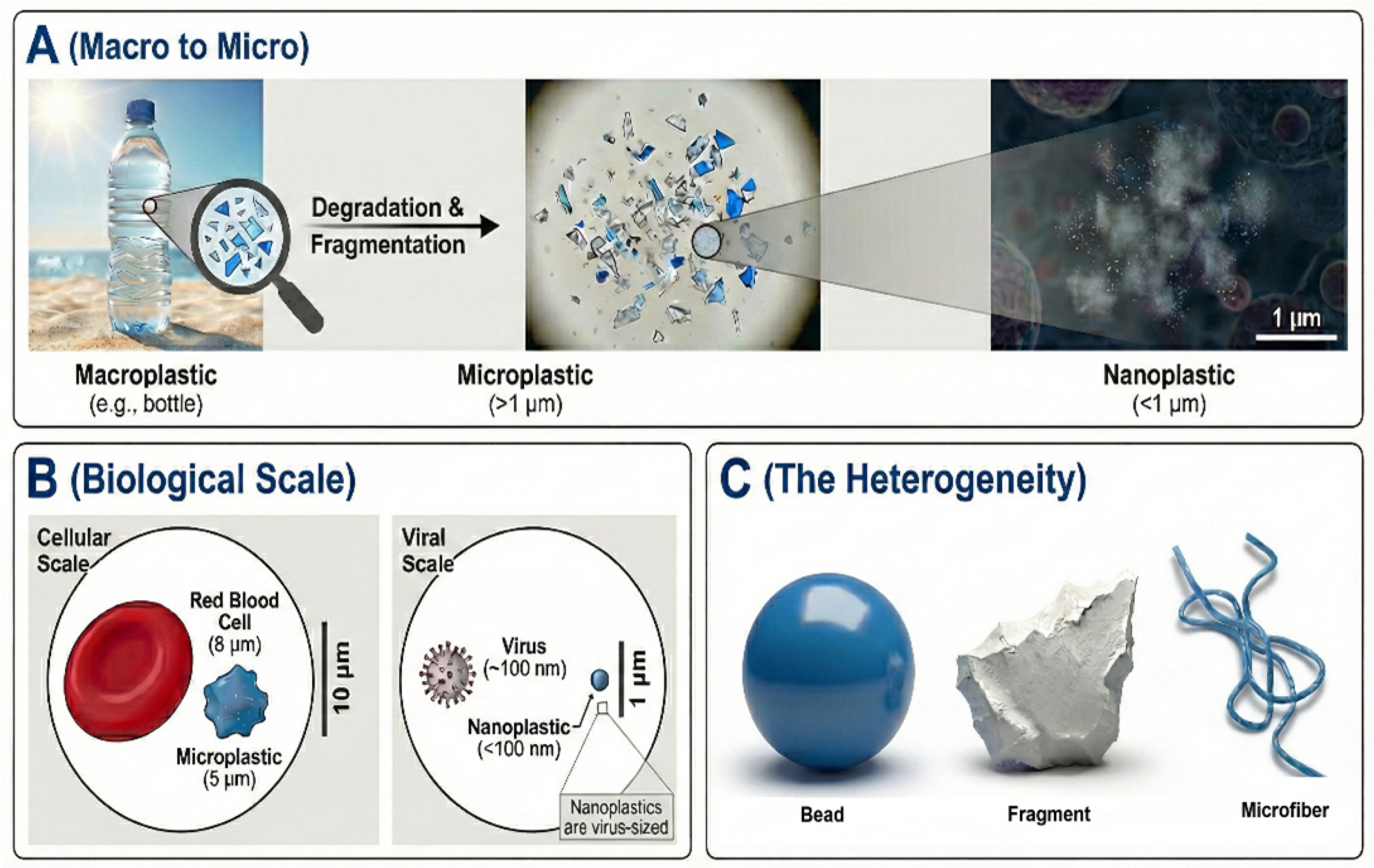
Microplastics (MPs) are defined as plastic particles spanning a size range from 0.1 μm to 5 mm, consistent with the European Food Safety Authority (EFSA) framework [4,17]. The defining upper limit of 5 mm is internationally accepted. Nanoplastics (NPs) constitute the subcategory of particles smaller than 1 μm (1000 nm), with particular concern attaching to those under 100 nm. Size matters considerably here. Nanoscale particles behave very differently from larger ones. They have more surface area relative to their volume, which makes them more reactive. More importantly, their diminutive dimensions permit traversal of biological barriers—intestinal epithelium, blood–brain barrier, placental membranes—that exclude larger particles, as depicted in Figure 2 [14,18]. Once past these barriers, they can spread through the body and accumulate in tissues [19].
2.2. Primary and Secondary Sources
Microplastics are classified by origin as either primary or secondary [20]. Primary ones are manufactured small intentionally microbeads in cosmetics, industrial pellets called nurdles, and fibers in synthetic clothing [20]. Secondary MPs, which constitute most environmental contamination, arise from fragmentation of larger plastic items. UV light, physical forces such as waves and abrasion, and some biological processes gradually weaken plastic until it fragments into countless smaller pieces [20].
Figure 2.
The Size Continuum of Plastic Pollution: From Visible Waste to Cellular Contaminants. (A) The environmental degradation pathway where macroplastics (e.g., bottles) fragment through weathering into microplastics (macro to nano size progression) and eventually into invisible nanoplastics (<1 µm). (B) A biological scale comparison shows that while microplastics are comparable in size to red blood cells (approximately 8 µm), nanoplastics penetrate the nanoscale (<100 nm), allowing them to breach cellular barriers. (C) The heterogeneity of plastic pollutants, illustrating the diverse morphologies—spherical beads, irregular fragments, and fibers—that influence toxicity and cellular uptake (created using Adobe Illustrator, Illustrae, and SciDraw).
2.3. Key Polymer Types and Their Prevalence
The types of plastic found in human tissue match what is produced most globally. Polyethylene (PE) is the most common plastic manufactured worldwide, used mainly for packaging. Polypropylene (PP), employed in packaging and textiles, reportedly leaches billions of nanoparticles from tea bags during steeping [21]. Polystyrene (PS) is used in disposable cups and foam packaging. Polyethylene terephthalate (PET) is used in most drink bottles and appears frequently in artery plaques [14]. Polyvinyl chloride (PVC) appears more toxic to cells than PE or PP, probably because of chemicals that leach from it [22]. Other plastics of concern include polyamide, polytetrafluoroethylene (PTFE), and polyurethane, all of which have been found in human reproductive fluids [23].
2.4. Physicochemical Properties Influencing Biological Interactions
Toxicity depends on more than just polymer type. The physical and chemical properties all interact (Table 1). Size is critical for biodistribution. Larger microplastics (>150 μm) typically remain confined within the gastrointestinal lumen, whereas smaller MPs and particularly NPs can penetrate into systemic circulation [24]. Shape matters too. Jagged fragments are taken up by cells more readily than smooth spheres and cause more inflammation [25]. Fiber-shaped microplastics deserve special attention as they act similarly to asbestos in the lungs [26]. Surface chemistry changes as plastic weathers in the environment, adding chemical groups that promote protein adsorption and immune recognition [18].
Table 1.
Classification and characteristics of MPs/NPs and their toxicological relevance.
2.5. The “Trojan Horse” Effect: MPs as Vectors for Co-Contaminants
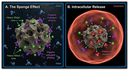
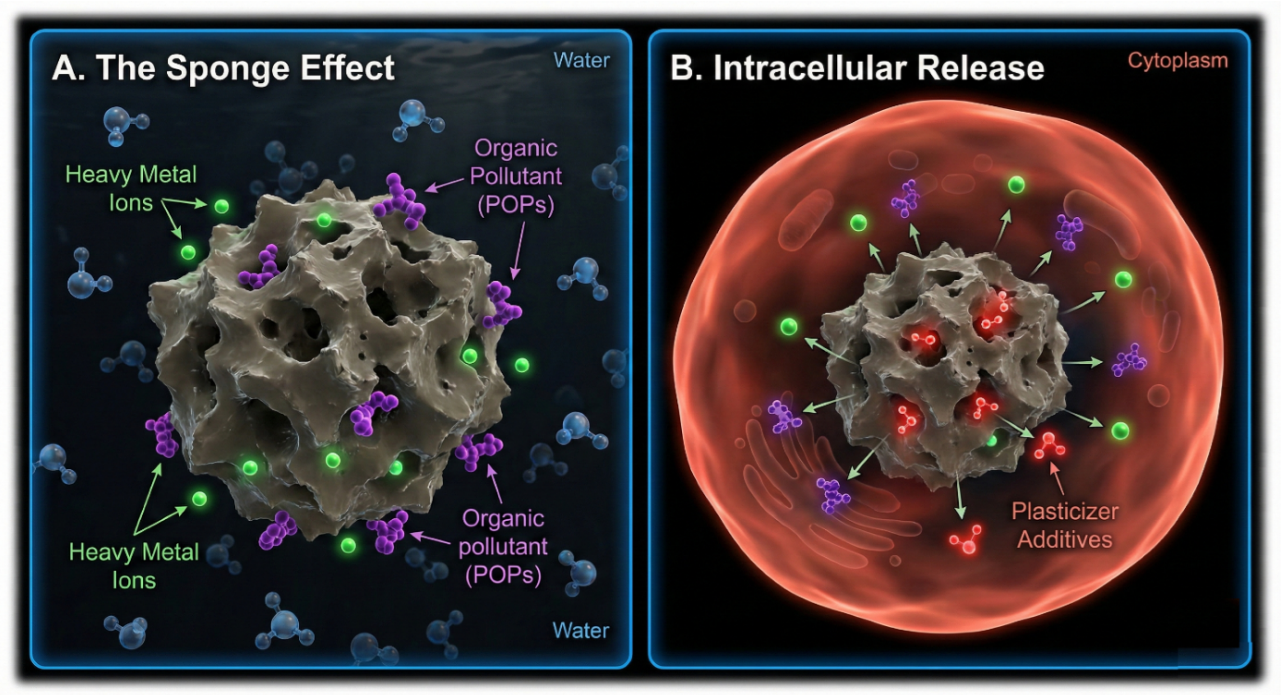
Besides their inherent toxicity, microplastics carry other pollutants. Their hydrophobic surfaces and high surface area enable adsorption of substantial quantities of co-contaminants [11]. These include persistent organic pollutants (POPs) such as polychlorinated biphenyls (PCBs) and polycyclic aromatic hydrocarbons (PAHs), heavy metals including lead and mercury, and endocrine disruptors including bisphenol A (BPA) and phthalates [11,14]. This “Trojan horse” mechanism creates synergistic hazard wherein combined toxicity exceeds that attributable to either particle or chemical alone, as illustrated in Figure 3 [9,10].
Figure 3.
The “Trojan Horse” Effect and Synergistic Toxicity. (A) The Sponge Effect: In the environment, the hydrophobic surface of weathered microplastics adsorbs and concentrates persistent organic pollutants (POPs) and heavy metal ions from the surrounding water. (B) Intracellular Release: Upon internalization by a human cell, the particle acts as a vector, releasing the concentrated environmental toxins alongside leached chemical additives (e.g., plasticizers, phthalates) into the cytoplasm, triggering a “chemical hit” that amplifies biological damage. (created using Adobe Illustrator, Illustrae, and SciDraw).
3. Human Exposure Routes
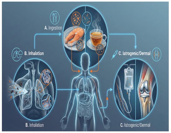
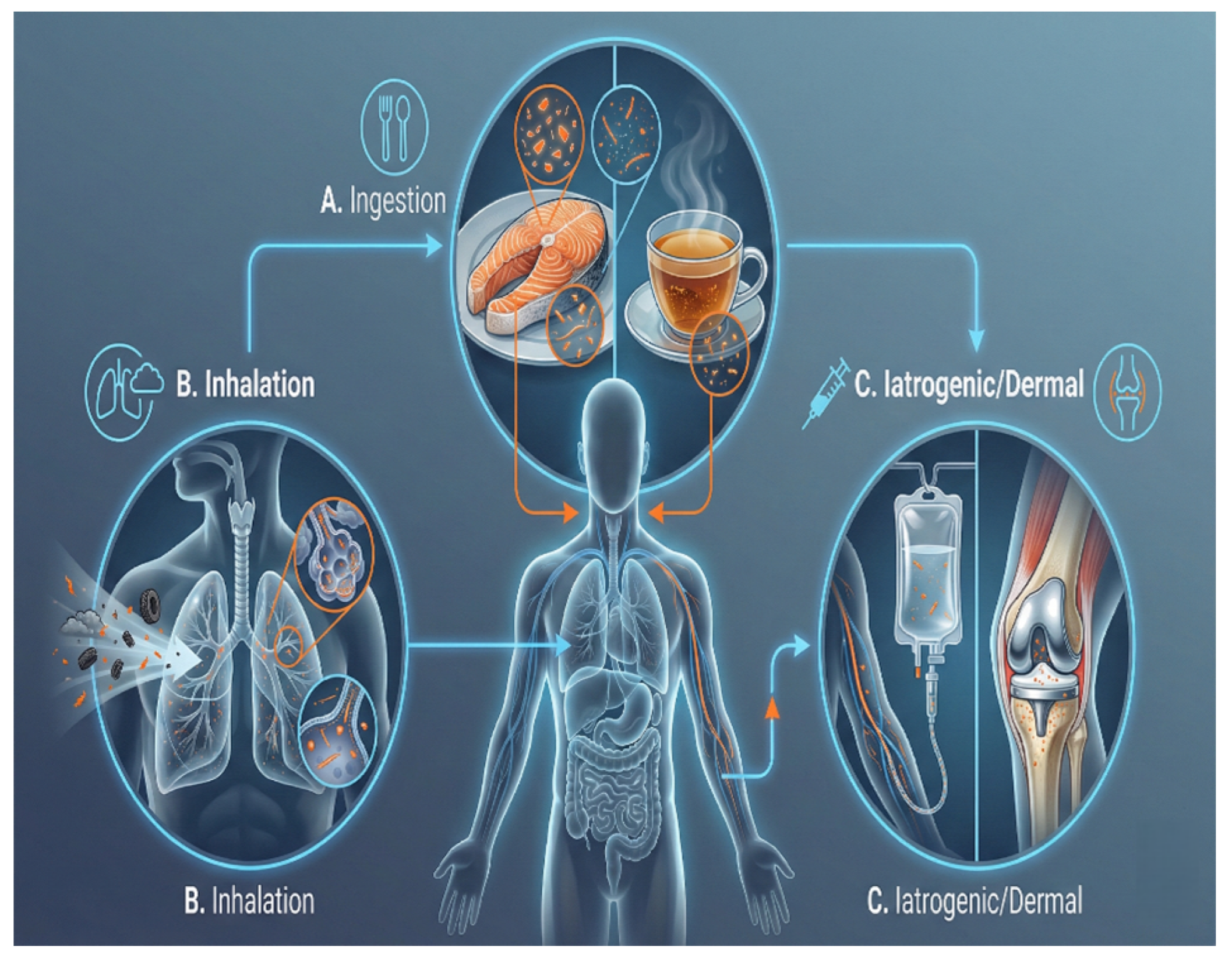
People are exposed to microplastics and nanoplastics through three main pathways: ingestion, inhalation, and dermal contact, as summarised in Figure 4. Estimates suggest people ingest somewhere between 39,000 and 52,000 particles per year, with inhalation adding to this burden [10].
Figure 4.
Comprehensive Pathways of Human Exposure to Micro- and Nanoplastics. (A) Ingestion: The alimentary route remains the dominant pathway, with particles entering via contaminated seafood (trophic transfer) and beverages (e.g., tea, bottled water). (B) Inhalation: An underestimated pathway where airborne fibers from textiles, urban dust, and tire wear particles are inhaled, lodging in the respiratory tract. (C) Iatrogenic and Dermal Contact: Emerging direct entry routes including medical interventions (e.g., IV fluid delivery, degradation of joint implants) and dermal penetration through damaged skin barriers. (created using Adobe Illustrator, Illustrae, and SciDraw).
3.1. Oral Ingestion
Ingestion is the primary route. Food becomes contaminated in several ways. Aquatic organisms fish, shellfish, bivalves accumulate MPs within their tissues via trophic transfer [24]. Bottled water contains substantially more particles than tap water, as bottles shed plastic [27]. Plastic tea bags release billions of particles during steeping [21]. Processing and packaging add further contamination. Sea salt, honey, beer, and numerous other foods contain detectable plastic [10,24].
3.2. Respiratory Inhalation
Airborne microplastics derive primarily from clothing fibers, urban dust, and tire wear [5]. City air contains tens to hundreds of plastic fibers per cubic meter [28]. Indoor air may be even more contaminated. Carpets, clothes, and house dust all contribute [29]. Researchers have found microplastics in lung tissue, sputum, and bronchoalveolar lavage fluid [10]. Particles smaller than 10 μm can reach deep into the lungs, where they cause inflammation and might enter the bloodstream [24].
3.3. Dermal Contact and Iatrogenic Routes
Skin exposure occurs from water, soil, or products like scrubs containing microbeads [24]. Available evidence indicates that nanoplastics smaller than 200 nm may penetrate compromised skin [24]. One often overlooked exposure source is medical devices. Plastic components wear down and release particles. Joint replacements, dental implants, and cosmetic implants all degrade over time and shed plastic fragments [24].
4. Evidence from Preclinical Toxicological Models
We do not yet have definitive proof that microplastics cause disease in humans. However, animal and cell studies demonstrate potential mechanisms. These studies establish that microplastics are not inert, they cause cell stress, inflammation, and organ damage (Table 2).
4.1. Insights from Animal Models
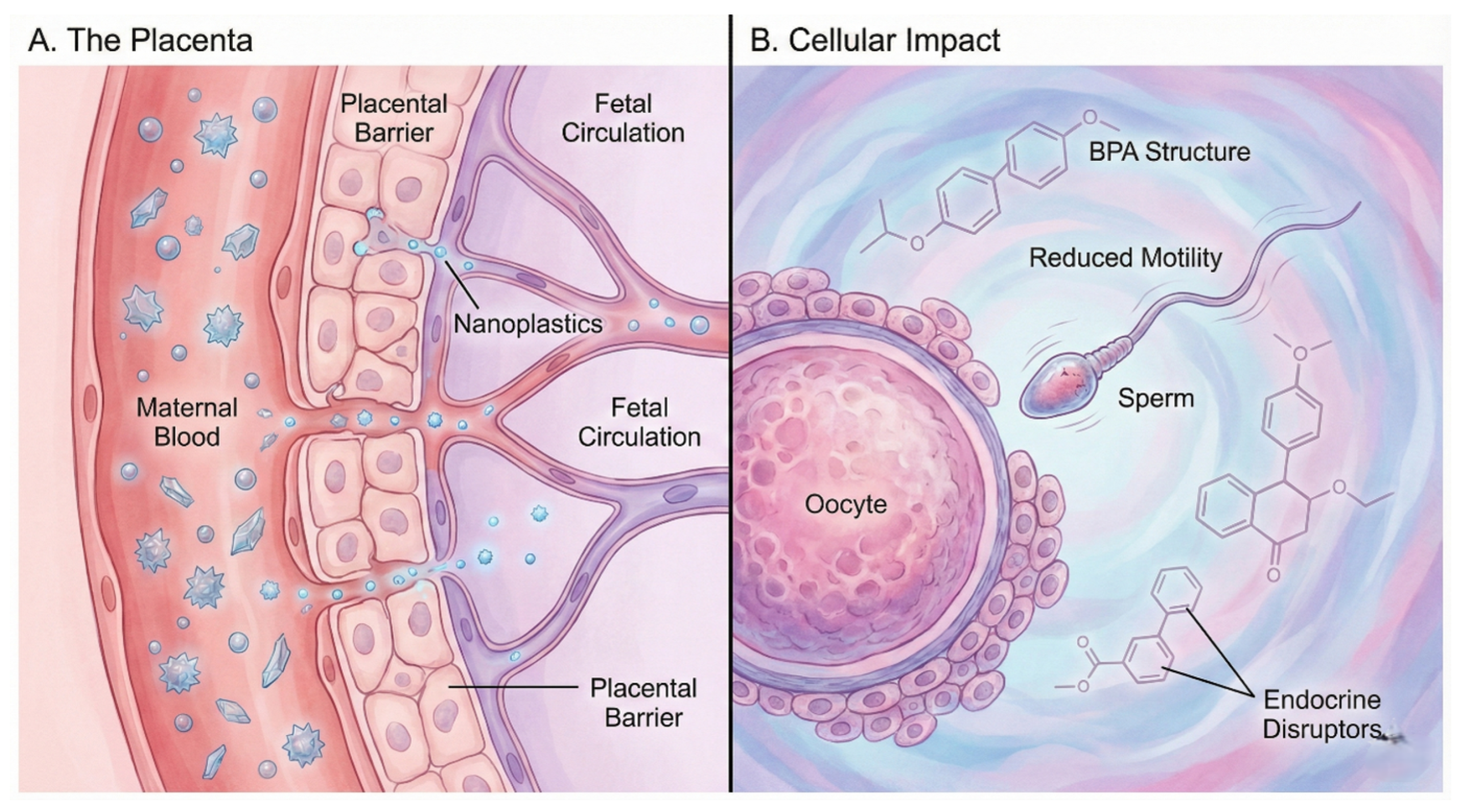
Fish exposed to microplastics accumulate particles in their gills, liver, and gut. They show oxidative stress, inflammation, and disrupted lipid metabolism [7]. Rodent investigations demonstrate that MP exposure produces biochemical and structural damage with functional impairment across intestine, liver, and reproductive systems [8]. Key findings include gut and metabolic effects (barrier damage and inflammation), nervous system effects (blood–brain barrier (BBB) damage and memory problems), reproductive effects (reduced male fertility, as illustrated in Figure 5), and systemic inflammation with elevated TNF-α and IL-1β [8,30,31].
A critical limitation of many animal studies is that the concentrations employed—often 0.1–100 mg/kg/day for oral exposures—substantially exceed estimated human dietary exposure, which ranges from approximately 0.1 μg to a few mg per day. Furthermore, most experiments use pristine, uniform-sized polystyrene spheres rather than the weathered, irregularly shaped, and chemically complex particles that humans actually encounter. Studies with higher doses may not translate directly to human health risk; findings should therefore be interpreted as mechanistic proof-of-concept rather than direct evidence of harm at current human exposure levels. Where dose–response data are available, several studies suggest that effects are concentration-dependent, reinforcing the importance of establishing no-observed-adverse-effect levels under environmentally relevant conditions [7,8].
Figure 5.
Reproductive and Developmental Toxicity Implications. (A) Transplacental Transfer: Nanoplastics are depicted crossing the placental barrier from maternal blood into the fetal circulation, thereby posing risks to in-utero development. (B) Gamete Toxicity: Exposure affects reproductive cells directly; sperm show reduced motility, while the local release of endocrine-disrupting chemicals (e.g., BPA analogs) creates a toxic microenvironment for oocytes, potentially impairing fertility (created using Adobe Illustrator, Illustrae, and SciDraw).
4.2. Mechanistic Clues from In Vitro Cell Culture
Studies with human cells have elucidated the main pathways of injury. Nanoplastics undergo ready internalization through multiple endocytic mechanisms [32]. Smaller particles under 100 nm are taken up more efficiently. Experiments with intestinal cells demonstrate that 50 nm particles can cross the gut lining. Across diverse human cell types—A549 lung epithelium, Caco-2 enterocytes, macrophages—a consistent triad of toxic mechanisms emerges: oxidative stress with elevated reactive oxygen species (ROS) production [33], inflammation through NLRP3 inflammasome activation, and cell death [34]. It should be noted that these studies frequently use particle concentrations and exposure durations that may not reflect chronic human exposure; findings represent mechanistic hypotheses that require validation at environmentally relevant doses.
4.3. Advanced Human-Relevant Models
Organoid cultures and organ-on-chip (OoC) platforms offer improved recapitulation of human physiology. Liver organoids derived from stem cells show disrupted lipid metabolism and liver damage when exposed to polystyrene at realistic concentrations [35]. Brain organoids demonstrate that nanoplastics disrupt neural development, kill cells, and impair connections between neurons. Multi-organ chip devices document translocation of polystyrene nanoparticles from gut to liver compartments, with subsequent hepatic injury [36].
Table 2.
Summary of key toxicological findings from preclinical models of microplastic exposure.
5. A Multi-Hit Pathophysiological Hypothesis of Microplastic Toxicity
We propose a conceptual multi-hit hypothesis to integrate the various toxicology findings; this framework requires future experimental and clinical validation. The hypothesis envisions MP-induced pathology as a sequential cascade wherein successive deleterious events build upon one another. It is important to note that while experimental and observational evidence supports each individual step, the complete causal chain in humans remains unestablished.
Hit 1: Exposure and Translocation. Particles enter through ingestion, inhalation, or skin contact. Smaller MPs and NPs penetrate biological barriers via endocytosis or paracellular transport, entering systemic circulation as evidenced by detection in human blood [12]. From the blood they distribute to organs including the brain [15,37].
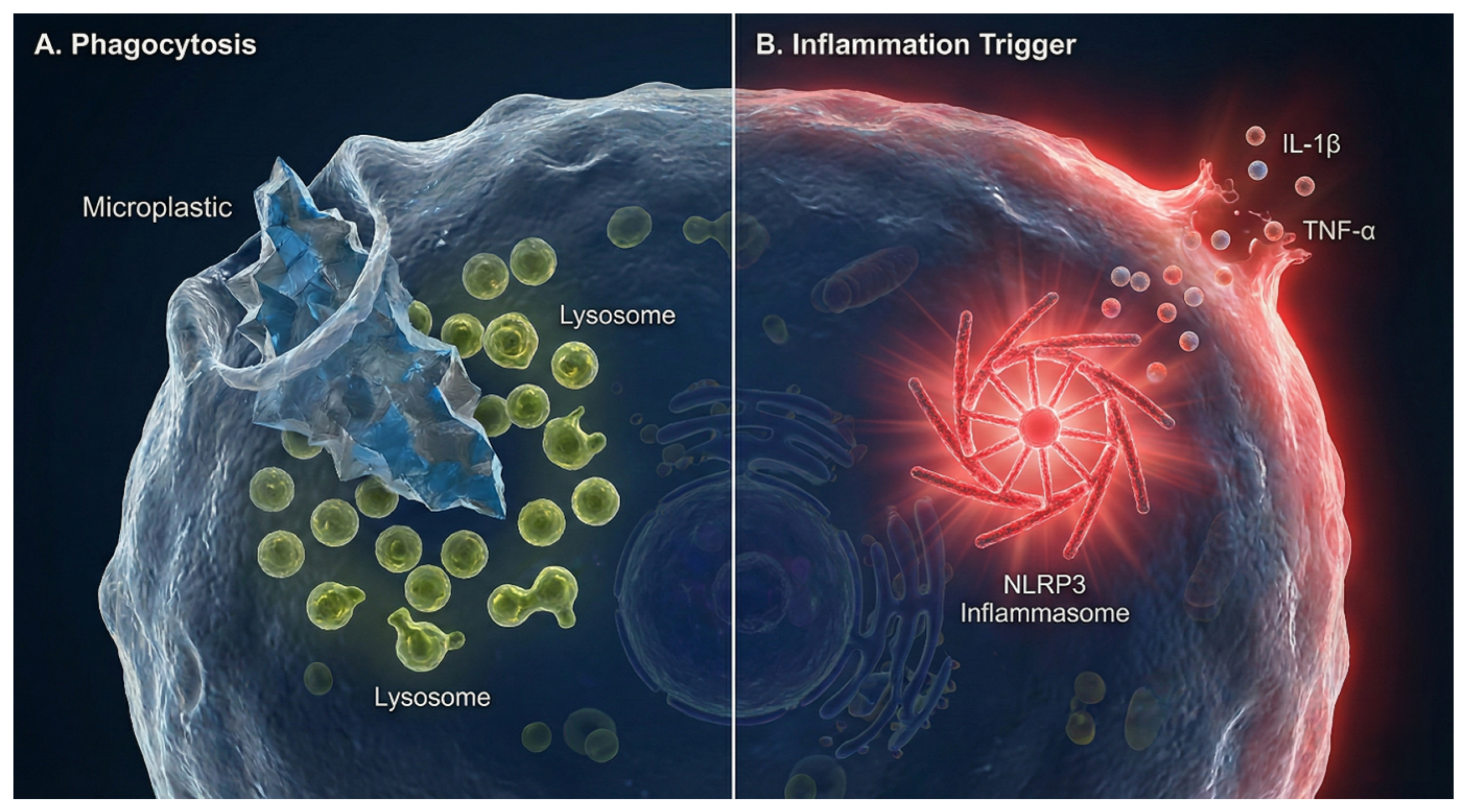
Hit 2: Physical Damage. Particles lodged in tissues act as irritants. Immune cells recognize these particles and trigger inflammation through the NLRP3 inflammasome, releasing IL-1β and TNF-α, as depicted in Figure 6. The body cannot break down plastic, so inflammation becomes chronic [25,26].
Hit 3: Chemical Insult. The “Trojan horse” mechanism transports environmental pollutants across biological barriers. Additionally, phthalates, BPA, and flame retardants are additives that leach from internalized particles. Several of these function as endocrine-disrupting chemicals (EDCs) and disrupt hormonal signaling at low concentrations, working synergistically with the physical insult [38].
Hit 4: Disease. Accumulated damage can ultimately lead to clinical illness. Microplastic-induced inflammation in the cardiovascular system disrupts plaques and facilitates clot formation [14]. In the brain, nanoplastics activate microglia, cause neuroinflammation, and might contribute to neurodegeneration [15]. Reproductive systems are affected by endocrine disruptors interfering with hormones. The gut develops microbiome imbalances and barrier dysfunction [39].
Figure 6.
Cellular Mechanisms of Sterile Inflammation (Conceptual Representation). (A) Phagocytosis: An immune cell (macrophage) engulfs a jagged microplastic fragment. The lysosomal machinery attempts to digest the bio-persistent material but fails, leading to lysosomal destabilization. (B) Inflammasome Activation: The persistent physical presence of the particle triggers intracellular stress, activating the NLRP3 inflammasome complex. This results in the maturation and burst release of pro-inflammatory cytokines (IL-1β, TNF-α), driving the chronic, sterile inflammation seen in affected tissues. Note: This figure represents a conceptual schematic based on experimental evidence primarily from in vitro and animal studies; causal mechanisms in humans remain under investigation (created using Adobe Illustrator, Illustrae, and SciDraw).
6. Bioaccumulation and Detection in Human Tissues and Fluids
One of the most significant recent discoveries is that these particles cross biological barriers and accumulate in human tissues. This understanding has emerged from improvements in detection methods capable of finding tiny particles in biological samples (Table 3).
6.1. Analytical Methodologies for Detection and Quantification
Finding microplastics in tissue requires careful sample preparation and sophisticated equipment [26]. The first step is removing the biological matrix using chemical digestion with KOH or H2O2 [40]. Laboratory procedures must rigorously avoid plastic contamination, which constitutes a major source of analytical uncertainty; this includes use of glass or stainless-steel equipment, positive-pressure clean-air filtration systems, procedural blanks, and field blanks in every analytical run [41]. Inter-laboratory variability remains a substantial challenge: round-robin exercises have demonstrated that recovery efficiencies for spiked samples can vary from below 30% to above 90% depending on the extraction protocol, matrix type, and particle size, underscoring the urgent need for standardized reference materials and validated protocols [41,42]. Spectroscopy then identifies the plastic type. Micro-Fourier-transform infrared spectroscopy (μ-FTIR) uses infrared absorption and has a detection threshold of approximately 10–20 μm; micro-Raman spectroscopy (μ-Raman) achieves better spatial resolution (approximately 1 μm); pyrolysis-gas chromatography/mass spectrometry (Py-GC/MS) is highly sensitive but destroys samples. Scanning electron microscopy with energy-dispersive X-ray spectroscopy (SEM-EDS) reveals shape and elemental composition [11,15].
Table 3.
Analytical techniques for MP/NP characterization in human tissues.
6.2. Evidence of Widespread Bioaccumulation
Researchers have found microplastics in many different human tissues and fluids (Table 4). A key 2022 study detected PET, PE, and PS in 77% of blood donors, averaging 1.6 μg/mL [12]. A 2024 study found MPs in 100% of 62 placental samples, with concentrations spanning 6.5–790 μg/g [37]. About 75% of breast milk samples contain microplastics [13]. Work published in Nature Medicine during 2025 documented brain MP levels 7–30 times greater than those in liver or kidney, with concentrations increasing approximately 50% between 2016 and 2024. Brain tissue from individuals with dementia contained 3–10 times more plastic than control specimens [15]. Polystyrene and PET have been found in artery plaques and blood clots [14]. PTFE, PET, and polyamide have been detected in semen and ovarian fluid [23,43]. Key methodological details for the five most pivotal biomonitoring studies are summarised in Table 5.
Table 4.
Summary of MP/NP detection in human tissues and fluids.
Table 5.
Structured extraction of key human biomonitoring studies: analytical methods, contamination controls, size detection thresholds, sample sizes, and key uncertainties.
7. Emerging Evidence of Organ-Specific Toxicity
The following subsections summarise key findings across organ systems; proposed mechanisms are consolidated in Table 6.
7.1. Cardiovascular System
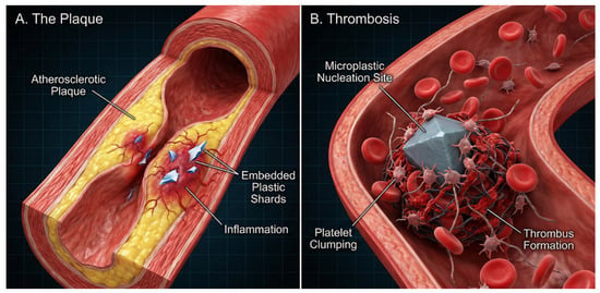
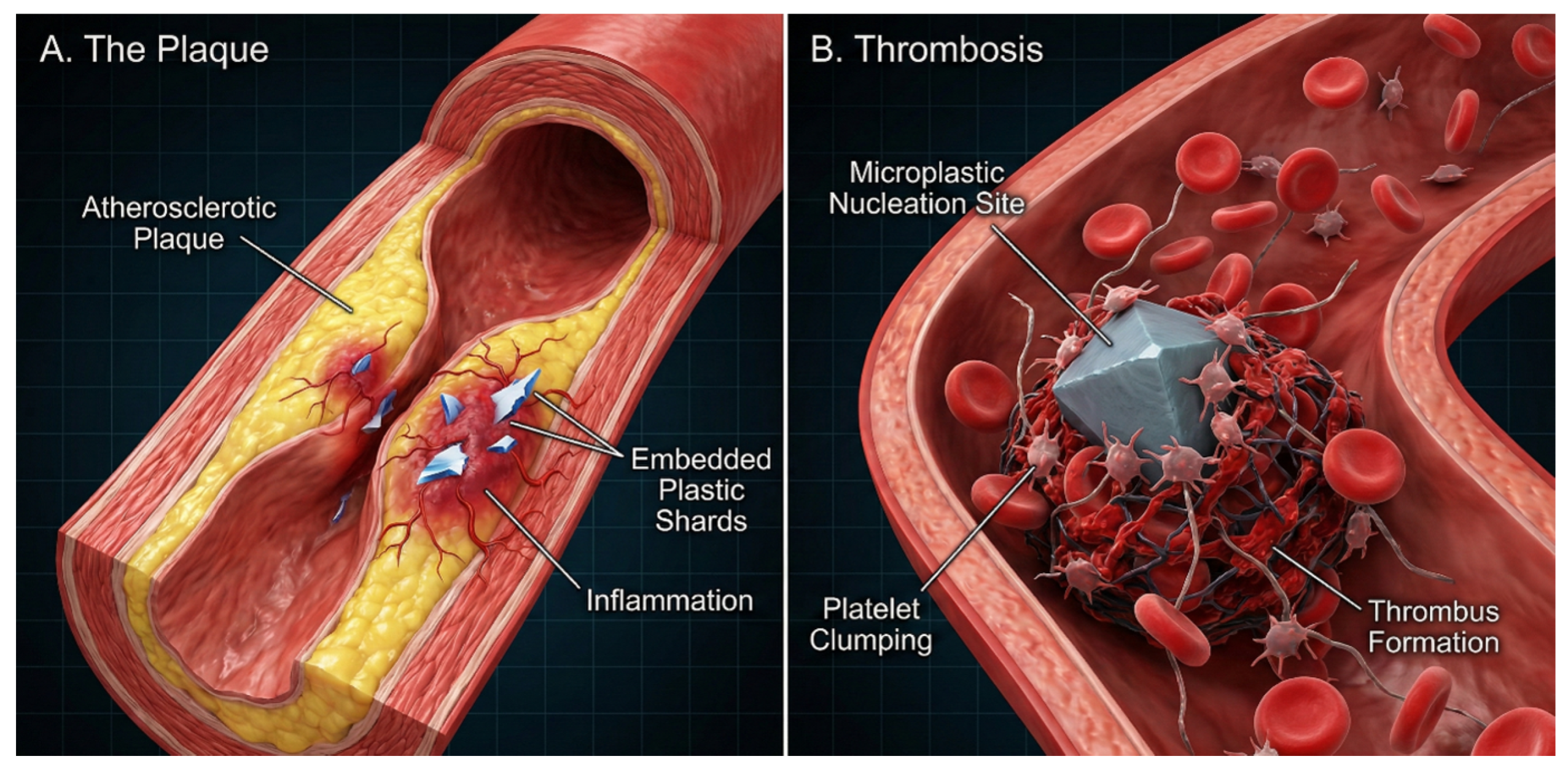
A 2024 multicenter Italian study enrolled patients undergoing carotid endarterectomy and analyzed excised atherosclerotic plaques. Polyethylene was found in 58.4% of patients, polystyrene in 12.1%, and PVC in 31.4%. Patients with plastic in their plaques had 4.5 times higher risk of heart attack, stroke, or death over the 34-month follow-up, as illustrated in Figure 7 [14]. This represents one of the earliest clinical investigations to report a potential association between measurable plastic burden within arterial tissue and adverse cardiovascular outcomes; the observational design precludes causal inference [14].
Figure 7.
Microplastics as a Cardiovascular Risk Factor. (A) Atherosclerotic Plaques: Cross-section of a diseased artery showing microplastic shards embedded within the fatty atherosclerotic plaque, contributing to local inflammation and plaque instability. (B) Thrombosis: A magnified view of a thrombotic event where a microplastic particle serves as a nucleation site for platelet aggregation, accelerating clot formation and increasing the risk of myocardial infarction or stroke (created using Adobe Illustrator, Illustrae, and SciDraw).
7.2. Neurological System
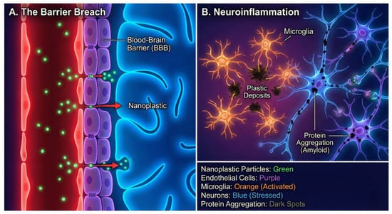
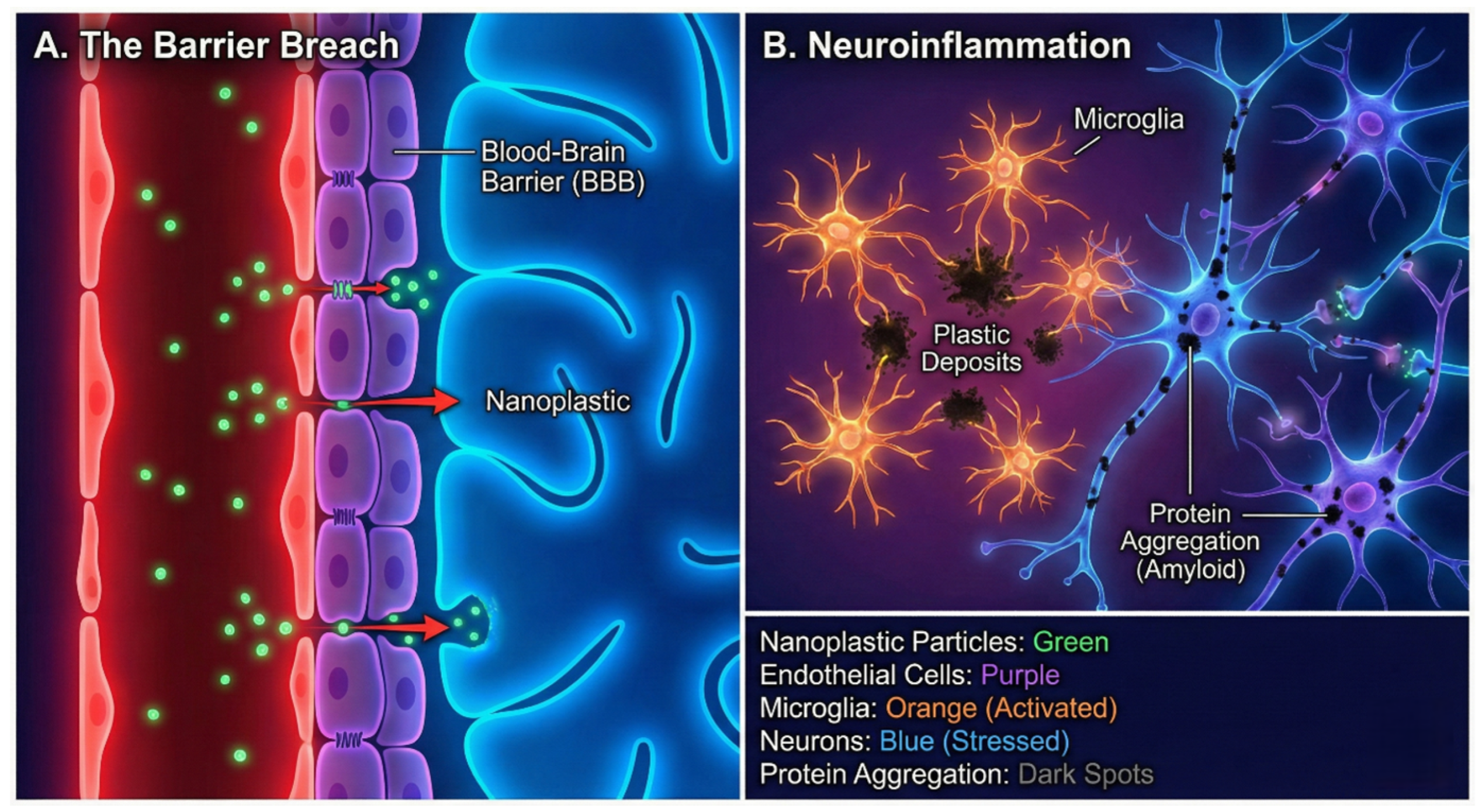
Brain tissue presents unique vulnerability to nanoplastic accumulation. A 2025 study analyzing postmortem brain samples found plastic concentrations 7–30 times higher than in liver or kidney. Brain plastic levels increased approximately 50% from 2016 to 2024. Other organs did not show this increase. Brain specimens from individuals diagnosed with dementia contained 3–10 times more plastic than those from cognitively intact controls [15]. These findings raise the possibility that accumulating plastic burden may contribute to neuroinflammatory processes and potentially to neurodegeneration, as depicted in Figure 8, though causality remains unestablished and the postmortem study design does not permit inference about clinical trajectories. The brain accumulation data are correlational; prospective studies with longitudinal biomonitoring are required before mechanistic or causal conclusions can be drawn.
Figure 8.
Nanoplastic Invasion of the Central Nervous System. (A) The Barrier Breach: Nanoplastics in the systemic circulation exploit transport mechanisms to traverse the endothelial tight junctions of the Blood-Brain Barrier (BBB), entering the brain parenchyma. (B) Neuroinflammation: Once within the CNS, plastic deposits trigger the activation of microglia (brain resident immune cells). This sustained immune response promotes oxidative stress in neurons and may accelerate pathological protein aggregation (e.g., amyloid plaques), linking pollution to neurodegenerative risks. (created using Adobe Illustrator, Illustrae, and SciDraw).
7.3. Gastrointestinal System
The gut is where ingested microplastics first arrive, resulting in direct exposure. A study comparing stool samples found substantially more microplastics in IBD patients than healthy people, with higher plastic content correlating with disease severity [39]. Animal studies demonstrate that microplastics damage the gut barrier, cause inflammation, and alter the microbiome [44].
7.4. Reproductive System
Microplastics have been found in semen and ovarian fluid, raising concerns about fertility and fetal development [23,43]. Placental studies document MP presence in 100% of samples analyzed, with particle concentrations varying widely [37]. Animal studies show that exposure during pregnancy adversely affects fetal development and can cause behavioral problems in offspring.
7.5. Hepatic and Renal Systems
Microplastics in cirrhotic livers suggest hepatic accumulation [44]. Animal studies demonstrate liver damage from microplastics including oxidative stress, inflammation, and lipid metabolism disruption [45]. In rodents, microplastics damage kidney structures. Chronic exposure may be nephrotoxic [44].
Table 6.
Key toxicological mechanisms of microplastic exposure.
8. Advances in Artificial Intelligence and Machine Learning for Microplastic Detection
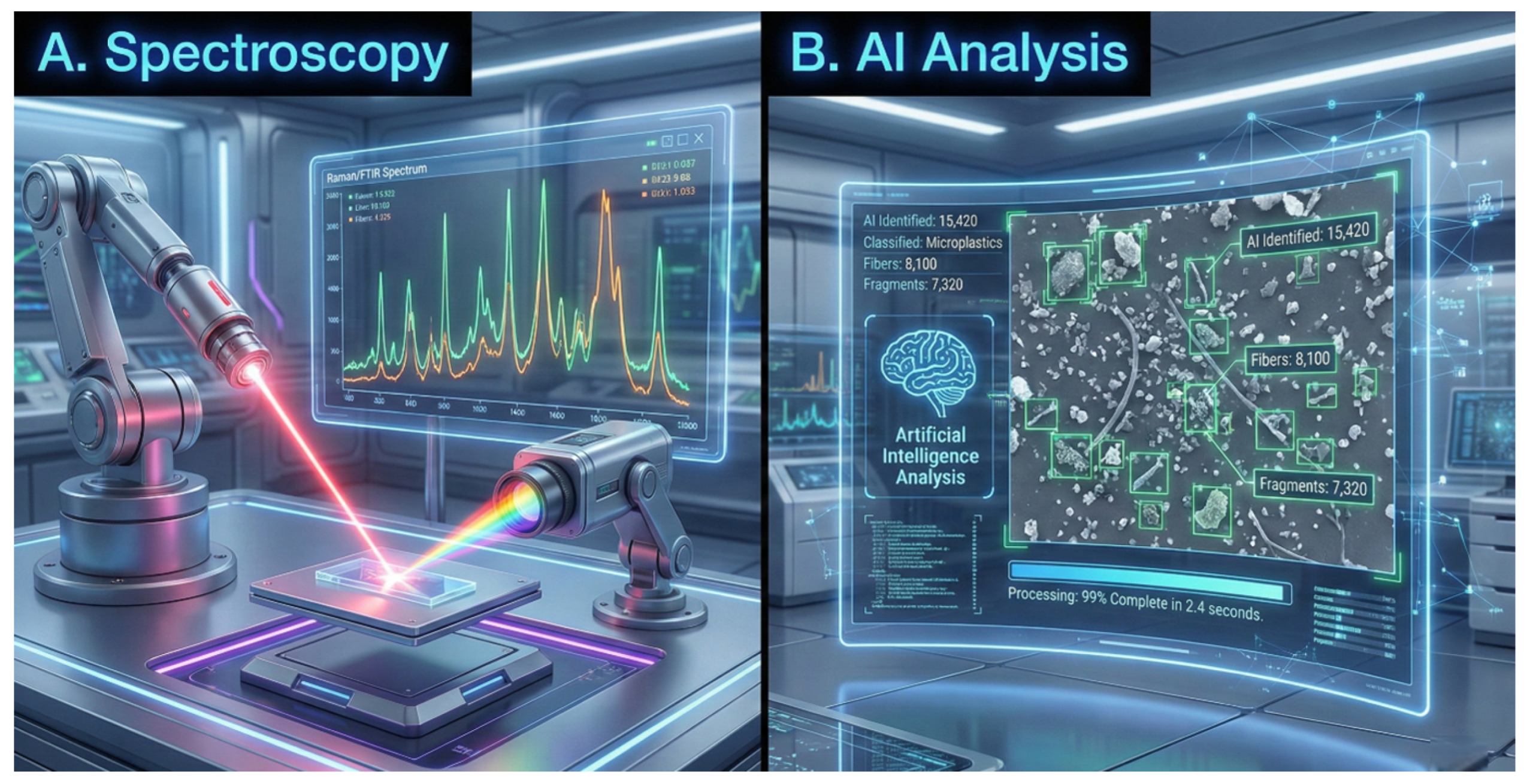
Artificial intelligence (AI) and machine learning (ML) are transforming how we detect and identify microplastics, as illustrated in Figure 9. Traditional methods work but they are slow and labor-intensive, requiring manual counting and classification of particles. By late 2024, over 1600 papers on microplastic detection had been published, with more than 100 examining AI applications [47,48]. Computers can now perform detection automatically, quickly, and accurately; the main approaches and their performance are summarised in Table 7.
Figure 9.
The Integration of Artificial Intelligence in Microplastic Analysis. (A) Advanced Spectroscopy: Automated Raman/FTIR spectroscopy setups generate complex chemical fingerprints for the identification of particles. (B) AI Analysis: Deep learning algorithms process microscopy data in real-time, automatically identifying, classifying, and quantifying thousands of particles (including nanoplastics) with speed and accuracy far exceeding manual human counting. (created using Adobe Illustrator, Illustrae, and SciDraw).
8.1. Deep Learning Architectures for Spectral Analysis
Deep learning, especially convolutional neural networks (CNNs), has substantially facilitated microplastic identification. These networks extract important features from spectral data autonomously [47]. Older ML methods such as support vector machines (SVMs) and random forests also perform well for differentiating PE, PP, and PET based on their FTIR spectra [49]. SVMs remain popular and improve accuracy over conventional analysis [50].
A 2025 paper from the Chinese Academy of Sciences proposed a dual-branch CNN with an attention mechanism named CBAM that achieved 98% accuracy on mixed microplastic samples using infrared spectroscopy [51]. This is significant since real samples tend to be mixtures, and varying plastic proportions alter the spectra and complicate identification. The attention mechanism captures both local and global patterns in the spectra, outperforming solutions that merely consider individual features.
A comparative study of CNN architectures (MobileNetV3Large, ResNet50V2, ResNet-101V2, and EfficientNetB7) using transfer learning for microplastic classification in beads, fibers, and fragments showed that all solutions performed very well on fiber detection but had difficulties with bead and fragment detection [52]. The focal plane array micro-FT-IR imaging-based PlasticNet deep learning model correctly identified over 11 common plastic types with accuracy exceeding 95% after training on more than 8000 spectra [53].
8.2. Automated Image Recognition and High-Throughput Analysis
CNNs and segmentation algorithms can identify microplastics automatically from microscopy images with accuracy over 85% [54]. These deep learning models extract morphological features particle size, shape, color, surface texture effectively distinguishing microplastics from natural organic particles and cellular debris. What previously required hours of manual analysis now takes minutes.
High-performance segmentation using U-Net and ResUNet architectures has been applied to SEM micrographs of microplastic particles (50 μm–1 mm range) and fibers (approximately 10 μm diameter), achieving good accuracy while proving substantially faster and less expensive than manual analysis [55]. SMACC is a deep learning system for counting and classifying microplastics in environmental samples at high throughput [56]. UAV-based monitoring combined with PlasticFinder software has achieved automatic detection and quantification of anthropogenic marine debris, representing the first deep learning application for large-scale environmental plastic monitoring [55].
8.3. Explainable AI and Holographic Imaging
Explainable AI is helping elucidate why classifiers make their decisions. A 2025 study presented a thorough XAI framework using SHAP (SHapley Additive exPlanations) to classify pristine and weathered microplastics versus biological materials [57]. Among seven supervised ML models benchmarked Decision Trees, Random Forest, k-Nearest Neighbours, Neural Networks, LightGBM, XGBoost, and Support Vector Machines k-NN and SVM achieved the highest accuracy (82.5%), with k-NN demonstrating the most balanced performance across precision and recall metrics [57].
Holographic imaging combined with ML can classify thousands of microplastics with over 99% accuracy, regardless of shape, size, or material [58]. A holographic flow cytometer on a chip can classify fibers as natural or plastic while they flow through, with ML handling the classification [58]. These tools work in real time on flowing samples, which is essential for environmental monitoring.
8.4. AI for Risk Assessment, Toxicity Prediction, and Quality Assurance
ML is also valuable for finding patterns in complex datasets [59,60]. Applications include predicting microplastic transport and accumulation, finding pollution hotspots, and predicting toxicity. Li and colleagues created an ensemble ML model predicting joint toxicity of micro/nanoplastics and pollutants across species with R2 exceeding 0.84 in validation and external tests [61]. Connecting AI to IoT sensors enables drones and buoys to monitor plastic in real time [62].
Researchers have even evaluated ChatGPT and Gemini for QA/QC screening in microplastic studies [63]. Using prompts constructed from established criteria for drinking water studies, LLMs evaluated 73 publications (2011–2024), efficiently extracting key information, judging study reliability, and closely matching human assessments. This could prove useful for accelerating QA/QC in regulatory settings.
Table 7.
Summary of AI and machine learning applications in microplastic detection.
9. Emerging Biosensor and Electrochemical Detection Technologies
While spectroscopic methods remain the gold standard for MP characterization, substantial advances in biosensor and electrochemical detection technologies offer promising alternatives for rapid, cost-effective, and field-deployable monitoring (Table 8). These platforms address critical limitations of conventional methods including high equipment costs, laboratory dependency, and low throughput [64,65].
Table 8.
Emerging biosensor and electrochemical technologies for MP/NP detection.
9.1. Electrochemical Sensors
Electrochemical sensors offer significant advantages in sensitivity, selectivity, and miniaturization for on-site monitoring [66]. Key strategies include nanomaterial incorporation, molecular imprinting, and surface modifications. Approaches including electrochemical impedance spectroscopy (EIS), particle-impact electrochemistry, and ML-enhanced systems have achieved detection limits as low as 10−11 M [66]. A groundbreaking CRISPR-based electrochemical aptasensor (CRISPR-MP) employing split gRNA with Cas12a-mediated cascade strand displacement achieves detection limits of 37 ng/mL for PVC and 45 ng/mL for PS [67]. Extracellular polymeric substance (EPS)-based biosensors derived from cyanobacteria can detect multiple polymer types including polystyrene sulfonate (PSS), polyamide (PA), polymethacrylic acid (PMA), and polyethylene (PE) [68].
9.2. Optical Biosensors and SPR Platforms
Optical biosensors, particularly surface plasmon resonance (SPR) platforms, offer low detection limits, high sensitivity, and multiplexed detection capacity [64]. An estrogen receptor (ER)-functionalized SPR platform on plastic optical fiber (ER-SPR-POF) provides a “smart” sensing interface enabling discrimination of both particle size (micro vs. nano) and material composition [69]. Polystyrene microplastics show the highest binding force attributable to larger surface charge, followed by PVC and PE. Gold nanograting platforms functionalized with ERs enable rapid nanoplastic detection in seawater [70].
9.3. Rapid Mass Spectrometry and Real-Time Monitoring
Flame ionization mass spectrometry (FIMS) enables MP/NP detection in seconds without time-intensive sample preparation [71]. Dried samples are directly combusted before the MS inlet, enabling rapid decomposition and ionization. FI-MS has detected PET in bottled water and apple juice, quantified MPs in soil (6.8% error), and identified 200 nm PS nanoplastics in mouse placentas without tedious preparation [71]. Real-time electrochemical sensing platforms monitor nanoplastic-induced toxicity at the exposure–organism interface, supporting “whole-process toxicity response monitoring” rather than endpoint profiling [72]. These miniature sensors detect NPL-induced toxicity with high sensitivity for rapid environmental response.
10. Critical Knowledge Gaps and Methodological Challenges
Research has expanded rapidly but major challenges remain for risk assessment. First, standardized methodologies for sampling, extraction, and quantification remain absent; studies vary in analytical techniques, size thresholds, classification systems, and reporting units [9,40,41,42]. Second, current models have significant limitations—many studies employ pristine, spherical polystyrene beads at high concentrations rather than environmentally relevant mixtures of shapes, sizes, and weathered surfaces [7,8,25,73]. Third, chronic, low-dose, and mixture-toxicity studies reflecting lifelong human exposure remain scarce [19,24]. Fourth, a nanoplastic blind spot persists—most methods detect only particles exceeding 1–10 μm, yet NPs may constitute approximately 90% of total particle number [30,42,74]. Current risk assessments are based upon the measurable “tip of the iceberg”.
Ubiquitous but Understudied Plastic Sources. An important category of plastic contaminants that has received insufficient attention encompasses particles that are ubiquitous in the global environment yet extraordinarily difficult to characterize definitively. The most illustrative example is tire wear particles (TWPs), which are generated continuously by the abrasion of rubber tires against road surfaces [75]. Tires are composite materials containing synthetic rubbers (styrene-butadiene, polybutadiene), carbon black, zinc compounds, and a suite of toxic additives including 6PPD-quinone, a transformation product linked to acute mortality in coho salmon and suspected endocrine disruption in mammals [76]. Globally, an estimated 6 million tonnes of TWPs are deposited annually onto roads and subsequently transported into waterways, soils, and air [75]. Despite this enormous release, TWPs are difficult to detect and quantify in environmental and biological samples because their heterogeneous chemical composition does not produce simple polymer-type spectral signatures, and most MP/NP analytical workflows are not optimized for rubber-based particles [41,75]. Other comparably understudied categories include synthetic textile microfibers—which are released in large quantities during washing of synthetic garments and enter waterways via wastewater effluent [77]—urban construction dust particles derived from polymer-modified building materials, and nano-scale degradation products of PTFE-coated cookware. Dedicated research efforts targeting these undercharacterized sources are needed to achieve a complete picture of the human plastic burden.
11. Future Directions: A Roadmap for Research and Policy
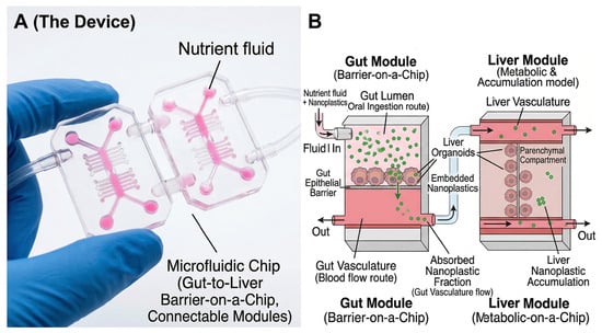
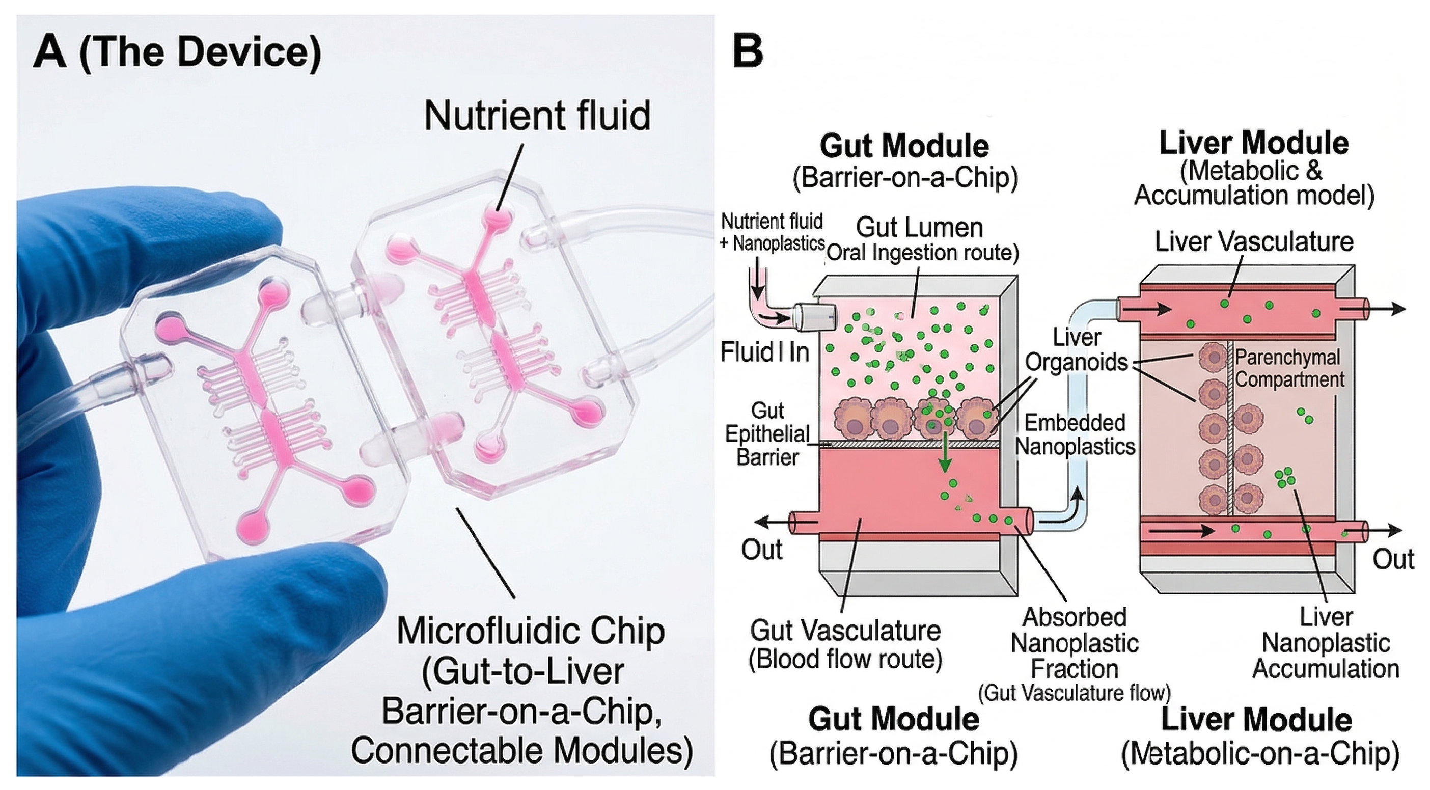
A concerted effort is required to answer urgent questions [35,78]. First, the development of human-relevant models in toxicology: organoids and organ-on-chip (OoC) platforms, illustrated in Figure 10, are physiologically relevant models that avoid animal use, and recent studies on brain, kidney, cardiac, and intestinal organoids have reported organ-specific toxicity [7,35]. Second, large-scale epidemiological research: what is most urgently needed is a Framingham Heart Study on microplastics which incorporates longitudinal human biomonitoring with clinical outcomes [14]. Third, developing quantitative risk assessment frameworks incorporating physiologically based pharmacokinetic (PBTK) models and identification of vulnerable populations [9]. Fourth, policy and public health interventions: supporting the UN Global Plastics Treaty, investing in biodegradable alternatives, and empowering individual risk reduction through water filtration, avoiding plastic food containers, and reducing indoor contamination [2,74].
Figure 10.
Next-Generation Toxicology: Organs-on-a-Chip. (A) The Device: A microfluidic “Organ-on-a-Chip” device designed to replicate human physiological fluid dynamics. (B) Microscopic Function: Within the chip’s channels, human-derived liver and gut organoids are co-cultured and exposed to a controlled stream of nanoplastics. This model allows for the study of particle translocation and organ-specific toxicity in a human-relevant system, reducing reliance on animal models. (created using Adobe Illustrator, Illustrae, and SciDraw).
The long-term health consequences of sustained MP/NP exposure represent one of the most critical unresolved questions in the field. Current evidence, predominantly derived from acute or subchronic animal studies, suggests that persistent low-grade inflammation, oxidative stress, and endocrine disruption are plausible chronic consequences of lifelong accumulation [8,25]. In cardiovascular biology, progressive plaque destabilization driven by plastic-induced inflammation is a biologically credible pathway linking decades of accumulation to increased risk of myocardial infarction and stroke; however, whether current human body burdens are sufficient to drive clinical events independently remains unknown [14]. In neurology, the parallel between rising brain plastic concentrations and the increasing prevalence of neurodegenerative conditions is hypothesis-generating but not causal; long-term cohort studies specifically enrolling participants with quantified plastic biomarkers and tracking cognitive outcomes are urgently needed [15,18]. Endocrine disruption from leached additives such as phthalates, BPA, and flame retardants represents a particularly insidious long-term threat given that sub-threshold chronic exposures to such agents are known to perturb hormonal axes involved in metabolism, reproduction, and neurodevelopment [11,38]. Finally, the accumulation of plastic particles in reproductive tissues raises concerns about multigenerational effects that transcend individual health outcomes [31,43]. Addressing these long-term dimensions will require substantially longer follow-up, better-standardized biomarker assays, and prospective cohort designs that treat plastic exposure as a measured exposure variable rather than an assumed constant [9,74].
12. Conclusions
The scientific knowledge on micro- and nanoplastics has developed at an accelerated pace, transforming the perception of a remote environmental threat into a substantive human health concern. Empirical detection across diverse human tissues and fluids leaves no doubt that these particles enter and persist within the body. They have been found in blood, lungs, placenta, and brain. Experimental evidence demonstrates they are not biologically inert—they are associated with oxidative stress, inflammatory responses, and organ-specific perturbations in animal and cell models. Importantly, observational data suggest a potential mechanistic basis for the reported association between plastic-laden arterial plaques and adverse cardiovascular events, though this association remains correlational and causality in humans has not been established. Similarly, elevated brain plastic concentrations raise hypotheses about neuroinflammatory contributions to neurodegeneration, but these require prospective validation.
There is encouraging development in detection technologies: AI and machine learning can identify plastics with 95–99% accuracy in automated mode using deep learning architectures such as dual-branch CNNs with attention mechanisms, and biosensor platforms can monitor in real-time with detection limits of 10−11 M. But gaps remain. Approaches are not standardized. Laboratory models are not realistic. There are few human lifelong studies. The inability to identify the smallest nanoplastics implies that body burden measurements likely represent gross underestimates.
Human-relevant models such as organoids and organ-on-chips are required in future research. We also require massive epidemiological trials that would monitor internal plastic quantities and health outcomes in the long run. Such evidence is required to develop multi-faceted public health policies and regulatory measures that can safeguard individuals against this long-term menace.
Author Contributions
Conceptualization, R.G. and S.G.; Writing—Original Draft Preparation, R.G. and R.C.; Visualization, S.G.; Writing—Review and Editing, S.G., M.R.D. and R.C.; Supervision, R.C. All authors have read and agreed to the published version of the manuscript.
Funding
This research did not receive any specific grant from funding agencies in the public, commercial, or not-for-profit sectors.
Institutional Review Board Statement
Not applicable.
Informed Consent Statement
Not applicable.
Data Availability Statement
All data come from the cited sources.
Acknowledgments
During the preparation of this manuscript, the authors used Adobe Illustrator, Illustrae, SciDraw, and Nano Banana 2 for the purposes of creating and refining scientific figures. The authors have reviewed and edited all outputs and take full responsibility for the content of this publication.
Conflicts of Interest
The authors declare no conflicts of interest.
Abbreviations
The following abbreviations are used in this manuscript:
| MPs | microplastics |
| NPs | nanoplastics |
| PE | polyethylene |
| PP | polypropylene |
| PS | polystyrene |
| PET | polyethylene terephthalate |
| PVC | polyvinyl chloride |
| PTFE | polytetrafluoroethylene |
| PA | polyamide |
| BBB | blood–brain barrier |
| FTIR | Fourier-transform infrared spectroscopy |
| μ-FTIR | micro-Fourier-transform infrared spectroscopy |
| μ-Raman | micro-Raman spectroscopy |
| Py-GC/MS | pyrolysis-gas chromatography/mass spectrometry |
| SEM-EDS | scanning electron microscopy with energy-dispersive X-ray spectroscopy |
| AI | artificial intelligence |
| ML | machine learning |
| DL | deep learning |
| CNN | convolutional neural network |
| SVM | support vector machine |
| EDCs | endocrine-disrupting chemicals |
| ROS | reactive oxygen species |
| IBD | inflammatory bowel disease |
| OoC | organ-on-chip |
| SPR | surface plasmon resonance |
| EIS | electrochemical impedance spectroscopy |
| XAI | explainable artificial intelligence |
| EPS | extracellular polymeric substance |
| PSS | polystyrene sulfonate |
| PMA | polymethacrylic acid |
| PBTK | physiologically based pharmacokinetic |
| TWPs | tire wear particles |
| EFSA | European Food Safety Authority |
| POPs | persistent organic pollutants |
| PCBs | polychlorinated biphenyls |
| PAHs | polycyclic aromatic hydrocarbons |
| BPA | bisphenol A |
References
- Geyer, R.; Jambeck, J.R.; Law, K.L. Production, Use, and Fate of All Plastics Ever Made. Sci. Adv. 2017, 3, e1700782. [Google Scholar] [CrossRef] [PubMed]
- Li, W.C.; Tse, H.F.; Fok, L. Plastic Waste in the Marine Environment: A Review of Sources, Occurrence and Effects. Sci. Total Environ. 2016, 566–567, 333–349. [Google Scholar] [CrossRef] [PubMed]
- Brandon, J.A.; Jones, W.; Ohman, M.D. Multidecadal Increase in Plastic Particles in Coastal Ocean Sediments. Sci. Adv. 2019, 5, eaax0587. [Google Scholar] [CrossRef] [PubMed]
- Eriksen, M.; Lebreton, L.C.M.; Carson, H.S.; Thiel, M.; Moore, C.J.; Borerro, J.C.; Galgani, F.; Ryan, P.G.; Reisser, J. Plastic Pollution in the World’s Oceans: More than 5 Trillion Plastic Pieces Weighing over 250,000 Tons Afloat at Sea. PLoS ONE 2014, 9, e111913. [Google Scholar] [PubMed]
- Gasperi, J.; Wright, S.L.; Dris, R.; Collard, F.; Mandin, C.; Guerrouache, M.; Langlois, V.; Kelly, F.J.; Tassin, B. Microplastics in Air: Are We Breathing It In? Curr. Opin. Environ. Sci. Health 2018, 1, 1–5. [Google Scholar] [CrossRef]
- Lusher, A.L.; McHugh, M.; Thompson, R.C. Occurrence of Microplastics in the Gastrointestinal Tract of Pelagic and Demersal Fish from the English Channel. Mar. Pollut. Bull. 2013, 67, 94–99. [Google Scholar] [CrossRef] [PubMed]
- Kayhan, S.; Yilmaz, E.; Tehli, O.; Izci, Y. Neurotoxicity of Microplastic Particles in the Human Brain: A Systematic Review. Turk. Neurosurg. 2025, 35, 817–829. [Google Scholar] [PubMed]
- Kadac-Czapska, K.; Ośko, J.; Knez, E.; Grembecka, M. Microplastics and Oxidative Stress—Current Problems and Prospects. Antioxidants 2024, 13, 579. [Google Scholar] [CrossRef] [PubMed]
- Koelmans, A.A.; Redondo Hasselerharm, P.E.; Mohamed Nor, N.H.; de Ruijter, V.N.; Mintenig, S.M.; Kooi, M. Risk Assessment of Microplastic Particles. Nat. Rev. Mater. 2022, 7, 138–152. [Google Scholar] [CrossRef]
- Amato-Lourenço, L.F.; Carvalho-Oliveira, R.; Júnior, G.R.; Dos Santos Galvão, L.; Ando, R.A.; Mauad, T. Presence of Airborne Microplastics in Human Lung Tissue. J. Hazard. Mater. 2021, 416, 126124. [Google Scholar] [CrossRef] [PubMed]
- Campanale, C.; Massarelli, C.; Savino, I.; Locaputo, V.; Uricchio, V.F. A Detailed Review Study on Potential Effects of Microplastics and Additives of Concern on Human Health. Int. J. Environ. Res. Public Health 2020, 17, 1212. [Google Scholar] [CrossRef] [PubMed]
- Leslie, H.A.; van Velzen, M.J.M.; Brandsma, S.H.; Vethaak, A.D.; Garcia-Vallejo, J.J.; Lamoree, M.H. Discovery and Quantification of Plastic Particle Pollution in Human Blood. Environ. Int. 2022, 163, 107199. [Google Scholar] [CrossRef] [PubMed]
- Ragusa, A.; Notarstefano, V.; Svelato, A.; Belloni, A.; Gioacchini, G.; Blondeel, C.; Zucchelli, E.; De Luca, C.; D’Avino, S.; Gulotta, A.; et al. Raman Microspectroscopy Detection and Characterisation of Microplastics in Human Breastmilk. Polymers 2022, 14, 2700. [Google Scholar] [CrossRef] [PubMed]
- Marfella, R.; Prattichizzo, F.; Sardu, C.; Fulgenzi, G.; Graciotti, L.; Spadoni, T.; D’Onofrio, N.; Scisciola, L.; La Grotta, R.; Frigé, C.; et al. Microplastics and Nanoplastics in Atheromas and Cardiovascular Events. N. Engl. J. Med. 2024, 390, 900–910. [Google Scholar] [CrossRef] [PubMed]
- Campen, M.; Nihart, A.; Garcia, M.; Liu, R.; Olewine, M.; El Hayek, E.; Howard, T.; Bleske, B.; Castillo, E.; Gonzalez-Estrella, J.; et al. Bioaccumulation of Microplastics in Decedent Human Brains. Nat. Med. 2025, 31, 1224–1231. [Google Scholar] [CrossRef]
- Prüst, M.; Meijer, J.; Westerink, R.H.S. The Plastic Brain: Neurotoxicity of Micro- and Nanoplastics. Part. Fibre Toxicol. 2020, 17, 24. [Google Scholar]
- European Food Safety Authority (EFSA). Presence of Microplastics and Nanoplastics in Food, with Particular Focus on Seafood. EFSA J. 2016, 14, e04501. [Google Scholar] [CrossRef] [PubMed]
- Ma, Y.; Yang, H.; Niu, S.; Guo, M.; Xue, Y. Mechanisms of Micro- and Nanoplastics on Blood–Brain Barrier Crossing and Neurotoxicity: Current Evidence and Future Perspectives. Neurotoxicology 2025, 109, 92–107. [Google Scholar] [PubMed]
- Chen, L.; Liu, Y.; Li, H.; Zhang, Y.; Wang, X.; Zhou, M.; Li, J.; Zhang, H. Size-Dependent Pulmonary Toxicity and Whole-Body Distribution of Inhaled Micro/Nanoplastic Particles in Male Mice from Chronic Exposure. Environ. Sci. Technol. 2025, 59, 6993–7003. [Google Scholar] [PubMed]
- Andrady, A.L. Microplastics in the Marine Environment. Mar. Pollut. Bull. 2011, 62, 1596–1605. [Google Scholar] [CrossRef] [PubMed]
- Hernandez, L.M.; Xu, E.G.; Larsson, H.C.E.; Tahara, R.; Maisuria, V.B.; Tufenkji, N. Plastic Teabags Release Billions of Microparticles and Nanoparticles into Tea. Environ. Sci. Technol. 2019, 53, 12300–12310. [Google Scholar] [CrossRef] [PubMed]
- Kudzin, M.H.; Piwowarska, D.; Festinger, N.; Chruściel, J.J. Risks Associated with the Presence of Polyvinyl Chloride in the Environment and Methods for Its Disposal and Utilization. Materials 2023, 17, 173. [Google Scholar] [CrossRef] [PubMed]
- Gómez-Sánchez, E.; Peñalver-Soler, R.M.; Almunia, N.; Pérez-Álvarez, M.C.; Luque, M.D.; Campillo, N.; Flores Monreal, A.; Arroyo-Manzanares, N.; Ruiz-Moreno, Y.; Jiménez, R.; et al. O-280 Unveiling the Hidden Danger: Detection and Characterisation of Microplastics in Human Follicular and Seminal Fluids. Hum. Reprod. 2025, 40, deaf097.280. [Google Scholar] [CrossRef]
- Wright, S.L.; Kelly, F.J. Plastic and Human Health: A Micro Issue? Environ. Sci. Technol. 2017, 51, 6634–6647. [Google Scholar] [CrossRef] [PubMed]
- Deng, Y.; Zhang, Y.; Lemos, B.; Ren, H. Tissue Accumulation of Microplastics in Mice and Biomarker Responses Suggest Widespread Health Risks of Exposure. Sci. Rep. 2017, 7, 46687. [Google Scholar] [CrossRef] [PubMed]
- Prata, J.C. Airborne Microplastics: Consequences to Human Health? Environ. Pollut. 2018, 234, 115–126. [Google Scholar] [CrossRef] [PubMed]
- Mason, S.A.; Welch, V.G.; Neratko, J. Synthetic Polymer Contamination in Bottled Water. Front. Chem. 2018, 6, 407. [Google Scholar] [CrossRef] [PubMed]
- Dris, R.; Gasperi, J.; Saad, M.; Mirande, C.; Tassin, B. Synthetic Fibers in Atmospheric Fallout: A Source of Microplastics in the Environment? Mar. Pollut. Bull. 2016, 104, 290–293. [Google Scholar] [CrossRef] [PubMed]
- Vianello, A.; Jensen, R.L.; Liu, L.; Vollertsen, J. Simulating Human Exposure to Indoor Airborne Microplastics Using a Breathing Thermal Manikin. Sci. Rep. 2019, 9, 8670. [Google Scholar] [CrossRef] [PubMed]
- Schwabl, P.; Köppel, S.; Königshofer, P.; Bucsics, T.; Trauner, M.; Reiberger, T.; Liebmann, B. Detection of Various Microplastics in Human Stool: A Prospective Case Series. Ann. Intern. Med. 2019, 171, 453–457. [Google Scholar] [PubMed]
- Zangene, S.; Morovvati, H.; Anbara, H.; Hye Khan, M.A.; Goorani, S. Polystyrene Microplastics Cause Reproductive Toxicity in Male Mice. Food Chem. Toxicol. 2024, 194, 115083. [Google Scholar] [CrossRef] [PubMed]
- Zytowski, E.; Mollavali, M.; Baldermann, S. Uptake and Translocation of Nanoplastics in Mono and Dicot Vegetables. Plant Cell Environ. 2025, 48, 134–148. [Google Scholar] [PubMed]
- Jin, Y.; Lu, L.; Tu, W.; Luo, T.; Fu, Z. Impacts of Polystyrene Microplastic on the Gut Barrier, Microbiota and Metabolism of Mice. Sci. Total Environ. 2019, 649, 308–317. [Google Scholar] [CrossRef] [PubMed]
- Gautam, R.; Jo, J.; Acharya, M.; Maharjan, A.; Lee, D.; Bahadur, K.C.P.; Kim, C.; Kim, K.; Kim, H.; Heo, Y. Evaluation of Potential Toxicity of Polyethylene Microplastics on Human Derived Cell Lines. Sci. Total Environ. 2022, 838, 156089. [Google Scholar] [CrossRef] [PubMed]
- Yuan, Q.; Liu, Y. Utilization of Intestinal Organoid Models for Assessment of Micro/Nano Plastic-Induced Toxicity. Front. Environ. Sci. 2023, 11, 1285536. [Google Scholar] [CrossRef]
- Kharaghani, D.; DeLoid, G.M.; He, P.; Swenor, B.; Bui, T.H.; Zuverza-Mena, N.; Tamez, C.; Musante, C.; Verzi, M.; White, J.C.; et al. Toxicity and Absorption of Polystyrene Micro-Nanoplastics in Healthy and Crohn’s Disease Human Duodenum-Chip Models. J. Hazard. Mater. 2025, 490, 137714. [Google Scholar] [CrossRef] [PubMed]
- Garcia, M.A.; Liu, R.; Nihart, A.; El Hayek, E.; Castillo, E.; Barrozo, E.R.; Suter, M.A.; Bleske, B.; Scott, J.; Forsythe, K.; et al. Quantitation and Identification of Microplastics Accumulation in Human Placental Specimens Using Pyrolysis Gas Chromatography–Mass Spectrometry. Toxicol. Sci. 2024, 199, 81–88. [Google Scholar] [PubMed]
- Hahladakis, J.N.; Velis, C.A.; Weber, R.; Iacovidou, E.; Purnell, P. An Overview of Chemical Additives Present in Plastics: Migration, Release, Fate and Environmental Impact during Their Use, Disposal and Recycling. J. Hazard. Mater. 2018, 344, 179–199. [Google Scholar] [CrossRef] [PubMed]
- Yan, Z.; Liu, Y.; Zhang, T.; Zhang, F.; Ren, H.; Zhang, Y. Analysis of Microplastics in Human Feces Reveals a Correlation between Fecal Microplastics and Inflammatory Bowel Disease Status. Environ. Sci. Technol. 2022, 56, 414–421. [Google Scholar] [CrossRef] [PubMed]
- Erni-Cassola, G.; Gibson, M.I.; Thompson, R.C.; Christie-Oleza, J.A. Lost, but Found with Nile Red: A Novel Method for Detecting and Quantifying Small Microplastics (1 mm to 20 μm) in Environmental Samples. Environ. Sci. Technol. 2017, 51, 13641–13648. [Google Scholar] [CrossRef] [PubMed]
- Hartmann, N.B.; Hüffer, T.; Thompson, R.C.; Hassellöv, M.; Verschoor, A.; Daugaard, A.E.; Rist, S.; Karlsson, T.; Brennholt, N.; Cole, M.; et al. Are We Speaking the Same Language? Recommendations for a Definition and Categorization Framework for Plastic Debris. Environ. Sci. Technol. 2019, 53, 1039–1047. [Google Scholar] [CrossRef] [PubMed]
- Silva, A.B.; Bastos, A.S.; Justino, C.I.L.; da Costa, J.P.; Duarte, A.C.; Rocha-Santos, T.A.P. Microplastics in the Environment: Challenges in Analytical Chemistry—A Review. Anal. Chim. Acta 2018, 1017, 1–19. [Google Scholar] [PubMed]
- Montano, L.; Raimondo, S.; Piscopo, M.; Ricciardi, M.; Guglielmino, A.; Chamayou, S.; Gentile, R.; Gentile, M.; Rapisarda, P.; Conti, G.O.; et al. First Evidence of Microplastics in Human Ovarian Follicular Fluid: An Emerging Threat to Female Fertility. Ecotoxicol. Environ. Saf. 2025, 291, 117868. [Google Scholar] [CrossRef] [PubMed]
- Zhou, L.; Ran, L.; He, Y.; Huang, Y. Mechanisms of Microplastics on Gastrointestinal Injury and Liver Metabolism Disorder (Review). Mol. Med. Rep. 2025, 31, 98. [Google Scholar] [CrossRef] [PubMed]
- Xie, P.; Li, P.; Zhu, X.; Chen, Y.; Zhang, L.; Wang, H.; Liu, J. Hepatotoxic of Polystyrene Microplastics in Aged Mice: Focus on the Role of Gastrointestinal Transformation and AMPK/FoxO Pathway. Sci. Total Environ. 2024, 917, 170471. [Google Scholar] [PubMed]
- Hirt, N.; Body-Malapel, M. Immunotoxicity and Intestinal Effects of Nano- and Microplastics: A Review of the Literature. Part. Fibre Toxicol. 2020, 17, 57. [Google Scholar]
- Khanam, M.; Uddin, M.K.; Kazi, J.U. Advances in Machine Learning for the Detection and Characterization of Microplastics in the Environment. Front. Environ. Sci. 2025, 13, 1573579. [Google Scholar] [CrossRef]
- Xie, L.; Ma, M.; Ge, Q.; Liu, Y.; Zhang, L. Machine Learning Advancements and Strategies in Microplastic and Nanoplastic Detection. Environ. Sci. Technol. 2025, 59, 8885–8899. [Google Scholar] [CrossRef] [PubMed]
- Enyoh, C.E.; Wang, Q. Automated Classification of Undegraded and Aged Polyethylene Terephthalate Microplastics from ATR-FTIR Spectroscopy Using Machine Learning Algorithms. J. Polym. Environ. 2024, 32, 4143–4158. [Google Scholar] [CrossRef]
- Choi, E.; Park, J.; Lim, H.; Song, Y. Development of a Machine-Learning Model for Microplastic Analysis in an FT-IR Microscopy Image. Bull. Korean Chem. Soc. 2024, 45, 379–384. [Google Scholar]
- He, M.; Tong, J.; Li, X.; Han, X.; Qin, Y.; Fang, R.; Chen, Z.; Gao, M. Research on Hybrid Microplastic Recognition Method Based on Dual-Branch Convolutional Neural Network Combined with Attention Mechanism. Microchem. J. 2025, 218, 115131. [Google Scholar] [CrossRef]
- Devipriya, K.; Tlija, M.; Chanumolu, K.; Kumar, V.; Jana, S.; Jana, C. Automated Micro-Plastic Detection and Classification Using Deep Convolution Neural Network Pre-Trained Models and Transfer Learning. AIP Adv. 2025, 15, 025207. [Google Scholar]
- Zhu, Z.; Parker, W.; Wong, A. Leveraging Deep Learning for Automatic Recognition of Microplastics (MPs) via Focal Plane Array (FPA) Micro-FT-IR Imaging. Environ. Pollut. 2023, 337, 122548. [Google Scholar] [PubMed]
- Tang, K.H.D. The Role of Artificial Intelligence in Microplastic Pollution Studies and Management. Recent Prog. Sci. Eng. 2025, 1, 016. [Google Scholar]
- Zhang, Y.; Zhang, D.; Zhang, Z. A Critical Review on Artificial Intelligence—Based Microplastics Imaging Technology: Recent Advances, Hot-Spots and Challenges. Int. J. Environ. Res. Public Health 2023, 20, 1150. [Google Scholar] [PubMed]
- Lorenzo-Navarro, J.; Castrillón Santana, M.; Santesarti, E.; De Marsico, M.; Martinez, I.; Raymond, E.; Gómez, M.; Herrera, A. SMACC: A System for Microplastics Automatic Counting and Classification. IEEE Access 2020, 8, 25249–25261. [Google Scholar] [CrossRef]
- Kalatzis, D.; Katsafadou, A.I.; Katsarou, E.I.; Chatzopoulos, D.C.; Kiouvrekis, Y. Explainable Artificial Intelligence for the Rapid Identification and Characterization of Ocean Microplastics. Microplastics 2025, 4, 51. [Google Scholar] [CrossRef]
- Bianco, V.; Memmolo, P.; Carcagnì, P.; Merola, F.; Paturzo, M.; Distante, C.; Ferraro, P. Microplastic Identification via Holographic Imaging and Machine Learning. Adv. Intell. Syst. 2020, 2, 1900153. [Google Scholar]
- Astray, G.; Soria-Lopez, A.; Barreiro, E.; Mejuto, J.C.; Cid-Samamed, A. Machine Learning to Predict the Adsorption Capacity of Microplastics. Nanomaterials 2023, 13, 1061. [Google Scholar] [CrossRef] [PubMed]
- Kida, M.; Pochwat, K.; Ziembowicz, S. Assessment of Machine Learning-Based Methods Predictive Suitability for Migration Pollutants from Microplastics Degradation. J. Hazard. Mater. 2024, 461, 132565. [Google Scholar] [PubMed]
- Li, J.; Jiang, Z.; Shu, L.; Li, X.; Wang, C.; Zhang, H. Machine Learning Models for Forecasting Microplastic Dynamics in China’s Coastal Waters. J. Hazard. Mater. 2025, 494, 138797. [Google Scholar] [CrossRef] [PubMed]
- Zhao, B.; Richardson, R.E.; You, F. Advancing Microplastic Analysis in the Era of Artificial Intelligence: From Current Applications to the Promise of Generative AI. Nexus 2024, 1, 100043. [Google Scholar] [CrossRef]
- Qiu, Y.; Mintenig, S.M.; Barchiesi, M.; Koelmans, A.A. Using Artificial Intelligence Tools for Data Quality Evaluation in the Context of Microplastic Human Health Risk Assessments. Environ. Int. 2025, 197, 109341. [Google Scholar] [CrossRef] [PubMed]
- Rivera-Rivera, D.M.; Quintanilla-Villanueva, G.E.; Luna-Moreno, D.; Sánchez-Álvarez, A.; Rodríguez-Delgado, J.M.; Cedillo-González, E.I.; Kaushik, G.; Villarreal-Chiu, J.F.; Rodríguez-Delgado, M.M. Exploring Innovative Approaches for the Analysis of Micro- and Nanoplastics: Breakthroughs in (Bio)Sensing Techniques. Biosensors 2025, 15, 44. [Google Scholar] [CrossRef] [PubMed]
- Daoutakou, M.; Kintzios, S. Biosensors for Micro- and Nanoplastics Detection: A Review. Chemosensors 2025, 13, 143. [Google Scholar] [CrossRef]
- Shabib, A.; Maraqa, M.; Mohammad, A.; Awwad, F. Design, Fabrication, and Application of Electrochemical Sensors for Microplastic Detection: A State-of-the-Art Review and Future Perspectives. Environ. Sci. Eur. 2025, 37, 38. [Google Scholar]
- Shi, K.; Chen, J.; Cheng, Y.; Song, J.; Li, Y.; Cheng, X.; Bai, X.; Chang, J.; Jiang, T. A Novel Label-Free Electrochemical Aptasensor for Sensitive and Selective Detection of Microplastics Based on Split gRNA with CRISPR/Cas12a-Mediated Cascade Strand Displacement. Sens. Actuators B Chem. 2025, 444, 138491. [Google Scholar]
- Gongi, W.; Touzi, H.; Sadly, I.; Ben Ouada, H.; Tamarin, O.; Ben Ouada, H. A Novel Impedimetric Sensor Based on Cyanobacterial Extracellular Polymeric Substances for Microplastics Detection. J. Polym. Environ. 2022, 30, 4738–4748. [Google Scholar] [CrossRef] [PubMed]
- Seggio, M.; Arcadio, F.; Radicchi, E.; Cennamo, N.; Zeni, L.; Bossi, A.M. Toward Nano- and Microplastic Sensors: Identification of Nano- and Microplastic Particles via Artificial Intelligence Combined with a Plasmonic Probe Functionalized with an Estrogen Receptor. ACS Omega 2024, 9, 18984–18994. [Google Scholar] [CrossRef] [PubMed]
- Arcadio, F.; Zeni, L.; Montemurro, D.; Eramo, C.; Di Ronza, S.; Perri, C.; D’Agostino, G.; Chiaretti, G.; Porto, G.; Cennamo, N. Biochemical Sensing Exploiting Plasmonic Sensors Based on Gold Nanogratings and Polymer Optical Fibers. Photonics Res. 2021, 9, 1397–1408. [Google Scholar] [CrossRef]
- Xiao, M.; Yang, Y.; Alahmadi, H.; Harbolic, A.; Moreno, G.M.; Yu, T.; Liu, J.; Guo, A.; Warner, G.R.; Stapleton, P.A.; et al. Rapid Detection of Microplastics and Nanoplastics in Seconds by Mass Spectrometry. J. Hazard. Mater. 2025, 493, 138322. [Google Scholar] [CrossRef] [PubMed]
- Zhou, H.; Vijver, M.G.; Peijnenburg, W.J.G.M. Electrochemical Sensing for Real-Time Monitoring of Nanoplastics-Induced Toxicity: Dynamic Measurements at the Exposure–Organism Interface. J. Hazard. Mater. 2025, 496, 139505. [Google Scholar] [PubMed]
- Hale, R.C.; Seeley, M.E.; La Guardia, M.J.; Mai, L.; Zeng, E.Y. A Global Perspective on Microplastics. J. Geophys. Res. Ocean. 2020, 125, e2018JC014719. [Google Scholar] [CrossRef]
- Senathirajah, K.; Attwood, S.; Bhagwat, G.; Carbery, M.; Wilson, S.; Palanisami, T. Estimation of the Mass of Microplastics Ingested—A Pivotal First Step towards Human Health Risk Assessment. J. Hazard. Mater. 2021, 404, 124004. [Google Scholar] [CrossRef] [PubMed]
- Kole, P.J.; Löhr, A.J.; Van Belleghem, F.G.A.J.; Ragas, A.M.J. Wear and Tear of Tyres: A Stealthy Source of Microplastics in the Environment. Int. J. Environ. Res. Public Health 2017, 14, 1265. [Google Scholar] [CrossRef] [PubMed]
- Tian, Z.; Zhao, H.; Peter, K.T.; Gonzalez, M.; Wetzel, J.; Wu, C.; Hu, X.; Prat, J.; Mudrock, E.; Hettinger, R.; et al. A Ubiquitous Tire Rubber-Derived Chemical Induces Acute Mortality in Coho Salmon. Science 2021, 371, 185–189. [Google Scholar] [PubMed]
- De Falco, F.; Gullo, M.P.; Gentile, G.; Di Pace, E.; Cocca, M.; Gelabert, L.; Brouta-Agnésa, M.; Rovira, A.; Escudero, R.; Villalba, R.; et al. Evaluation of Microplastic Release Caused by Textile Washing Processes of Synthetic Fabrics. Environ. Pollut. 2018, 236, 916–925. [Google Scholar] [CrossRef] [PubMed]
- Abdessalam, S.; Hardy, T.J.; Pershina, D.; Yoon, J.Y. A Comparative Review of Organ-on-a-Chip Technologies for Micro- and Nanoplastics versus Other Environmental Toxicants. Biosens. Bioelectron. 2025, 281, 117476. [Google Scholar]

Disclaimer/Publisher’s Note: The statements, opinions and data contained in all publications are solely those of the individual author(s) and contributor(s) and not of MDPI and/or the editor(s). MDPI and/or the editor(s) disclaim responsibility for any injury to people or property resulting from any ideas, methods, instructions or products referred to in the content. |
© 2026 by the authors. Licensee MDPI, Basel, Switzerland. This article is an open access article distributed under the terms and conditions of the Creative Commons Attribution (CC BY) license.